ORIGINAL ARTICLE
PLASMA APELIN LEVEL IS DECREASED IN PATIENTS WITH CORONARY ARTERY DISEASE
Hiroaki Yokoyama
1),Shin Saito
1),Takumi Higuma
1),Hiroyuki Hanada
1),Tomohiro Osanai
1), Kazuyuki Daitoku
2),Ikuo Fukuda
2)and Ken Okumura
1)Abstract Adipose tissue secretes various bioactive molecules (adipokines), and apelin is one kind of adipokines.
Recently, it was shown that plasma apelin level is decreased in patients with chronic heart failure, and apelin might play an important role in the pathogenesis of cardiovascular disease. However, plasma apelin level in coronary artery disease (CAD) or other heart disease such as valvular heart disease (VHD) has not been elucidated. We enrolled 31 patients with CAD and 14 patients with VHD who underwent elective cardiac surgery. We also examined plasma apelin level in 20 healthy subjects (Control). Blood samples were obtained before the surgery. Paired samples of visceral and subcutaneous adipose tissues were harvested during surgery. Plasma apelin level was lower in both CAD and VHD than in Control. When compared between CAD and VHD, it was lower in CAD than in VHD, and was not aff ected by treatment with HMG-CoA reductase inhibitors (statins) which was shown to increase adiponectin level.
Left ventricular ejection fraction (LVEF) was lower in CAD than in VHD. There was no correlation between plasma apelin level and LVEF. Gene expression of apelin in visceral adipose tissue was higher than that in subcutaneous adipose tissue, but it was similar between two groups. These suggest that plasma apelin level was decreased in patients with cardiac diseases, especially in those with CAD. Its role in the pathophysiology of CAD remains to be elucidated.
Hirosaki Med.J. 61:58―64,2010
Key words: apelin; coronary atherosclerosis; adipose tissue; chronic heart failure
原 著
冠動脈疾患における血中アペリン濃度の減少
横 山 公 章
1)齋 藤 新
1)樋 熊 拓 未
1)花 田 裕 之
1)長 内 智 宏
1)大 徳 和 之
2)福 田 幾 夫
2)奥 村 謙
1)抄録 脂肪細胞は様々な生理活性をもつアディポカインを分泌するが,アペリンはその一つである.血中アペリン濃 度は心不全患者で低下することが報告され,心血管疾患で重要な役割を演じている可能性がある.しかし,冠動脈疾 患(CAD)や弁膜疾患(VHD)などの心疾患における血中アペリン濃度は解明されていない.我々は待機的手術を行った CAD 患者31例と VHD 患者14例を対象に検討した.心疾患のない健常例20例においても検討した.術前に採血を行い,
術中に内臓・皮下脂肪を採取した.血中アペリン濃度は健常例に比して CAD と VHD では低下していた.アペリン濃度 を CAD と VHD 間で比較すると,CAD で有意に低下しており,HMG-CoA 還元酵素阻害薬(スタチン)の影響を受けな かった.左室収縮能(LVEF)は CAD で低下していた.血中アペリン濃度と LVEF の相関関係は認めなかった.内臓脂 肪のアペリン mRNA 発現は皮下脂肪より増加していたが,2 群間に差を認めなかった.以上より,血中アペリン濃度は CAD で低下するが,その病態生理学的意義については今後の検討を要する.
弘前医学 61:58―64,2010
キーワード:アペリン;冠動脈の動脈硬化;脂肪細胞;慢性心不全
1)Department of Cardiology Hirosaki University Graduate School of Medicine, Hirosaki, Japan
2)Department of Thoracic and Cardiovascular Surgery, Hirosaki University Graduate School of Medicine, Hirosaki, Japan
Correspondence: T. Osanai
Received for publication, December 14, 2009 Accepted for publication, January 4, 2010
1)弘前大学大学院医学研究科循環呼吸腎臓内科学
2)弘前大学大学院医学研究科胸部心臓血管外科学
別刷請求先:長内智宏 平成21年12月14日受付 平成22年 1 月 4 日受理
1. Introduction
It is now widely recognized that adipose tissue is not only a reservoir for energy storage but also an endocrine tissue. Adipose tissue secretes various bioactive molecules (adipokines)
including adiponectin, tumor necrosis factor
(TNF)-
α, and interleukin (IL)-6. Apelin, onekind of adipokines, is a novel peptide identified as the endogenous ligand to the angiotensin receptor-like 1 (APJ) receptor, which resembles the angiotensin receptor, but does not bind to angiotensin II
1). It is expressed in the human heart, lung, kidney, adrenal gland, adipose tissue, and large conduit vessels including coronary artery
2). In contrast to angiotensin, a potent vasopressor and anti-diuretic hormone, apelin lowers blood pressure via a nitric oxide- dependent mechanism, produces diuresis by inhibition of arginine vasopressin activity and release, and has a positive inotropic eff ect
1,3-8). Recently, it was shown that plasma apelin level is decreased in patients with chronic heart failure (CHF)
3,9), indicating that apelin might play an important role in the pathogenesis of cardiovascular disease. There are many studies suggesting the relation between plasma apelin and CHF, but there are only few studies suggesting the relation between plasma apelin and atherosclerosis. Hashimoto et al. reported that APJ might play an important role in the progression of oxidative stress-linked atherosclerosis in mice, and might be a therapeutic target for atherosclerosis
10). To our knowledge, plasma apelin level in coronary artery disease (CAD) or other heart disease such as valvular heart disease (VHD) , and the gene expression of apelin in the visceral and subcutaneous adipose tissues have not been investigated. The aim of this study is to investigate the plasma apelin level in the patients with CAD and VHD, and the gene expression in the visceral and subcutaneous adipose tissues, and to clarify the relationship between atherosclerosis
and apelin.
2. Methods
2.1. Patient’s profi le
All patients gave written, informed consent before the study. The study protocol was approved by the Ethics Committee on Human Research at our institution (2008-151). This study enrolled 45 patients undergoing cardiac surgery at Hirosaki University Hospital. There were 31 patients with CAD (mean age 66 years, ranging from 50 to 81) who underwent elective coronary artery bypass graft surgery (CABG)
(CAD group), and 14 patients with VHD (mean age 62 years, ranging 42 to 77) who underwent elective valve replacement or plasty (VHD group). The VHD patients were confirmed to have no CAD by coronary angiography. No patients had severe renal or hepatic dysfunction, or any history of neoplastic or autoimmune diseases. We further enrolled 20 healthy subjects
(mean age 39 years, ranging from 27 to 61)
who had no risk factors such as hypertension, diabetes mellitus and dyslipidemia, and were treated with no medications (Control subjects).
All control subjects gave written, informed consent before the study.
Patients treated with antihypertensive agents or those with blood pressure > 140/90 mmHg were diagnosed as having hypertension. Diabetes mellitus was diagnosed according to the criteria of the World Health Organization
11). Body mass index (BMI) was calculated and was used to stratify obesity degree. Insulin sensitivity was assessed by homeostasis model assessment
(HOMA) index. Serum total cholesterol, low-
density lipoprotein (LDL) cholesterol, high-
density lipoprotein (HDL) cholesterol, triglyceride,
and fasting blood sugar concentrations were
measured using standard methods. The plasma
level of B-type natriuretic peptide (BNP) was
measured by immunoradiometric assay (Shionoria
BNP RIA kit, Tokyo, Japan).
2.2. Blood sampling and measurement of plasma apelin
Blood samples were obtained from the peripheral vein before the induction of general anesthesia and the infusion of any perioperative metabolic substrates at surgery. After cen- trifugation at 3000 × g for 15 min at 4 °C, the supernatants were stored at -80 °C until use.
Plasma apelin level was determined using a commercially available enzyme immunoassay
(Phoenix Pharmaceuticals, CA, USA) according to the manufacturerʼs instruction.
2.3. Adipose tissue sampling
Paired samples of the visceral and subcutaneous adipose tissues were harvested during surgery.
In this study, adipose tissue beneath the sternum including the thymus tissue replaced by adipose tissue was used as the visceral adipose tissue
12). To measure the gene expression, the sample was immediately submerged in RNAlater
TMfor storage to stabilize and protect cellular RNA. These samples were stored at -30 ° C until use.
2.4. Measurement of gene expression of apelin in adipose tissues
Adipose tissue RNA was extracted using a RNeasy Lipid Tissue minikit (Qiagen, GmbH, Hiden, Germany) , according to the manufacturerʼs instruction. A two -step rea l time reverse transcription-polymerase chain reaction (RT- PCR) was carried out according to the instruction supplied with the TaqMan Gold RT-PCR kit
(Applied Biosystems Foster City, CA, USA) . All oligonucleotide primers and TaqMan probes for human apelin and glyceraldehydes-3-phosphate dehydrogenase (GAPDH) were purchased from Applied Biosystems. Values were averaged from duplicate data and normalized with human GAPDH.
2.5. Statistical analysis
All data were expressed as mean ± SEM.
The data were analyzed with unpaired Studentʼs
t test between two groups. One way analysis ofvariance (ANOVA) was used for the comparison of 3 or more variables followed by Tukey- Kramerʼs post hoc test. Probability values < 0.05 were considered to be statistically signifi cant.
3. Results
3.1. Clinical characteristics of subjects
None of age, gender, BMI, and risk factors such as hypertension, diabetes mellitus, smoking, and medications including angiotensin converting enzyme inhibitor (ACE-I) , angiotensin II type 1 receptor blocker (ARB) , and diuretics differed between two groups (Table 1) . Aspirin,
β-blockers, and HMG-CoA reductase inhibitors (statins) were less administered to VHD patients compared with CAD patients. The incidence of dyslipidemia was
Table 1 Comparison of clinical profi les
CAD VHD
(n=31) (n=14)
Age 66± 2 62± 3
Male (n, %) 28 ( 90%) 12 (86%)
BMI (kg/m2) 24.4± 0.6 23.8± 0.8 Risk factor (n, %)
hypertension 11 ( 87%) 10 (85%)
diabetes mellitus 13 ( 42%) 3 (21%)
dyslipidemia 29 ( 94%)** 2 (14%)
smoking 16 ( 52%) 8 (57%)
Medication (n, %)
aspirin 31 (100%)** 1 ( 7%)
ACE-I 4 ( 13%) 4 (29%)
ARB 20 ( 65%) 6 (43%)
β-blocker 26 ( 84%)** 4 (29%)
statin 19 ( 61%)** 0 ( 0%)
diuretics 14 ( 45%) 8 (57%)
LVEF (%) 48± 2* 57± 3
CAD, patients with coronary artery disease; VHD, patients with valvular heart disease
ACE-I, angiotensin converting enzyme inhibitor; ARB, angiotensin II type 1 receptor blocker; BMI, body mass index; LVEF, left ventricular ejection fraction; statin, HMG-CoA reductase inhibitor
Variables are shown as mean ± S.E.M.
* P<0.05 vs VHD, ** P<0.01 vs VHD
higher in CAD patients than in VHD patients.
Left ventricular ejection fraction (LVEF) assessed by left ventriculography (LVG) was lower in CAD than in VHD. There were no diff erences in the serum levels of total cholesterol, triglyceride, blood sugar, HbA1c, HOMA index, BNP and high sensitive C-reactive protein (hs-CRP) between two groups (Table 2) . Serum levels of LDL- cholesterol and HDL-cholesterol in CAD patients were lower than those in VHD patients.
3.2. Level of plasma apelin
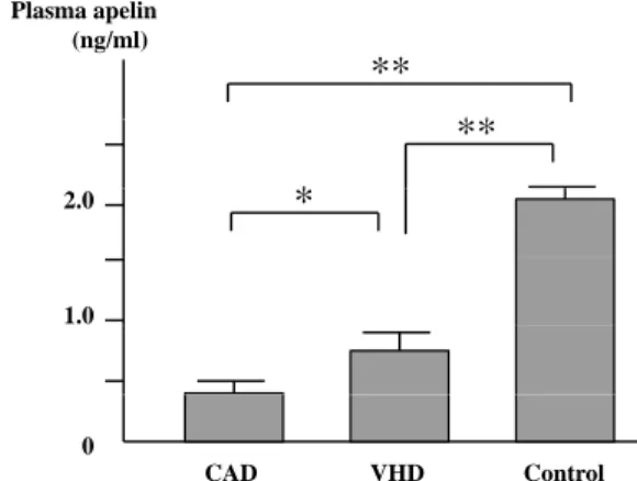
As shown in Figure 1, the plasma apelin level was significantly lower in CAD and VHD patients compared with Control subjects, respectively (p<0.01). Moreover, its level was significantly lower in CAD patients compared with VHD patients (p<0.05). There was no difference in plasma apelin level between CAD patients with and without
β-blockers (
β-blockers
(+); 0.41±0.06 vs (-); 0.45±0.06 ng/ml). Statins also did not affect plasma apelin level in CAD patients (Statin (+); 0.41±0.06 vs (-); 0.43±0.11 ng/ml).
3.3. Gene expression of apelin in the visceral and subcutaneous adipose tissues
As shown in Figure 2, apelin gene expression was higher in visceral adipose tissue than in subcutaneous adipose tissue in VHD patients
(P<0.05). The gene expression in visceral ad ipose t issue tended to be h igher t ha n that in subcutaneous tissue in CAD patients
(P=0.1). There was no diff erence in apelin gene
Table 2 Comparison of biochemical parameters
CAD VHD
(n=31) (n=14)
TC (mg/dl) 167± 6 189± 11
TG (mg/dl) 147± 12 112± 14
LDL-C (mg/dl) 93± 4* 113± 11
HDL-C (mg/dl) 42.6± 1.9** 54.0± 3.4
FBS (mg/dl) 114± 8 100± 4
HbA1c (%) 6.0± 0.2 5.7± 0.4
HOMA index 2.3± 0.5 1.5± 0.4
BNP (pg/ml) 116± 32 163± 43
hs-CRP (μg/ml) 275± 77 137± 52
CAD, patients with coronary artery disease; VHD, patients with valvular heart disease
BNP, B-type natriuretic peptide; FBS, fasting blood sugar;
HDL-C, high-density lipoprotein cholesterol; HOMA index, homeostasis model assessment index; hs-CRP, high sensitive C-reactive protein; LDL-C, low-density lipoprotein cholesterol; TC, total cholesterol; TG, triglyceride
Variables are shown as mean ± S.E.M.
* P<0.05 vs VHD, ** P<0.01 vs VHD
Plasma apelin (ng/ml) ( g )
**
**
*
**
*
2.0
1.0
0
CAD VHD Control
Figure 1 Comparison of the plasma apelin level among the patients with coronary artery disease
(CAD) and valvular heart disease (VHD), and healthy subjects (Control). The plasma apelin level was lower in CAD and VHD patients than that in Control subjects respectively, and its level was lower in CAD patients than that in VHD. *P<0.05, **P<0.01.
sue) (%)
*
A eral tis 200 ( )
*
*
H mRNA he visce
*
GAPDH up in th 100 100 into G HD grouof apel the VH
0 CAD VHD
Ratioo (% of
CAD VHD (visceral tissue) (subcutaneous tissue) (visceral tissue) (subcutaneous tissue) Figure 2 Gene expressions of apelin normalized by human
glyceraldehydes-3-phosphate dehydrogenase
(GAPDH) in the visceral and subcutaneous adipose tissues. Boxes show medians and interquartile ranges, with whiskers representing the 10th/90th percentiles. *P<0.05.
expression in visceral and subcutaneous adipose tissue between two groups, respectively.
3.4. Relationship between left ventricular function and plasma apelin level
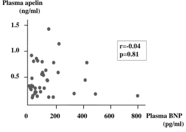
There were no significant correlations between plasma apelin level and plasma BNP level (Figure 3) or LVEF (Figure 4).
4. Discussion
4.1. Plasma apelin level and coronary atherosclerosis The major findings of this study were that the plasma apelin level was significantly lower in CAD and VHD patients than that in Control
subjects respectively, and that its level was lower in CAD patients than that in VHD patients.
Our result that BNP level was increased in CAD and VHD patients respectively supports the previous fi ndings that plasma apelin level is decreased in patients with CHF
3,9). We also found that LVEF in CAD patients was lower than that in VHD patients. A recent study showed a positive correlation between LVEF and plasma apelin level in patients with CHF
3). It seems reasonable to presume that the diff erence of left ventricular function explains the finding that plasma apelin level in CAD patients was lower than that in VHD patients, although there was no significant correlation between apelin level and LVEF. The disparity of the relation between apelin level and LVEF may be dependent on the small number of subjects and the administration of medicines for CHF. Second, the decrease in plasma apelin level in CAD patients is likely to be dependent on coronary atherosclerosis.
Recently, Li et al. showed that the plasma apelin level in patients with stable angina pectoris was lower than in controls
13). Since VHD patients were confirmed to have no CAD by coronary angiography, the difference in the degree of coronary atherosclerosis between CAD and VHD patients might be associated with the plasma apelin level. Our result that hs-CRP tended to be higher in CAD patients than in VHD patients also supports this idea. Overall, plasma apelin level appears to be a useful predictor of the progression of coronary atherosclerosis.
The difference of medication between two groups might affect the plasma apelin level.
However, there was no difference in plasma apelin level between CAD patients with and without
β-blockers. Statins also did not affect plasma apelin level in CAD patients. Recent study has shown that LDL-cholesterol lowering in healthy subjects with isolated dyslipidemia results in an increase in plasma apelin level
14). Therefore, the decrease in plasma apelin level is
Plasma apelin ( / ) 1.5
(ng/ml)
r= 0 04
1.0 r=-0.04
p=0.81 0.5
200 400 600 800 Plasma BNP ( / l) 0
(pg/ml)
i 3
Plasma apelin (ng/ml) 1 5
(ng/ml)
r=0 065
1.5 r=0.065
p=0.870 1.0
0.5
0
20 30 45 60 LVEF (%)
Figure 3 Correlation between plasma apelin level and plasma B-type natriuretic peptide (BNP) in the combination of coronary artery disease (CAD)
and valvular heart disease (VHD) patients.
Figure 4 Correlation between plasma apelin level and left ventricular ejection fraction (LVEF) in the combination of coronary artery disease (CAD)
and valvular heart disease (VHD) patients.
independent of the diff erence in medication.
4.2. Gene expression of apelin in adipose tissue We investigated apelin gene expression in visceral and subcutaneous adipose tissues, but no difference was found between two groups.
Therefore, the decrease in plasma apelin level is independent of apelin gene expression in adipose tissue. Apelin was identifi ed from bovine stomach extracts for the first time and was known existing in various tissues such as central nervous system, heart, lung, stomach, pancreas, adrenal gland and adipose tissue. Boucher et al. showed that the gene expression of apelin in the heart and the kidney is higher than that in adipose tissue
15). Moreover, recent study showed that apelin gene expression is 200-fold higher in atrial tissue than in ventricular tissue, suggesting that the heart may be the major source of circulating apelin in humans
9). There was no difference in apelin gene expression in visceral and subcutaneous adipose tissue between two groups, respectively. However, to our knowledge, this is the fi rst report indicating that gene expression of apelin in visceral adipose tissue is higher than that in subcutaneous adipose tissue in humans.
4.3. Apelin as the therapeutic target of chronic heart failure
The plasma apelin level in CAD patients was decreased compared with that in VHD patients, and LVEF in CAD patients was also lower than that in VHD patients. In rats with CHF due to post-myocardial infarction, apelin infusion restores EF and increases cardiac output
16). Therefore, apelin might be useful for not only the predictor of coronary atherosclerosis but also the therapeutic target of CHF. Further studies are needed to establish the effectiveness and safety of apelin infusion for the therapy of CHF in humans.
4.4. Study limitations
We showed that plasma apelin level was decreased in patients with CA D, but the major source of circulating apelin still remains unclear. Since the heart is a possible candidate for the major source, further studies for apelin expression in the heart are needed.
5. Conclusions
Plasma apelin level in CAD patients was lower than that in VHD patients, and it may be associated with coronary atherosclerosis. Gene expression of apelin in visceral adipose tissue was higher than that in subcutaneous adipose tissue in humans. However, adipose tissue might not be the major source of plasma apelin.
References
1)Tatemoto K, Hosoya M, Habata Y, Fujii R, Kakegawa T, Zou MX, Kawamata Y, et al. Isolation and characterization of a novel endogenous peptide ligand for the human APJ receptor. Biochem Biophys Res Commun 1998;251:471-6.
2)Kleinz MJ, Davenport AP. Emerging roles of apelin in biology and medicine. Pharmacol Ther 2005;107:198-211.
3)Chong KS, Gardner RS, Morton JJ, Ashley EA, McDonagh TA. Plasma concentrations of the novel peptide apelin are decreased in patients with chronic heart failure. Eur J Heart Fail 2006;8:355- 60.
4)Seyedabadi M, Goodchild AK, Pilowsky PM. Site- specific effects of apelin-13 in the rat medulla oblongata on arterial pressure and respiration.
Auton Neurosci 2002;101:32-8.
5)Tatemoto K, Takayama K, Zou MX, Kumaki I, Zhang W, Kumano K, Fujimiya M. The novel peptide apelin lowers blood pressure via a nitric oxide-dependent mechanism. Regul Pept 2001;99:
87-92.
6)Cheng X, Cheng XS, Pang CC. Venous dilator eff ect of apelin, an endogenous peptide ligand for the orphan APJ receptor, in conscious rats. Eur J Pharmacol 2003;470:171-5.
7)Ishida J, Hashimoto T, Hashimoto Y, Nishiwaki S, Iguchi T, Harada S, Sugaya T, et al. Regulatory roles for APJ, a seven-transmembrane receptor related to angiotensin-type 1 receptor in blood pressure in vivo. J Biol Chem 2004;279:26274-9.
8)De Mota N, Reaux-Le Goazigo A, El Messari S, Chartrel N, Roesch D, Dujardin C, Kordon C, et al. Apelin, a potent diuretic neuropeptide counter- acting vasopressin actions through inhibition of vasopressin neuron activity and vasopressin release.
Proc Natl Acad Sci USA 2004;101:10464-9.
9)Foldes G, Horkay F, Szokodi I, Vuolteenaho O, Ilves M, Lindstedt KA, Mayranpaa M, et al.
Circulating and cardiac levels of apelin, the novel ligand of the orphan receptor APJ, in patients with heart failure. Biochem Biophys Res Commun 2003;308:480-5.
10)Hashimoto T, Kihara M, Imai N, Yoshida S, Shimoyamada H, Yasuzaki H, Ishida J, et al.
Requirement of apelin-apelin receptor system for oxidative stress-linked atherosclerosis. Am J Pathol 2007;171:1705-12.
11)Prevention of diabetes mellitus. Report of WHO
Study Group. World Health Organ Tech Rep Ser 1994;844:1-100.
12)Saito S, Fujiwara T, Matsunaga T, Minagawa K, Fukui K, Fukuda I, Osanai T, et al. Increased adiponectin synthesis in the visceral adipose tissue in men with coronary artery disease treated with pravastatin: A role of the attenuation of oxidative stress. Atherosclerosis 2008;199:378-83.
13)Li Z, Bai Y, Hu J. Reduced apelin levels in stable angina. Intern Med 2008;47:1951-5.
14)Tasci I, Erdem G, Ozgur G, Tapan S, Dogru T, Genc H, Acikel C, et al. LDL-cholesterol lowering increases plasma apelin in isolated hypercholes- terolemia. Atherosclerosis 2009;204:222-8.
15)Boucher J, Masri B, Daviaud D, Gesta S, Guigne C, Mazzucotelli A, Castan-Laurell I, et al. Apelin, a newly identifi ed adipokine up-regulated by insulin and obesity. Endocrinology 2005;146:1764-71.
16)Berry MF, Pirolli TJ, Jayasankar V, Burdick J, Morine KJ, Gardner TJ, Woo YJ. Apelin has in vivo inotropic eff ects on normal and failing hearts.
Circulation 2004;110(11 Suppl 1):II187-93.