NK-ACTIVATING DENDRITIC CELLS ARE ELICITED BY STIMULATION WITH TOLL-LIKE RECEPTOR 3
Tsukasa Seya
1,2),Misako Matsumoto
1,2),Takashi Ebihara
1)and Takashi Akazawa
2)Abstract Double-stranded (ds)RNA-recognition receptors reside in the cytoplasm and membranes of cells. These receptors are implicated in the differential screening of microbes by the host. Myeloid dendritic cells (mDCs)
recognize and respond to polyI:C, an analog of dsRNA, by endosomal TLR3 and cytoplasmic MDA5. NK cells are induced in vivo by the administration of polyI:C to mice and in vitro are reciprocally activated by mDCs, although the molecular mechanisms as yet undetermined. Here, we show that the TLR adapter TICAM-1 (TRIF) participates in mDC-derived antitumor NK activation. In a syngeneic mouse tumor implant model, intraperitoneal administration of polyI:C led to the retardation of tumor growth, which eff ect relied largely on NK activation. This NK-dependent tumor regression did not occur in TICAM-1-/- or IFNAR-/- mice, while a normal NK antitumor response was induced in PKR-/-, MyD88-/-, IFN-β-/- and wild-type mice. IFNAR was a prerequisite for the induction of IFN-α/β and TLR3. The lack of TICAM-1 did not aff ect IFN production but resulted in unresponsiveness to IL-12 production, mDC maturation and polyI:C-mediated antitumor activity. This NK activation required NK-mDC contact in in vitro transwell analysis. NK- antitumor activity was successfully introduced into tumor-implanted mice by transferring mDCs expressing TICAM-1.
Implanted tumor growth in IFNAR-/- mice was retarded by adoptively transferring polyI:C-treated TICACM-1-positive mDCs but not TICAM-1-/- mDCs. Thus, TICAM-1 rather than MDA5 in mDCs critically facilitated mDC-NK contact and activation of antitumor NK, resulting in the regression of low MHC-expressing tumors
Hirosaki Med.J. 59, Supplement:S43―S51,2007
Key words: natural killer cells
(NK) ; dendritic cells; Toll-like receptor (TLR) ; TICAM-1 (TRIF) ; adjuvant
1)Department of Microbiology and Immunology, Hokkaido University Graduate School of Medicine, Kita-15, Nishi-7, Kita-ku Sapporo 060-8638 Japan,
2)Department of Immunology, Osaka Medical
Center for Cancer and Cardiovascular Diseases, Nakamichi 1-3-2, Higashinari-ku, Osaka 537-8511 Japan
Email: [email protected]
Introduction
Microbial components that activate the host immune system have been designated as adjuvants
1). Infections usually raise potent immune responses in hosts because pathogens have pattern molecules that serve as adjuvants.
They enhance antibody (Ab) production, cytotoxic T cells (CTL) and NK cell activation.
Spontaneously occurring tumor lacks adjuvancy since the tumor is intrinsic origin with no pathogenic pattern molecules.
Enhancing host immunity and increasing tumor antigenicity have long been goals of immunotherapy. Rosenberg et al.
2), summarized the results of 440 cancer patients who received a peptide vaccine. The overall objective response
rate for all vaccine treatments was just 2.6%
2). Although their criteria for clinical objective response were strict, the results clearly refl ect a limited effectiveness of the sole peptide vaccine therapy approach. Tumor cells have often diminished MHC class I
3), thereby circumventing the host immune surveillance system. They suggested further technical exploration would be required to est abl ish ef fect ive tumor immunotherapy. Adjuvants are known to largely serve as ligands for Toll-like receptors
(TLRs) on myeloid dendritic cells (mDCs) , which engage antigen presentation. Thus, supplement of adjuvants to tumor antigen may have the potential to compensate for the weakness of the peptide vaccine therapy.
We have studied the profiles of effector
induction in mDCs stimulated with adjuvants
4). CTL mainly eliminate tumors with high levels of MHC class I proteins, whereas NK cells target tumors with low MHC levels. In human and mouse the BCG-cell-wall skeleton (CWS) acts as a TLR2/4 agonist and induces mDC maturation followed by tumor-specific CTL in a syngeneic tumor implantation model if the tumors express MHC class I
5). Tumor regression and CTL induction largely relied on the MyD88 adapter- dependent pathway of TLR
5). Exogenously- added tumor antigen (Ag) and adjuvant induce the mDCs cross-priming which is required for MHC class I-mediated Ag presentation and CTL induction
6). However, NK cells are barely activated in wild-type mice via the TLR2/4- MyD88 responses
5). Thus, MyD88-independent cellular responses are crucial in NK-mediated MHC-negative population of tumor cytotoxicity.
The TLR3 agonist polyI:C and TLR3 signaling in mDCs are able to regress tumors
6,7). TLR3 links TICAM-1 (or TRIF) in the cytoplasmic domain TIR (Toll-IL-1R homology domain) for signal transmission
8,9). This adapter is unique in possessing two arrays of signals that activate two transcription factors, NF-κB and IRF-3
8-10). The latter is a strong inducer of type I IFN
(interferon), particularly IFN-β
10 -12). Recent reports suggest an important role for type I IFN in p53 induction
13)and cancer immunoediting
14). Type I IFN has been reported to be an inducer of various NK functions in vitro
15). Amplifi cation of type I IFN production by IFNAR governs the expression of a number of IFN-inducible genes
16). These genes may be crucial for the functional modulation of mDCs.
Here we have investigated the mechanism whereby exogenously administered polyI:C induces tumor regression in gene-disrupted mice. We noticed that the retardation in the growth of MHC-negative tumors largely depended on the NK activity induced by polyI:C-stimulated mDCs.
Using TICAM-1 (TRIF)
-/-C57BL/6 mice
17)and a
syngeneic tumor implant model, this NK activation was found to depend on direct contact of NK cells to mDCs, where NK activation is controlled by the TICAM-1 pathway of TLR in mDCs.
Retardation of tumor growth by NK cells in polyI:C-treated mice
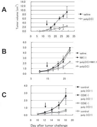
Using a C57BL/6-B16 syngeneic tumor-implant model, we evaluated the antitumor activity of polyI:C, which was injected intraperitoneally twice a week (Figure 1A) . Tumor growth was suppressed in the group that received polyI:
Figure 1 PolyI: C induces NK-medaited MHC class I-negative tumor regression. Panel A: Tumor- implantation model for evaluation of polyI:
C a nt it u mor a ct iv it y. PolyI : C (2 5 0 μg intraperitoneally injected twice a week) caused antitumor effect on B16D8 cells (MHC class I-negative) implanted into C57BL/6 mice.
Arrow indicates the start point of polyI:C treatment (tumor average size>0.8 cm3). Panel B, C: NK is an eff ecter for poly(I:C)-mediated antitumor activity. Mice were challenged with B16D8 cells and intraperitoneally injected with anti-NK1.1 (B) or anti-CD8β ascites (C). Antibody and polyI:C treatment was repeated twice a week from day 10.
C when the tumor cells barely expressed MHC class I.
PolyI:C-mediated suppression of tumor growth was terminated when NK activity in mice was blocked by an injection of NK1.1 Ab (Figure 1B)
or asialo-GM1 Ab (data not shown) . Suppression of B16 tumor growth was not significantly changed by the treatment of mice with anti-CD8 Ab that eliminates CTL (Figure 1C) . Thus, the polyI:C-mediated tumor suppression largely relies on the eff ector NK cells.
TICAM-1 and IFNAR participate in NK activation in mice
We then assessed the tumor cytotoxicity of NK cells isolated from polyI:C-administered C57BL/6 mice. PolyI:C was administrated i.p.
to mice with implant tumor. In vivo tumor implant study using these KO mice revealed the retardation of B16 tumor growth by polyI:
C in MyD88
-/-and IFN-β
-/-mice at a level comparable to the control wild-type mice
(Figure 2A, C). The antitumor activity of polyI:
C diminished in TICAM-1
-/-mice (Figure 2B).
Tumor regression by polyI:C was not aff ected by depletion of IFN-β, suggesting the participation of other IFNs (especially IFN-α) in the IFNAR- mediated antitumor response. These results infer that the antitumor activity by polyI:C is elicited through TICAM-1 in these mice. IFNAR is additionally required for expression of TLR3 by polyI:C in bone-marrow derived mDCs (data not shown); therefore, the TLR3-TICAM-1 pathway is essential for mDCs to elicit tumoricudal NK cells in this mouse tumor-implant model. The results were confirmed with the spleen NK cells isolated from tumor-bearing mice (data not shown) . A signifi cant decrease of cytotoxicity was observed in spleen cells prepared from IFNAR
-/-or TICAM-1
-/-mice. The polyI:C-mediated B16 cell cytotoxicity was not signifi cantly reduced in spleen cells of MyD88
-/-mice.
Mechanisms of NK activation by polyI:
C-stimulated mDCs
PolyI:C functions through multiple pattern- recognition receptors, i.e., PKR, RIG -I and MDA5, in addition to TLR3
18 ,19). All these receptors are induced by IFN-α/β in mDCs to recognize polyI:C in vitro
20). Although RIG-I and MDA5 were normally induced in TICAM-1
-/-mDCs, polyI:C-mediated NK activation was abolished in TICAM-1
-/-mice (Figure 2A,C).
Figure 2 Antitumor activity of PolyI: C depends on TICAM-1/IFNAR in vivo. Antitumor eff ect of polyI:C on various KO mice were evaluated using in vivo mouse tumor implant model.
B16 tumor cells were inoculated on day 0.
Each point represents tumor size average±
S.E (n=4-6). The disrupted genes in mice are shown over the panels. WT, wild-type.
Implant tumor growth was suppressed in wild- type and TICAM-1
-/-mice by adoptive transfer of mDCs over-expressing TICAM-1 (Figure 3A) . In vitro cytotoxic assay showed NK-mediated B16 killing by function of TICAM-1-transduced mDCs
(Figure 3B) . Thus, mDCs expressing TLR3- TICAM-1 contribute to antitumor NK activation.
We found that the level of NKG2D ligand Rae-1 has nothing to do with the TICAM-1- mediated NK activation. Next, we measured the
cytokine levels in the culture supernatant (sup)
of polyI:C-stimulated mDCs prepared from gene- disrupted mice (data not shown). TICAM-1
-/-mice had normal serum levels of IFN-α and decreased levels of IL-12p40. These cytokines, however, barely affected the activation state of NK cells for the criteria of tumor killing. The results were confirmed with tumor implant model using IL-12 depleted mice (Figure 4A).
B16 killing due to NK cells activated by polyI:
Figure 3 TICAM-1 in mDCs is essential for antitumor activity of polyI:C. Panel A: Adoptive transfer of TICAM-1- transduced mDCs confers tumor growth suppression in mice. The lentiviral system was employed for gene transfection into mDCs. mDCs were prepared from bone marrow cells and transfected with TICAM-1
(TICAM-1 DC) or empty vector (vector DC). Wild-type mice were implanted with B16 cells on day 0. On day 12, 16 and 19, the mice with tumor burden were intraperitoneally injected with TICAM-1-expressing mDCs (1x10⁶ cells) (arrows). Retardation of tumor growth was measured in the mouse groups. Panel B: B16 killing by NK cells co-cultured with mDCs. TICAM-1-expressing mDCs (TICAM-1 DC) and vector DC were prepared as in panel A. Poly I:C-stimulated mDCs (polyI:C DC) were prepared by incubation of mDCs with poly I:C for 4h. The mDCs were co-cultured with NK cells (DC: NK=1: 2) for 24h. NK cytotoxicity against B16 was measured by 51Cr release assay. Pannel C: TICAM-1 in mDCs is required for polyI:C-mediated tumor regression. mDCs were prepared from wild-type and TICAM-1 KO mice. mDCs (3x10⁶ cells) either from wild-type (WT DC) or TICAM-1 KO mice (TICAM-1 KO DC) and polyI:C (250μg) were injected into the peritoneal cavity of IFNAR-/- mice which had the tumor burden. Tumor growth retardation in response to polyI:C was measured in the mDC-injected mice. The arrows indicate the time points at which the mDCs were administered.
C-primed mDCs was impaired if spleen NK cells were cultured with polyI:C-primed mDCs in the transwell (Figure 4B), suggesting that tumoricidal activity NK cells acquire via mDCs is largely attributable to cell-cell contact.
R IG -I and MDA5 as well as TLR3 and IFN-α/β are IFN-inducible genes which were up-regulated in the response of mDCs to polyI:
C within 6 h. The up-regulation was impaired in IFNAR
-/-mice but not TICAM-1
-/-mice (Figure 5) . We te sted whet her T ICA M -1
- /-m D C s stimulated with polyI:C still retain the activity to induce antitumor NK cells (Figure 4C).
Only weak antitumor NK activity was detected when the spleen NK cells were preincubated with TICAM-1
-/-mDCs. Co-stimulator CD86
Figure 4 TICAM-1 of mDCs activate NK cells via cell-cell contact. mDCs prepared from wild-type or TICAM-1 KO mice were incubated with poly I:C for 24 h, and NK cells were added to the culture at an mDC/NK ratio of 1: 2. After 24h, NK cells were incubated with B16 cells for 5 h at the indicated E/T ratio. Cytotoxicity of NK against B16 cells was examined by 51Cr release assay. NK cells were co-cultured with mDCs in the presence of 5 μg/ml anti-IL12 antibody (A) or in the transwell system (B). Pannel C: NK cells co-cultured with mDCs derived from TICAM-1 KO mice.
up-regulation in response to polyI:C was also impaired in the mDCs prepared from TICAM-1
-/-as well as IFNAR-/- mice (Figure 6).
Thus, NK cell-binding molecules, rather than cytokines, are induced in mDCs through the TLR3-TICAM-1 pathway in response to exogenic dsRNA stimuli, which may explain the functional link between mDC TICAM-1 and NK activation.
Discussion
Here we have demonstrated that a unique TICAM-1-dependent polyI:C response occurs in mDCs which elicits antitumor NK immunity. The NK-activating mDC subset is induced through the TICAM-1 pathway by the administration of polyI:
C. NK cells and mDCs are reciprocally activated
via cytokines/IFNs and/or cell-to-cell contact
21). The molecular mechanism by which mDCs activate NK has not been clearly addressed.
This study is the first to demonstrate that the TICAM-1 pathway in mDCs serves to modulate mDCs to have NK-activating properties via cell- cell contact with NK cells.
While TICAM-1 and IFNAR are not IFN- inducible, RIG-I, MDA5, TLR3 and IFN-α are IFN-inducible genes (Figure 5). They are rapidly up-regulated in response to polyI:C. Thus, IFNAR is an indispensable factor for inducing TLR3 to complete the TLR3-TICAM-1 pathway and stimulate RIG-I/MDA5 to amplify IFN-α/
β production. However, exogenously-added IFN-α/β does not elicit NK-activating mDCs.
Furthermore, adoptive transfer of TICAM-1- positive mDCs into IFNAR
-/-mice with a tumor burden elicits an antitumor response. Thus, the Type I IFN robustly produced by lymphocyte
IFNAR barely participates in the elicitation of antitumor NK activity. Although MDA5 and RIG-I are normally induced by polyI:C in TICAM-1
-/-mice with the ability to induce type I IFN
19,20), mDCs in TICAM-1
-/-mice have lost NK-activating activity in response to polyI:C. In fact, in vitro NK-mediated tumor-killing activity is minimally induced via co-culture of NK cells with polyI:C-treated TICAM-1
-/-mDCs (Figure 4C). IFN-α/β and/or IL-12 p40 have been reported to be associated with NK activation in vitro
22). A primary role of pDCs in IFN-α/
β
23)and NK activation
22)has been reported.
However, these are not the case in in vivo NK- mediated tumor suppression. Cell-cell contact between mDCs and NK cells rather than IL-12 or IFN-α/β is closely associated with provoking NK-mediated tumor cytotoxicity (Figure 4B) . Hence, the NK-activating property depends only partly on cytokines or RIG-I/MDA5, but is rooted
Figure 5 Poly I:C-inducible genes of mDCs derived from various KO mice. mDCs were stimulated with 50 μg/ml of poly I:C for specifi ed times, and RNAs were isolated for RT-PCR analysis. PCR was run as follows: denature for 30 sec at 95℃, anneal for 30 sec at indicated individual optimal temperature and extend for 1 min at 72℃. Samples were extended for an additional 5 min at 72℃. PCR products were analyzed on 1% TAE-agarose gel. W: Wild type, M: MyD88 KO, T: TICAM-1 KO, IR: IFNAR KO. IPS1 and MDA5 show non-specifi c band presumably a primer dimmer. Arrows indicate the objective bands. IFN-inducible genes, TLR3, RIG-I and MDA5, were induced 6 h after poly I:C stimulation. These genes were not induced in IFNAR KO mice.
in the factors secondary to TICAM-1 in mDCs.
Moderately successful targeting of TLR3 by dsRNA in breast cancer has been reported with certain side effects
24). These investigations have suggested the TLR3 on tumor cells is a specific trigger of apoptosis signaling. Thus, the main focus in those studies was on polyI:C as an inducer of apoptosis in tumor cells
25). Other in vitro studies have suggested that the TLR3 pathway in mDCs is involved in cross-priming and CTL induction
6,26). In this study, we show that the TICAM-1 pathway in mDCs induces mDC maturation that in turn directs NK activation.
dsRNA acts on mDCs and tumor cells and in mDCs has multiple targets, resulting in the
Figure 6 PolyI:C-mediated CD86 up-regulation in mDCs of KO mice. Levels of CD86 in mDCs were measured by FACS 24 h after stimulation with polyI:C. As a positive control, LPS was used instead of polyI:C (bottom panels) and as a negative control, saline was added (top panels). CD86 up-regulation was examined as a marker of mDC maturation. Wild-type and MyD88 -/- mDCs normally exhibited the up-regulation of CD86, while TICAM-1 -/- or IFNAR -/- mDCs allowed far less or no CD86 up-regulation (center panels). The expected results were obtained with the controls.
exerting of CTL induction and/or NK activation, both of which are pivotal in antitumor immunity.
Ultimately, polyI:C-mediated NK activation
can be assigned to certain NK-activating ligands
induced through the TLR3-TICAM-1 pathway in
mDCs. Target recognition by NK is determined
by the balance of NK-activating and -inhibitory
receptors
2 7). These receptors are variably
expressed on mDCs
28)as well as tumor cells
29).
Identifi cation of the pathway and NK-activating
molecules participating in the induction of NK-
contact in mDCs by dsRNA stimulation should
lead to novel antitumor strategies against a
variety of tumors.
References
1)Seya T, Akazawa T, Matsumoto M, Begum NA, Azuma I, Toyoshima K. Role of toll-like receptors and their adaptors in adjuvant immunotherapy for cancer. Anticancer Res 2002;23:4369-76.
2)Rosenberg SA, Yang JC, Restifo NP. Cancer immunotherapy: moving beyond current vaccines.
Nat Med 2004;10:909-15.
3)Seliger B, Maeurer MJ, Ferrone S. TAP off-- tumors on. Immunol Today 1997;18:292-9.
4)Medzhitov R, Janeway CA Jr. Innate immunity:
the virtues of a nonclonal system of recognition.
Cell 1997;91:295-8.
5)Akazawa T, Masuda H, Saeki Y, Matsumoto M, Takeda K, Akira S, Azuma I, Toyoshima K, Seya T. Adjuvant-mediated tumor regression and tumor-specifi c cytotoxic response are impaired in MyD88-defi cient mice. Cancer Res 2004;64:757-64.
6)Schulz O, Diebold SS, Chen M, Naslund TI, Nolte MA, Alexopoulou L, Azuma YT, Flavell RA, Liljestrom P, Reis e Sousa C. Toll-like receptor 3 promotes cross-priming to virus-infected cells.
Nature 2005;433:887-92.
7)Datta SK, Redecke V, Prilliman KR, Takabayashi K, Corr M, Tallant T, DiDonato J, Dziarski R, Akira S, Schoenberger SP, Raz E. A subset of Toll-like receptor ligands induces cross- presentation by bone marrow-derived dendritic cells. J Immunol 2003;170:4102-10.
8)Oshiumi H, Matsumoto M, Funami K, Akazawa T, Seya T. TICAM-1, an adaptor molecule that participates in Toll-like receptor 3 -mediated interferon-beta induction. Nat Immunol 2003;4:161-7.
9)Yamamoto M, Sato S, Hemmi H, Hoshino K, Kaisho T, Sanjo H, Takeuchi O, Sugiyama M, Okabe M, Takeda K, Akira S. Role of adaptor TRIF in the MyD88-independent toll-like receptor signaling pathway. Science 2003;301:640-3.
10)Fitzgerald KA, McWhirter SM, Faia KL, Rowe DC, Latz E, Golenbock DT, Coyle AJ, Liao SM, Maniatis T. IKKepsilon and TBK1 are essential components of the IRF3 signaling pathway. Nat Immunol 2003;4:491-6.
11)Mori M, Yoneyama M, Ito T, Takahashi K, Inagaki F, Fujita T. Identification of Ser-386 of interferon regulatory factor 3 as critical target for inducible phosphorylation that determines activation. J Biol Chem 2004;279:9698-702.
12)tenOever BR, Servant MJ, Grandvaux N, Lin R, Hiscott J. Recognition of the measles virus nucleocapsid as a mechanism of IRF-3 activation.
J Virol 2002;76:3659-69.
13)Takaoka A, Yanai H, Kondo S, Duncan G, Negishi H, Mizutani T, Kano S, Honda K, Ohba Y, Mak TW, Taniguchi T. Integral role of IRF-5 in the gene induction programme activated by Toll-like receptors. Nature 2005;434:243-9.
14)Dunn GP, Bruce AT, Sheehan KC, Shankaran V, Uppaluri R, Bui JD, Diamond MS, Koebel CM, Arthur C, White JM, Schreiber RD. A critical function for type I interferons in cancer immunoediting. Nat Immunol 2005;6:722-9.
15)Hamerman JA, Ogasawara K, Lanier LL. NK cells in innate immunity. Curr Opin Immunol 2005;17:29-35.
16)Taniguchi T, Takaoka A. The interferon-alpha/
beta system in antiviral responses: a multimodal machinery of gene regulation by the IRF family of transcription factors. Curr Opin Immunol 2002;14:111-6.
17)Akazawa T, Ebihara T, Okuno M, Okuda Y, Shingai M, Tsujimura T, Takahashi T, Ikawa M, Okabe M, Inoue N, Okamoto-Tanaka M, Ishizaki H, Miyoshi J, Matsumoto M, Seya T. Antitumor NK activation induced by the Toll-like receptor 3-TICAM-1 (TRIF) pathway in myeloid dendritic cells. Proc Natl Acad Sci USA 2007;104:252-7.
18)Yoneyama M, Kikuchi M, Natsukawa T, Shinobu N, Imaizumi T, Miyagishi M, Taira K, Akira S, Fujita T. The RNA helicase RIG-I has an essential function in double-stranded RNA-induced innate antiviral responses. Nat Immunol 2004;5:730-7.
19)Gitlin L, Barchet W, Gilfi llan S, Cella M, Beutler B , F l a v e l l R A , D i a m o n d M S , C o l o n n a M . Essential role of mda-5 in type I IFN responses to polyriboinosinic:polyribocytidylic acid and encephalomyocarditis picornavirus. Proc Natl Acad Sci USA 2006;103:8459-64.
20)Kato H , Takeuchi O, Sato S , Yoneyama M, Yamamoto M, Matsui K, Uematsu S, Jung A, Kawai T, Ishii KJ, Yamaguchi O, Otsu K, Tsujimura T, Koh CS, Reis e Sousa C, Matsuura Y, Fujita T, Akira S. Diff erential roles of MDA5 and RIG-I helicases in the recognition of RNA viruses. Nature 2006;441:101-5.
21)Fernandez NC, Lozier A, Flament C, Ricciardi- Castagnoli P, Bellet D, Suter M, Perricaudet M, Tursz T, Maraskovsky E, Zitvogel L. Dendritic cells directly trigger NK cell functions: cross-talk relevant in innate anti-tumor immune responses in vivo. Nat Med 1999;5:405-11.
22)Degli-Esposti MA, Smyth MJ. Close encounters of diff erent kinds: dendritic cells and NK cells take centre stage. Nat Rev Immunol 2005;5:112-24.
23)Honda K, Yanai H, Negishi H, Asagiri M, Sato M, Mizutani T, Shimada N, Ohba Y, Takaoka A, Yoshida N, Taniguchi T. IRF-7 is the master regulator of type-I interferon-dependent immune responses. Nature 2005;434:772-7.
24)Khan AL, Richardson S, Drew J, Larsen F, Campbell M, Heys SD, Ah- See AK, Eremin
O. Polyadenylic-polyuridylic acid enhances the natural cell-mediated cytotoxicity in patients with breast cancer undergoing mastectomy. Surgery 1995;118:531-8.
25)Salaun B, Coste I, Rissoan MC, Lebecque SJ, Renno T. TLR3 can directly trigger apoptosis in human cancer cells. J Immunol 2006;176:4894-901.
26)Iwasaki A, Medzhitov R. Toll-like receptor control of the adaptive immune responses. Nat Immunol 2004;5:987-95.
27)Cerwenka A, Lanier LL. Natural killer cells, viruses and cancer. Nat Rev Immunol 2001;1:41-9.
28)Dull T, Zuff erey R, Kelly M, Mandel RJ, Nguyen M, Trono D, Naldini L. A third-generation lentivirus vector with a conditional packaging system. J Virol 1998;72:8463-71.
29)Masuda H, Saeki Y, Nomura M, Hirahashi T, Matsumoto M, Ui M, Lanier LL, Seya T. High levels of RAE-1 isoforms on mouse tumor cell lines assessed by anti-"pan" RAE-1 antibody confer tumor susceptibility to NK cells. Biochem Biophys Res Commun 2002;290:140-5.