ORIGINAL ARTICLE
IN VIVO FUNCTIONAL ADAPTIVE IMMUNE RESPONSES IN MICE SUBJECTED TO ALLOGENEIC UMBILICAL CORD BLOOD CELL
TRANSPLANTATION
Hiroshi Maeda
1),Hideaki Sato
1), 2),Mika Araya
3),Kyoko Ito
1),Ken-ichiro Terui
4), Eiki Tsushima
5), Manabu Nakano
1)and Koichi Ito
1)Abstract Using a murine model, we investigated whether allogeneic umbilical cord blood cell (UCBC)
transplantation facilitates immune reconstitution with functional maturity in comparison with bone marrow cell
(BMC) transplantation. UCBCs and BMCs prepared from green fluorescent protein (GFP)-transgenic C57BL/6 (H- 2b) mice were transferred into BALB/c (H-2d) mice that had been subjected to lethal total body X-ray irradiation.
Although UCBC-transplanted mice showed poorer survival than BMC-transplanted mice for the same number of transplanted cells, increasing the number of transplanted UCBCs improved survival with an increase of donor- derived GFP+ cell engraftment. Flow cytometric analysis revealed development of GFP+ immune cells of donor origin, including T cells, B cells, monocytes, and granulocytes, in the peripheral blood of both sets of recipient mice.
Furthermore, the functional maturity of T and B cells involved in adaptive immunity following allogeneic UCBC- and BMC-transplantation was assessed in T- and B-deficient RAG2-/-BALB/c mice. Both sets of recipients showed complete rejection of third-party C3H (H-2k) skin grafts and antibody responses to the T cell-dependent antigen, 2,4,6-trinitrophenyl-keyhole limpet hemocyanin, with immunoglobulin class switching. These results suggest that UCBCs have essentially the same ability as BMCs to reconstitute a functional adaptive immune system, even in an allogeneic environment.
Hirosaki Med.J. 66:162―175,2016
Key words: umbilical cord blood cell; allogeneic transplantation; hematopoietic stem cell;
adaptive immune system; murine model.
1) Department of Biomedical Sciences, Hirosaki University Graduate School of Health Sciences, Hirosaki, 036-8564, Japan
2) StemCell Institute Inc., Minato-ku, Tokyo, 105-0004, Japan
3) Department of Medical Technology, Hirosaki University School of Health Sciences, Hirosaki, 036-8564, Japan
4) Department of Cellular Biochemistry and Molecular
Biology, Hirosaki University Graduate School of Health Sciences, Hirosaki, 036-8564, Japan
5) Department of Development and Aging, Hirosaki University School of Health Sciences, Hirosaki, 036- 8564, Japan
Correspondence: K. Ito
Received for publication, November 26, 2015 Accepted for publication, December 11, 2015
Introduction
For hematopoietic stem cell (HSC) trans- plantation with both self-renewal capacity and multipotentiality for treatment of various hematological diseases, bone marrow cell (BMC)
transplantation has been mainly employed.
Since human cord blood is known to contain HSCs with the same abilities as bone marrow, an increasing number of clinical trials have demonstrated the usefulness of cord blood as a
source of HSCs for treatment of hematopoiesis- abnormalities
1-3). Umbilical cord blood cell
(UCBC) transplantation has several advantages over BMC transplantation, including a much larger available donor pool, a rich proportion of hematopoietic progenitor cells
4), a low risk of cytomegalovirus infection
5), and a low content of mature T cells that would cause graft-versus- host disease (GVHD)
6, 7).
In clinical situations, perfect human leukocyte
antigen (HLA) matching cannot be expected in
163 Immune Reconstitution By Allo-UCBC Transplantation
UCBC transplantation, which is based essentially on non-related donor-recipient combinations.
Although the lower content of mature T cells allows the use of even HLA-mismatched UCBC
7,8)
, it is questionable whether HLA-mismatched UCBC transplantation can achieve complete recovery of immune function in such clinical cases. So far, analyses of the hematopoietic system after UCBC transplantation have been limited to postoperative clinical observations
1,2, 9)
, in vitro studies
10, 11),and xenogeneic in vivo experiments
12, 13). To obtain more substantive information on the adaptive immune functions of allogeneic UCBC-derived T and B cells in the whole body, verification using an animal experimental system is mandatory. Such studies are essential for ensuring the safety and efficacy of allogeneic UCBC transplantation.
Recently, our group have established a murine UCBC transplantation system utilizing green fluorescent protein (GFP)-transgenic UCBC and demonstrated the development of functional T and B cells in vitro and in vivo after syngeneic UCBC transplantation
14, 15). To resolve the issue of allogeneic transplantation, fully major histocompatibility complex (MHC)-mismatched UCBC transplantation was performed using the same murine model system. In order to reflect the hematopoietic performance of HSCs in recipients, T-cell depleted UCBCs and BMCs were used as a transplant source, and additionally natural killer
(NK) cells that cause host-versus-graft disease in the recipient were depleted before allogeneic transplantation. UCBCs differentiated into GFP
+blood cells of all lineages, such as T cells, B cells, monocytes, and granulocytes, even in allogenic recipients. In comparison with BMC-HSCs, slow/
low engraftment of UCBC-HSCs was noteworthy, particularly in allogeneic recipients relative to syngeneic recipients. The newly developed allogeneic GFP
+T and B cells showed functional maturity as evidenced by third-party skin graft rejection and antibody production, respectively,
in T cell- and B cell-deficient RAG2-knockout BALB/c recipient mice, indicating reconstitution of a functional hematopoietic system by alloge- neic UCBC transplantation.
Materials and Methods
Animals
Eight-week-old female C57BL6 (B6; H-2
b), BALB/c (H-2
d), and C3H/HeN (C3H; H-2
k) mice were purchased from CLEA Japan Inc. (Tokyo).
B6-TgN (act-EGFP) OsbY01 (green fluorescent protein transgenic mice on a B6 background, H-2
b: referred to as GFP
+/+B6 in this study)
16)and RAG2-knockout BALB/c (T cell- and B cell-deficient mice on a BALB/c background, H-2
d: referred to as RAG2
-/-BALB/c in this study) were kindly provided by Dr. Masaru Okabe (Osaka University) and Dr. Nobukata Shinohara (Kitasato University), respectively.
Mice were maintained in a specific pathogen-free facility at Hirosaki University. The experimental procedure was approved by the Animal Research Committee of Hirosaki University and all animal experiments were conducted in accordance with the Committee guidelines.
Culture medium
High-glucose Dulbecco’s Modified Eagle Medium (DMEM) (Sigma-Aldrich) supplemented with 5% heat-inactivated fetal bovine serum, 200 mM non-essential amino acids, 10 mM HEPES buffer (pH 7.2), penicillin/streptomycin (100 units/ml), and 2 mM extra L-glutamine was used for preparation of UCBCs and BMCs. Assays of cellular response were performed in RPMI1640
(Sigma-Aldrich) containing 10% heat-inactivated fetal bovine serum, 10 mM HEPES buffer (pH 7.2), penicillin/streptomycin (100 units/ml), 2 mM extra L-glutamine and 50 µM 2-mercaptoethanol.
Preparation of UCBCs and BMCs
UCBCs were collected from (GFP
+/+B6
male × B6 female) F1 fetuses, as described previously
15). Briefly, at 18.5 days after overnight mating, uteri were removed from pregnant females immediately following sacrifice, and fetuses with the umbilical cord and placenta were prepared in ice-cold phosphate-buffered saline (PBS). The umbilical cord connected to the fetus and placenta was then cut in warmed complete DMEM containing 100 IU/ml heparin at 37 °C, and blood was allowed to flow out completely from the fetus. BMCs were obtained from the femurs and pelvic bones of (GFP
+/+B6 male × B6 female) F1 adults. Red blood cells
(RBCs) present in UCBCs and BMCs were removed with RBC lysis buffer (0.85% NH
4Cl, pH 7.2). Then, mature T cells present in the UCBCs and BMCs were depleted by induction of complement-dependent cytotoxicity with anti- CD4 (GK1.5) and anti-CD8 (83-12-5) monoclonal antibodies in complete DMEM as described previously
17). These anti-CD4 and -CD8 an- tibodies were purified from the cell culture supernatant of hybridoma cells GK1.5 and 83-12- 5, respectively, in our laboratory.
Transplantation
To deplete NK cells in allogeneic recipients, both normal BALB/c and RAG2
-/-BALB/c recipients were intraperitoneally administered an anti-asialo GM1 antiserum obtained by inoculation of asialo GM1 (Sigma-Aldrich)
into a JW rabbit (Oriental Yeast) one day before transplantation. On the following day, the recipients were subjected to lethal total- body irradiation using an MBR-1505R2 X-ray irradiator (Hitachi Medico Co.) with a filter
(Cu: 0.5 mm, Al: 2 mm), followed immediately by transplantation of the prepared T-cell- depleted B6 GFP
+UCBCs (1.0 × 10
6or 2.5 × 10
6/ recipient) or B6 GFP
+BMCs (1.0 × 10
6/ recipient) via a tail vein. Normal B6 mice untreated with the anti-asialo GM1 serum also received transplants using the same protocol as
syngeneic recipients.
Colony assay
Five independent samples of UCBCs and BMCs before T-cell depletion were plated separately at 2 × 10
4/dish in 1 mL of methylcellulose-based medium containing recombinant human (rh)
insulin (10 µg/mL), rh transferrin (200 µg/mL), rh IL-6 (10 ng/mL), rh erythropoietin (EPO) (3 U/mL), recombinant mouse (rm) stem cell factor
(SCF) (50 ng/mL), and rm IL-3 (10 ng/mL) as growth factors (StemCell Technologies; #03434)
in 35-mm culture dishes and cultured at 37 °C in a 5% CO
2atmosphere. On day 12, the numbers of hematopoietic colonies were identified as the numbers of colony-forming units (CFUs) by counting under a microscope.
Flow cytometric analysis
Cells were stained in, and washed with, ice- cold PBS solution containing 0.25% bovine serum albumin and 0.05% sodium azide. Before staining, all cells were blocked with anti- murine FcγRII/III antibody prepared from 2.4G2 hybridomas. For staining, biotinylated lineage-specific antibodies (anti-CD3e for T cells, anti-CD45/B220 for B cells, anti-CD11b for monocytes, anti-Ly-6G and Ly-6C for granu- locytes, and anti-TER119 for erythrocytes)
followed by PE-labeled streptavidin were purchased from BD Biosciences. After washing, the stained cells were analyzed on a Cytomics FC500 flow cytometer using the CXP software package ver.2 (Beckman Coulter).
Skin grafting
Dermis for skin grafting was harvested from
the tails of anesthetized BALB/c, B6 and C3H
mice and placed on the shaved backs of RAG2
-/-BALB/c recipients that had survived for 16
weeks after UCBC or BMC transplantation. The
grafts were protected immediately by wrapping
them with a bandage. After 7 days, the bandage
165 Immune Reconstitution By Allo-UCBC Transplantation
was removed carefully to allow observation.
Rejection time was determined as the day on which the grafted skin became completely detached.
Cytotoxic T lymphocyte (CTL) assay
Splenocytes were obtained from UCBC- or BMC-transplanted RAG2
-/-BALB/c recipients after C3H skin graft rejection. The cells (3
× 10
6) were stimulated with mitomycin-C- treated C3H splenocytes (2 × 10
6) in 2 ml of complete RPMI1640 per well of 24-well culture plates at 37 °C in a 5% CO
2atmosphere. On day 6, the effector cells were cultured with 3
× 10
5 51Cr-labeled concanavalin A-blasted C3H splenocytes as target cells in 200 µl of complete RPMI1640 per well of 96-well round-bottomed plates for 4 h at 37 °C in a 5% CO
2atmosphere.
The supernatant was then harvested, and the radioactivity was measured using a gamma counter (Aloka, ARC-380). Specific lysis was calculated as: percentage specific lysis = 100 × [(experimental release – spontaneous release)
/ (maximum release – spontaneous release)].
Spontaneous release or maximum release was defined as the mean count per minute (cpm)
released from 3 × 10
5labeled cells incubated in medium alone or in medium containing 1%
Nonidet-P40, respectively.
Immunization and antibody detection
At 16 weeks after UCBC and BMC trans- plantation, the RAG2
-/-BALB/c recipients were immunized with 2 biweekly intraperitoneal in- jections of 100 µ g of 2,4,6-trinitrophenyl-keyhole limpet hemocyanin (TNP-KLH), initially with complete Freund’s adjuvant and the second time without the adjuvant. The immunized mice were bled from the tail vein 2 weeks after the second immunization. Anti-TNP antibody production was measured by an enzyme-linked immunosorbent assay (ELISA) on TNP-bovine serum albumin (BSA)-coated 96-well plates.
Both TNP-KLH and TNP-BSA conjugates were purchased from Biosearch Technilogies. Two- fold serial serum dilutions were added to TNP- BSA-coated plates, and bound TNP-specific immunoglobulins (Igs) were detected using horseradish peroxidase (HRP)-conjugated rabbit anti-mouse IgM, IgG1, IgG2a, IgG2b, and IgG3 antibodies (Invitrogen), followed by addition of O-phenylenediamine dihydrochloride. The color development was stopped by adding 1N H
2SO
4solution, and the level of antibody production in sera was expressed in terms of optical density
(OD) units at 490 nm.
Results
Characterization of UCBCs and BMCs as a hema- topoietic stem cell source
Phenotypic analysis of UCBC and BMC before and after T-cell depletion was performed by cell staining with biotinylated antibodies against CD3e (T cells), CD45R/B220 (B cells), CD11b
(monocytes), Ly-6G and Ly-6C (granulocytes), and TER119 (erythrocytes), followed by PE- labeled streptavidin. The percentages of cells of each lineage were determined by flow cytometry (Table 1). Essentially, UCBCs did not include mature T cells (before T-cell depletion:
0.6 ± 0.1%; after T-cell depletion: 0.5 ± 0.2%).
The percentage of T cells among BMCs de- creased to a level similar that of UCBCs after T-cell depletion (before T-cell depletion: 3.4
± 0.2%; after T-cell depletion: 0.5 ± 0.0%). In addition, relative to BMCs, UCBCs contained approximately 6 times as many lineage-negative cells after T-cell depletion with a mixture of lineage antibodies (UCBCs: 32.8 ± 4.6%; BMCs:
5.8 ± 0.1%). In spite of the presence of many
immature cells among UCBCs, colony assay
demonstrated a significantly lower number
of CFUs among UCBCs relative to BMCs,
indicating a low frequency of hematopoietic
stem and progenitor cells (HSPCs) in the UCBC
population (Figure 1).
Survival rate after UCBC and BMC transplantation Recipients were lethally irradiated and then subjected to transplantation with T-cell- depleted UCBCs or BMCs obtained from GFP transgenic B6 mice. As a control, mice that had been irradiated but not given a transplant were prepared. In syngeneic transplantation
(UCBCs or BMCs from GFP.B6 > B6), although the survival rate for UCBC transplantation was slightly lower than that for BMC transplantation, both sets of recipients maintained a high sur- vival rate up to 16 weeks after transplantation
(80% for UCBCs; n=10, 100% for BMCs; n=10)
(Figure 2, left). In allogeneic transplantation
(UCBCs or BMCs from GFP.B6 > BALB/c), survival was greatly reduced in the UCBC recipients relative to the BMC recipients for the same number of transplanted cells (1 × 10
6)
(13.3% for UCBCs; n=15, 86.7% for BMCs; n=15);
however, increasing the number of transplanted UCBCs (2.5 × 10
6) improved survival to a level comparable to that of the BMC-transplanted mice (77.8%; n=9) (Figure 2, right). This suggested that the difference in hematopoietic system recovery was attributable to the number of HSCs in the transplanted cell population.
Notably, BMCs showed a high survival rate in both syngeneic and allogeneic transplants.
Engraftment of donor HSC-derived GFP
+cells
Table 1 Phenotypic profile of UCBCs and BMCs used for transplantation
UCBC BMC
T-cell depletion
Before After Before After
Lineage- cells 24.9 ± 3.2 % 32.8 ± 4.6 % 6.2 ± 0.4 % 5.8 ± 0.1 % T cells 0.6 ± 0.1 0.5 ± 0.2 3.4 ± 0.2 0.5 ± 0.0 B cells 6.1 ± 1.6 8.1 ± 0.9 23.9 ± 0.4 22.0 ± 1.6 Macrophages 29.8 ± 4.9 27.5 ± 1.8 16.2 ± 3.5 14.5 ± 1.3 Granulocytes 38.9 ± 3.5 32.6 ± 4.7 50.7 ± 3.2 58.1 ± 0.7 Erythlocytes 7.0 ± 0.7 4.8 ± 0.9 0.4 ± 0.1 0.2 ± 0.0
Figure 1 Methylcellulose colony assay. To identify the number of CFUs in 2 × 10⁴ UCBCs and BMCs before T-cell depletion, colony assay was performed in methylcellulose-based medium containing human or mouse recombinant growth factors (insulin, transferrin, SCF, IL-3, IL-6, and EPO).
The number of CFUs in UCBCs and BMCs were identified as the number of resulting colonies.
Bar graphs show mean number of CFUs ± SD in each of five independent samples of UCBCs and BMCs. **p <0.01 by Student’s t test; UCBCs versus BMCs.
30 0
20 40 60 80 100 120 140 160
UCBC BMC
CFU / 2.0 × 10
4cells
Figure 1 **
×
167 Immune Reconstitution By Allo-UCBC Transplantation
The development of GFP
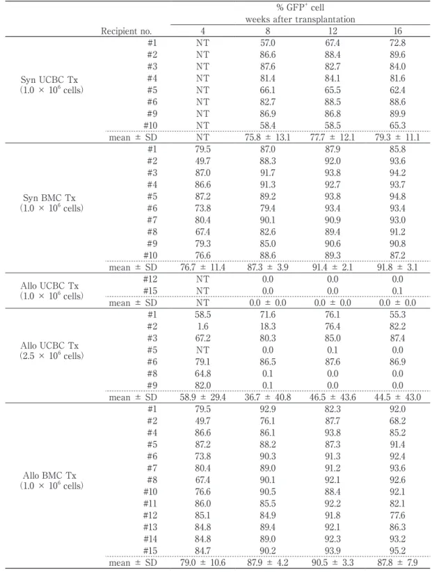
+donor cells was evaluated by flow cytometry every 4 weeks in the peripheral blood of recipients that survived for up to 16 weeks after transplantation (Table 2). The non-GFP
+cell population represents that derived from the host. In syngeneic UCBC and BMC transplantation that showed a higher survival rate in Figure 2 (left), engraftment of GFP
+cells was saturated at 12 weeks after transplantation. At 16 weeks after transplantation, UCBCs showed a similar high GFP
+cell engraftment in the recipients in comparison with BMCs (UCBCs, 79.3 ± 11.1%;
BMCs, 91.8 ± 3.1%). In allogeneic transplanta- tion, as well as syngeneic transplantation, BMCs achieved high levels of GFP
+cell engraftment in the peripheral blood of all recipients by 4 weeks (79.0 ± 10.6%) after transplantation, and were maintained for up to 16 weeks (87.8
± 7.9%), indicating that the survival of the recipients in Figure 2 (right) was dependent on the development of donor-derived GFP
+cells. At the same number of transplanted
cells (1 × 10
6), UCBCs were unable to induce GFP
+cell engraftment for up to 16 weeks after transplantation in all two surviving recipients.
However, an increase in the number of trans- planted UCBCs (2.5 × 10
6) resulted in GFP
+cell engraftment in 4 (#1, #2, #3, and #6) of 7 recipients, with an associated increase in survival rate. The percentage GFP
+cell engraftment in the four recipients was 78.0 ± 15.3% at 16 weeks after transplantation. These results suggested that the low engraftment of donor cells in allogeneic UCBC transplantation was improved to a degree by increasing the number of transplanted cells.
Reconstitution of immune cells
Reconstitution of immune cells in the GFP
+cell population at 16 weeks after transplantation was detected by flow cytometry with lineage- specific antibodies (Figure 3). Recipients with no GFP
+cell engraftment were excluded from the study. Although the total numbers of reconstituted immune cells were relatively
Figure 2 Survival rate after transplantation. Recipients (B6 and BALB/c) were lethally irradiated and then received a transplant of UCBCs or BMCs, which had been depleted of mature T cells, obtained from GFP-transgenic B6 donors. The survival curves for recipients of (a) syngeneic
(UCBCs or BMCs from GFP. B6 > B6) and (b) allogeneic (UCBCs or BMCs from GFP. B6 >
BALB/c) transplants were plotted for up to 120 days after transplantation.
**p <0.01 by log-rank test; UCBC (1.0 × 10⁶) versus BMC (1.0 × 10⁶).
##p <0.01 by log-rank test; UCBC (1.0 × 10⁶) versus UCBC (2.5 × 10⁶).
31
0 20 40 60 80 100
0 30 60 90 120
% Survival
Days after transplantation
UCBC 1.0 × 10⁶ (n=10) BMC 1.0 × 10⁶ (n=10) Rad. Cont (n=3)
0 20 40 60 80 100
0 30 60 90 120
% Survival
Days after transplantation
Rad. Cont (n=6) UCBC 2.5 × 10⁶ (n=9) BMC 1.0 × 10⁶ (n=15) UCBC 1.0 × 10⁶ (n=15)
Figure 2
Syngeneic Tx Allogeneic Tx
##**
lower for UCBCs relative to BMCs in both syngeneic and allogeneic transplantation, the transplanted UCBCs and BMCs developed into T cells, B cells, monocytes, and granulocytes with similarly reconstituted proportions in
individuals receiving any type of transplant, suggesting that UCBC-HSCs and BMC-HSCs have the potential to differentiate into all types of major immune cells, even in an allogeneic environment.
Table 2 Engraftment of donor HSC-derived GFP+ cells in peripheral blood of recipients
% GFP+ cell weeks after transplantation
Recipient no. 4 8 12 16
Syn UCBC Tx
(1.0 × 10⁶ cells)
#1 NT 57.0 67.4 72.8
#2 NT 86.6 88.4 89.6
#3 NT 87.6 82.7 84.0
#4 NT 81.4 84.1 81.6
#5 NT 66.1 65.5 62.4
#6 NT 82.7 88.5 88.6
#9 NT 86.9 86.8 89.9
#10 NT 58.4 58.5 65.3
mean ± SD NT 75.8 ± 13.1 77.7 ± 12.1 79.3 ± 11.1
Syn BMC Tx
(1.0 × 10⁶ cells)
#1 79.5 87.0 87.9 85.8
#2 49.7 88.3 92.0 93.6
#3 87.0 91.7 93.8 94.2
#4 86.6 91.3 92.7 93.7
#5 87.2 89.2 93.8 94.8
#6 73.8 79.4 93.4 93.4
#7 80.4 90.1 90.9 93.0
#8 67.4 82.6 89.4 91.2
#9 79.3 85.0 90.6 90.8
#10 76.6 88.6 89.3 87.2
mean ± SD 76.7 ± 11.4 87.3 ± 3.9 91.4 ± 2.1 91.8 ± 3.1 Allo UCBC Tx
(1.0 × 10⁶ cells)
#12 NT 0.0 0.0 0.0
#15 NT 0.0 0.0 0.1
mean ± SD NT 0.0 ± 0.0 0.0 ± 0.0 0.0 ± 0.0
Allo UCBC Tx
(2.5 × 10⁶ cells)
#1 58.5 71.6 76.1 55.3
#2 1.6 18.3 76.4 82.2
#3 67.2 80.3 85.0 87.4
#5 NT 0.0 0.1 0.0
#6 79.1 86.5 87.6 86.9
#8 64.8 0.1 0.0 0.0
#9 82.0 0.1 0.0 0.0
mean ± SD 58.9 ± 29.4 36.7 ± 40.8 46.5 ± 43.6 44.5 ± 43.0
Allo BMC Tx
(1.0 × 10⁶ cells)
#1 79.5 92.9 82.3 92.0
#2 49.7 76.1 87.7 68.2
#4 86.6 86.1 93.8 85.2
#5 87.2 88.2 87.3 91.4
#6 73.8 90.3 91.3 92.4
#7 80.4 89.0 91.2 93.6
#8 67.4 90.1 92.1 92.6
#10 76.6 90.5 88.4 92.1
#11 86.0 85.5 92.2 82.1
#12 85.1 84.9 91.8 77.6
#13 84.8 89.4 92.1 86.3
#14 84.8 89.0 92.3 93.2
#15 84.7 90.2 93.9 95.2
mean ± SD 79.0 ± 10.6 87.9 ± 4.2 90.5 ± 3.3 87.8 ± 7.9
169 Immune Reconstitution By Allo-UCBC Transplantation
Development of T and B cells in RAG2
-/-BALB/c recipients after allogeneic transplantation
To evaluate the functional maturity of newly developed T and B cells responsible for adaptive immunity induced by allogeneic transplantation, B6 GFP
+UCBC- or GFP
+BMC-transplanted RAG2
-/-BALB/c recipients were prepared for study. At 16 weeks after transplantation, GFP
+T and B cells were observed in the peripheral blood of both sets of recipients (Figure 4).
These T- and B-developed RAG2
-/-BALB/c chimeric recipients were used for the functional studies described below.
Function of allogeneic UCBC-derived adaptive immunity
To examine the functional maturity of allogeneic UCBC- or BMC-derived adaptive immunity, chimeric RAG2
-/-BALB/c recipients
were engrafted with skin from BALB/c, B6, and C3H mice. Both sets of chimeric recipients rejected the third-party skin graft from C3H mice (Figure 5A), whereas the skin grafts from BALB/c and B6 mice were accepted (data not shown) in the absence of host-derived T cells. Although the rejection time for UCBC chimeric recipients was slightly longer than that for BMC chimeric recipients and normal BALB/c mice, the UCBC recipients completely eliminated the third-party C3H grafts within 13 days, indicating reconstitution of allogeneic UCBC-derived functional CD8
+killer T cells and CD4
+helper T cells that activated the killer T cells via interleukin-2 (IL-2) secretion and the ability of both types of T cells to discriminate between self and non-self antigens in vivo.
Furthermore, using the chromium release assay, the in vitro cytolytic activity of CD8
+T-cells against C3H-splenocytes was also examined
Figure 3 Immune reconstitution. Peripheral blood cells of recipients at 16 weeks after transplantation were analyzed by flow cytometry using biotin-labeled antibodies against lineage markers (CD3e for T cells, CD45R/B220 for B cells, CD11b for monocytes, Ly-6G and Ly-6C for granulocytes), followed by addition of streptavidin-PE. In addition, nucleated cells in peripheral blood were stained with Turk solution and counted under a microscope. The proportions of each lineage among the GFP+ cells were calculated on the basis of cell counting.
32
0 5000 10000 15000 20000 25000 30000
#1 #2 #3 #4 #5 #6 #9 #1 0 #1 #2 #3 #4 #5 #6 #7 #8 #9 #1 0 #1 #2 #3 #6 #1 #2 #4 #5 #6 #7 #8 #1 0 #1 1 #1 2 #1 3 #1 4 #1 5
Num ber of cells (/µL)
Recipient no.
Granulocyte Macrophage B cell T cell Figure 3
1.0 × 10
6Syn UCBC
1.0 × 10
6Syn BMC
2.5 × 10
6Allo UCBC
1.0 × 10
6Allo BMC
using splenocytes obtained from UCBC- and BMC-chimeric and normal BALB/c mice that had rejected C3H-skin (Figure 5B). The activity of UCBC and BMC chimeras was significantly lower than that of normal BALB/c mice, but
reduction of the activity did not markedly impact on the C3H skin graft rejection time, as shown in Figure 5A. Thus, T cells reconstituted as a result of allogeneic UCBC transplantation are immunologically competent in vivo for the
Figure 4 Development of T and B cells in RAG2-/-BALB/c recipients after allogeneic UCBC and BMC transplantation. At 16 weeks after transplantation, newly developed T and B cells in the recipients were detected by flow cytometry. Peripheral blood cells were stained with biotin-labeled antibodies against CD3e (for T cells) or CD45R/B220 (for B cells), followed by addition of streptavidin-PE.
Similar results were observed in each of other 2 chimeric recipients.
33
GFP T cell
B cell
UCBC Tx BMC Tx
CD3e CD45R/B220
Figure 5 Functional maturity of T cells developing after allogeneic transplantation. (A) The T-cell response to skin grafts was examined in allogeneic UCBC (△)- or BMC (◇)-transplanted chimeric RAG2-/-BALB/c recipients.
Dermis for skin grafting was harvested from the tails of BALB/c, B6, and C3H mice, and placed on the shaved backs of chimeric recipients that had survived for more than 16 weeks after transplantation. Both chimeric recipients completely rejected the third-party skin graft from C3H as well as normal BALB/c mice (□). (B)
In vitro cytolytic activity of splenic T-cells in skin-grafted UCBC (△)- and BMC (◇)-chimeras against C3H- splenocytes was evaluated by the 51Cr-release assay at the indicated E/T ratios. Normal BALB/c mice (□)
were used as a control. The results are expressed as the mean percentages of specific lysis ± SD in each group.
*p <0.05 by Tukey’s test; UCBC versus normal BALB/c mice.
#p <0.05 by Tukey’s test; BMC versus normal BALB/c mice.
34
0 20 40 60 80 100
1 3 5 7 9 11 13 15
% Skin engraf tm ent
Days after skin grafting
-100 10 20 30 40 50 60
1 : 60 1 : 30 1 : 15 1 : 7.5
% Specific lysis
E/T ratio
Normal BALB/c (n=5) BMC (n=5)
UCBC (n=5)
Normal BALB/c (n=3) BMC (n=3)
UCBC (n=3)
Figure 5
(A) (B)
# # * #
*
171 Immune Reconstitution By Allo-UCBC Transplantation
recognition and elimination of non-self antigens.
Another important function of adaptive immunity is antibody production. Therefore, the functional maturity of allogeneic UCBC- and BMC-derived B cells in chimeric RAG2
-/-BALB/c recipients was investigated by intraperitoneal injection of TNP-KLH, a T-cell- dependent antigen (Figure 6). In the absence of host-derived B cells, specific IgM and IgG
(IgG1 and IgG2b) antibody responses to TNP were successfully induced in both chimeric recipients and control BALB/c mice at a level similar to the magnitude and spectrum of the Ig classes, whereas TNP-specific antibody responses were not detected in the non-chimeric RAG2
-/-BALB/c mice used as negative controls.
These findings suggested that B cells derived from UCBC-HSCs in allogeneic RAG2
-/-BALB/
Figure 6 Functional maturity of B cells developing after allogeneic transplantation. More than 16 weeks after allogeneic UCBC or BMC transplantation, the chimeric RAG2-/-BALB/c recipients were immunized intraperitoneally with TNP-KLH, and TNP-specific IgM and IgG (IgG1, IgG2a, IgG2b, and IgG3)
antibody titers in serial 2-fold dilutions of serum samples were determined using ELISA. The values were expressed as mean OD ± SD at 490 nm in each group.
*p <0.05 by Tukey-Kramer test; UCBC versus normal BALB/c mice.
35
0.0 0.5 1.0 1.5 2.0
5 6 7 8 9 10 11 12 13 14 15 16 17 18 19
OD (490 nm)
Serum dilution 1/n TNP-specific IgM
BMC (n=6) UCBC (n=8)
RAG2(-/-) (n=6) Normal BALB/c (n=6)
0.0 0.5 1.0 1.5 2.0
5 6 7 8 9 10 11 12 13 14 15 16 17 18 19
OD (490 nm)
Serum dilution 1/n TNP-specific IgG1
0.0 0.5 1.0 1.5 2.0
5 6 7 8 9 10 11 12 13 14 15 16 17 18 19
OD (490 nm)
Serum dilution 1/n TNP-specific IgG2a
0.0 0.5 1.0 1.5 2.0
5 6 7 8 9 10 11 12 13 14 15 16 17 18 19
OD (490 nm)
Serum dilution 1/n TNP-specific IgG2b
0.0 0.5 1.0 1.5 2.0
5 6 7 8 9 10 11 12 13 14 15 16 17 18 19
OD (490 nm)
Serum dilution 1/n TNP-specific IgG3 25 28 211 214 217
25 28 211 214 217 25 28 211 214 217
25 28 211 214 217 25 28 211 214 217
* * *
Figure 6
c recipients had normal ability to produce TNP- specific antibodies, similar to those derived from BMC-HSCs. Furthermore, Ig class switching from IgM to IgG strongly confirmed that interaction between helper T cells and B cells was complete, because Ig class switching should be accomplished by helper T cell-mediated activation of B cells through CD40/CD40 ligand
(CD40L) interaction
18, 19)and cytokines. All of these immune functions were active in the UCBC chimeric recipients.
Discussion
UCBCs are now widely used instead of BMCs for treatment of hematopoietic diseases.
However, information on reconstitution of immunological competence by allogeneic UCBC transplantation has been limited to clinical observations
1, 2, 9), and in vitro and xenogeneic in vivo experiments
10-13). The immune system that develops in such a chimeric environment is thought to form through competitive MHC restriction, due to the co-existence of two types of MHC-mismatched selection system derived from the donor and the host in the chimera.
Thus, it is questionable whether the immune system reconstituted in such a complex situation would be functional. This issue can be resolved only by in vivo studies of recipients. Our group have previously established a murine model of GFP
+UCBC transplantation and demonstrated successful reconstitution of the hematopoietic system in a syngeneic setting
14, 15). In the present study, we used this murine model system to evaluate the degree of hematopoietic recovery following allogeneic UCBC transplantation.
By using T cell-depleted UCBCs and BMCs, both types of syngeneic transplantation achieved a high survival rate at 120 days after transplan- tation (Figure 2, left). In this case, the observed difference in the CFU count did not greatly reflect the survival rate. In contrast, the survival
rate following allogeneic transplantation was significantly reduced for UCBCs relative to BMCs when 1 × 10
6cells were transplanted
(Figure 2, right), whereas an increase in the number of transplanted UCBCs (2.5 × 10
6) improved the survival rate to a degree comparable with that for BMCs, indicating that the number of transplanted cells was a factor for successful allogeneic UCBC transplantation.
This result probably reflects those of previous studies suggesting differences in the numbers of potent HSCs among UCBCs (1 cell per 4
× 10
4)
15)and BMCs (1 cell per 2.5 × 10
4)
14)
that contribute to long-term engraftment.
Controversially, one study using a similar murine model has suggested that there was no correlation between the increase in the number of transplanted allogeneic UCBCs and improvement of survival
20).
As shown in Table 2, total lineages derived from HSCs of donor origin were clearly discriminated as GFP
+cells in the peripheral blood of recipients. As reflected in the survival rate, similar high engraftment of GFP
+cells after syngeneic transplantation occurred for both UCBCs and BMCs until 16 weeks after transplantation. In contrast, no engraftment of GFP
+cells of donor HSC origin was induced in 2 surviving recipients of 1 × 10
6allogeneic UCBCs, indicating that the recipient's own HSCs had facilitated recovery of the hematopoietic system. However, transplantation using 2.5 × 10
6UCBCs achieved GFP
+cell engraftment in more than half of the surviving recipients.
Although it is not known whether a further increase in the number of transplanted UCBCs would lead to high GFP
+cell engraftment in allogeneic recipients, reconstitution of the hematopoietic system by the recipient's own HSCs would mean recurrence of a hematopoietic disease such as leukemia in a clinical situation.
Successful reconstitution of the hematopoietic
system following allogeneic UCBC transplantation
173 Immune Reconstitution By Allo-UCBC Transplantation
is an issue that needs to be considered in order to make this form of transplantation reliable. Unlike BMC-HSCs, UCBC-HSCs showed especially low engraftment in allogeneic recipients. This suggests that UCBC-HSCs are more easily eliminated than BMC-HSCs by the radiation-resistant host immune system. BMCs may contain the components needed to protect HSCs from the immune system. In fact, a recent study has suggested that co-transplantation of mesenchymal stem cells expanded from BMCs led to enhanced hematopoietic recovery in allogeneic UCBC transplantation
21). Furthermore, Blanc et al. have proposed the use of myeloid- derived suppressor cells as a therapeutic tool for improving the efficacy of allogeneic HSC transplantation
22, 23). With the exception of these engraftment issues, GFP
+cells that developed in allogeneic recipients contained mature T cells, B cells, monocytes, and granulocytes after both UCBC and BMC transplantation, indicating normal in vivo development of HSCs in an allogeneic environment (Figure 3).
Allogeneic UCBC transplantation has al- ready been performed clinically for various hematopoietic diseases. In this situation, the most important issue is to assess the in vivo functional maturity of the reconstituted T and B cells involved in adaptive immunity after transplantation. For this purpose, T- and B-deficient RAG2
-/-BALB/c mice were used as allogeneic recipients because examination of the in vivo immune response of UCBC-derived T and B cells is not informative when host
(recipient) BMC-derived T and B cells are also present. Both UCBC and BMC chimeric RAG2
-/-BALB/c recipients strictly rejected third- party skin grafts from C3H mice (Figure 5A), but accepted skin grafts from B6 and BALB/c mice (data not shown). These results indicate that the repertoire of T cell receptors (TCRs)
was formed through double negative selection by MHC molecules from both B6 and BALB/
c mice in these chimeric recipients, and that their narrowed TCR repertoire depleted by the double MHC selection might have caused low cytolytic activity against in vitro C3H targets in comparison with normal mice (Figure 5B). This hypothesis has been supported by Chen et al.
20). However, the low cytolytic activity was not correlated with any serious delay in rejection of C3H skin grafts in vivo in both chimeras
(Figure 5A), indicating the immunocompetence of the reconstituted killer T cells throughout the whole body. This functional reconstitution also includes IL-2 production from helper T cells involved in killer T cell activation
24). On the other hand, TNP-KLH immunization successfully induced antigen-specific antibody responses in both sets of chimeric recipients.
The magnitude and the spectrum of Ig classes of the antibodies produced were quite similar among both chimeras and normal mice (Figure 6). Production of antibody against TNP-KLH is T-cell-dependent, and Ig class switching requires interaction between helper T cells and B cells.
This cellular interaction includes presentation of antigen by activated B cells to helper T cells, recognition of the presented antigen by helper T cells, and helper T-cell-mediated activation of B cells by CD40/CD40L interaction
18, 19)and cytokine production. Immunologically, IgG production by class switching contributes to innate immunity through opsonization
25). These observations indicate that reconstituted T cells and B cells are not only functionally competent, but also that their cellular interaction is effective in RAG2
-/-BALB/c recipients.
Overall, our present results suggest that UCBC-HSCs and BMC-HSCs have essentially the same ability to differentiate into cells of all hematopoietic lineages with mature immunological function, even in an allogeneic environment, indicating the effectiveness of allogeneic UCBC transplantation for hematopoietic reconstitution.
Further work will be required in order to achieve
higher engraftment of HSCs after allogeneic UCBC transplantation to establish a more practical treatment strategy using this murine model.
Acknowledgments
The authors thank Dr. Nobukata Shinohara and Dr. Koji Eshima (Kitasato University) for helpful comments and discussion about the experiments. This work was supported by JSPS KAKENHI Grant Number 25461868.
References
1)Gluckman E, Broxmeyer HE, Auerbach AD, Friedman HS, Douglas GW, Devergie A, Esperou H, et al. Hematopoietic reconstitution in a patient with Fanconi's anemia by means of umbilical-cord blood from an HLA-identical sibling. N Engl J Med. 1989;321:1174-8.
2)Gluckman E, Rocha V, Boyer-Chammand A, Locatelli F, Arcese W, Pasquini R, Ortega J, et al. Outcome of cord-blood transplantation from related and unrelated donors. N Engl J Med.
1997;337:373-81.
3)Rubinstein P, Carrier C, Scaradavou A, Kurtzberg J, Adamson J, Migliaccio AR, Berkowitz RL, et al.
Outcomes among 562 recipients of placental-blood transplants from unrelated donors. N Engl J Med.
1998;339:1565-77.
4)Wang JCY, Doedens M, Dick JE. Primitive human hematopoietic cells are enriched in cord blood compared with adult bone marrow or mobilized peripheral blood as measured by the quantitative in vivo SCID-repopulating cell assay. Blood.
1997;89:3919-24.
5)Rubinstein P, Rosenfield RE, Adamson JW, Stevens CE. Stored placental blood for unrelated bone marrow reconstitution. Blood. 1993;81:1679- 90.
6)Bofill M, Akber AN, Salmon M, Robinson M, Burford G, Janossay G. Immature CD45RAlowROlow T cells in the human cord blood. J Immunol.
1994;152:5613-23.
7)Madrigal JA, Cohen SBA, Gluckman E, Charron DJ. Does cord blood transplantation result in lower graft-versus host disease? Hum Immunol.
1997;56:1-5.
8)Wagner JE, Rosenthal J, Sweetman R, Shu XO, Davies SM, Ramsay NK, McGlave PB, et al.
Successful transplantation of HLA-matched and HLA-mismatched umbilical cord blood from unrelated donors: analysis of engraftment and acute graft-versus host disease. Blood. 1996;88:795- 802.
9)Broxmeyer HE, Douglas GW, Hangoc G, Cooper S, Bard J, English D, Arny M, et al. Human umbilical cord blood as a potential source of transplantable hematopoietic stem/progenitor cells. Proc Natl Acad Sci USA. 1989;86:3828-32.
10)Erices A, Conget P, Minguell JJ. Mesenchymal progenitor cells in human umbilical cord blood. Br J Haematol. 2000;109:235-42.
11)Rutella S, Bonanno D, Marone M, De Ritis D, Mariotti A, Voso MT, Scambia G, et al.
Identification of a novel subpopulation of human cord blood CD34-CD133-CD7-CD45+lineage-cells capable of lymphoid/NK cell differentiation after in vitro exposure to IL-15. J Immunol. 2003;171:2977- 88.
12)Yahata T, Ando K, Nakamura Y, Ueyama Y, Shimamura K, Tamaoki N, Kato S, et al. Func- tional human T lymphocyte development from cord blood CD34+ cells in nonobese diabetic/Shi- scid, IL-2 receptor gamma null mice. J Immunol.
2002;169:204-9.
13)Traggiai E, Chicha L, Mazzucchelli L, Bronz L, Piffaretti JC, Lanzavecchia A, Manz MG.
Development of a human adaptive immune system in cord blood cell-transplanted mice. Science.
2004;304:104-7.
14)Migishima F, Oikawa A, Kondo S, Ema H, Morita Y, Nakauchi H, Yokoyama M, et al.
Full reconstitution of hematopoietic system by murine umbilical cord blood. Transplantation.
2003;75:1820-6.
15)Oikawa A, Ito K, Seguchi H, Okabe M, Migishima F, Eshima K, Azuma S, et al. Development of immunocompetent lymphocytes in vivo from murine umbilical cord blood cells. Transplantation.
175 Immune Reconstitution By Allo-UCBC Transplantation
2007;84:23-30.
16)Okabe M, Ikawa M, Kominami K, Nakanishi T, Nishimune Y. 'Green Mice' as a source of ubiquitous green cells. FEBS Lett. 1997;407:313-9.
17)Park SY, Kojima M, Suzuki H, Shinohara N.
Effective blocking of natural cytotoxicity of young rabbit serum on murine thymocytes by high concentration of glucose in complement-dependent cytotoxicity method. J Immunol Methods.
1992;154:109-19.
18)Aruffo A, Farrington M, Hollenbaugh D, Li X, Milatovich A, Nonoyama S, Bajorath J, et al. The CD40 ligand, gp39, is defective in activated T cells from patients with X-linked hyper-IgM syndrome.
Cell. 1993;72:291-300.
19)Xu J, Foy TM, Laman JD, Elliott EA, Dunn JJ, Waldschmidt TJ, Elsemore J, et al. Mice deficient for the CD40 ligand. Immunity. 1994;1:423-31.
20)Chen BJ, Cui X, Sempowski GD, Gooding ME, Liu C, Haynes BF, Chao NJ. A comparison of murine T-cell-depleted adult bone marrow and full-term fetal blood cells in hematopoietic engraftment and immune reconstitution. Blood. 2002;99:364-71.
21)Li ZY, Wang CQ, Lu G, Pan XY, Xu KL. Effects of bone marrow mesenchymal stem cells on hematopoietic recovery and acute graft-versus- host disease in murine allogeneic umbilical cord blood transplantation model. Cell Biochem Biophys.
2014;70:115-22.
22)Le Blanc K, Jitschin R, Mougiakakos D. Myeloid- derived suppressor cells in allogeneic hematopoietic stem cell transplantation: A double-edged sword?
Oncoimmunology. 2013;2:e25009.
23)Jitschin R, Braun M, Büttner M, Dettmer-Wilde K, Bricks J, Berger J, Eckart MJ, et al. CLL-cells induce IDOhi CD14+HLA-DRlo myeloid-derived suppressor cells that inhibit T-cell responses and promote Tregs. Blood. 2014;124:750-60.
24)Bellgrau D. Induction of cytotoxic T cell precursors in vivo. Role of T helper cells. J Exp Med.
1983;157:1505-15.
25)Stendahl O, Tagesson C, Magnusson KE, Edebo L. Physiochemical consequences of opsonization of Salmonella typhimurium with hyperimmune IgG and complement. Immunology. 1977;32:11-8.