Circulation Journal
Official Journal of the Japanese Circulation Society http://www.j-circ.or.jp
concentration using a BCA Protein Assay (Thermo) and added to 2×SDS sample buffer. Samples were then boiled at 60°C for 10 min, or 95°C for 5 min. These samples were separated by SDS-PAGE and transferred to a PVDF membrane, and blocked with 5% skim milk in TBS-T. Antibodies used for immunoblotting were V2R (AB1797P; Millipore), phospho- eNOS (#9571S; Cell signaling technology, Tokyo, Japan), endothelin-1 (ab2786; Abcam, Tokyo, Japan), vascular endo- thelial growth factor (sc-7269; Santa Cruz), TGF-
β1 (sc-146;
Santa Cruz) and collagen tissue growth factor (ab6992; Abcam) for overnight at 4°C. The membranes were then washed and incubated with anti-rabbit (conformation specific #5127, CST) or anti-mouse horseradish peroxidase-conjugated IgG (GE Healthcare, Buckinghamshire, England), and visualized using an ECL system. The membranes were stripped and re-incu- bated with total-eNOS (610297; BD transduction Laboratories, Franklin Lakes, NJ, USA) and
β-actin antibody (ab8227;
Abcam). Laser densitometry (ImageQuant LAS 4000; Fuji, Tokyo, Japan) was used to quantitate the bands.
cAMP Assay
After reaching confluence in 12-well dishes, rat PASMCs incubating with the DMEM containing 10% FBS were stimu- lated with dDAVP (10
–6M, for each 0–60 min). The reaction was stopped by aspiration of medium and the cells were rinsed with ice-cold PBS. The cAMP was extracted from cells by adding 400
μl of 0.1 N HCL. These samples were sent to aclinical laboratory testing service (SRL, Tokyo, Japan) for the measurement of cAMP with a Yamasa cyclic AMP RIA assay kit (YAMASA Shoyu, Chiba, Japan).
Proliferation Assays
Rat PASMCs (5
×10
3cells/well) were seeded into 96-well in DMEM supplemented with 10% FBS. For each experiment, cells were incubated in 10% FBS media containing dDAVP (10
–6M) and/or tolvaptan (10
–9to 10
–6M) for 72 h, and then cell proliferation was determined by a Promega Cell Titer 96 MTT assay.
Methods
Cell Preparation and Culture, and Chemicals
Human pulmonary artery smooth muscle cells (PASMCs) were purchased from Lonza and grown by using a SmGM-2 BulletKit. Rat PASMCs were prepared from 16-week-old male Sprague-Dawley (SD) rats. The isolated pulmonary arte- rial branches (3
rd–4
thdivision) were incubated in HBSS con- taining collagenase type II (1 mg/ml) and elastase (0.025 mg/ml) for 30 min. After incubation, a thin layer of adventitia was carefully stripped off. The remaining medial wall was digested with the above same solution until each cell was separated.
Cells were cultured with DMEM containing 10% FBS, peni- cillin and streptomycin (each 100 mg/ml) and maintained at 37°C in an atmosphere of 5% CO
2and 95% O
2. Tolvaptan was purchased from Sigma (St Louis, MO, USA), and dDAVP was purchased from Prospec-Tany Technogene, Ltd (East Brunswick, NJ, USA).
Immunofluorescence Staining
For immunofluorescence analysis, rat PASMCs were grown on glass slides and fixed in 4% paraformaldehyde for 10 min and was followed by permeabilization with 0.1% NP-40 in phosphate-buffered saline (PBS) for 5 min.
After blocking, cells were incubated overnight at 4°C with anti- vasopressin V2 receptor (anti-V2R; 1:250, Millipore, Temecula, CA, USA) or normal rabbit IgG (Santa Cruz, Dallas, TX, USA) and α-smooth muscle actin (α-SMA, 1:20,000, A5228, SIGMA) diluted with Can Get Signal Immunostain (TOYOBO, Osaka, Japan). Slides were incubated with anti- rabbit IgG-Alexa 488, anti-mouse IgG-Alexa 568 (Thermo, Rockford, IL, USA), and DAPI.
Semiquantitative Immunoblotting
The lung tissues of rats, and PASMCs from humans and rats were individually homogenized in ice-cold buffer RIPA buf- fer. The homogenates were centrifuged at 4,000 g for 15 min at 4°C. The supernatant was adjusted to the same final protein
Detrimental Impact of Vasopressin V2 Receptor Antagonism in a SU5416/Hypoxia/Normoxia-Exposed Rat Model of
Pulmonary Arterial Hypertension
Itaru Goto, MD; Kaoru Dohi, MD, PhD; Yoshito Ogihara, MD; Ryuji Okamoto, MD, PhD;
Norikazu Yamada, MD, PhD; Yoshihide Mitani, MD, PhD; Masaaki Ito, MD, PhD
——— Supplementary File 1 ———
Supplementary File
Advance Publication by-J-STAGE
Figure S1. (A) Rat pulmonary artery smooth muscle cells (PASMCs) immunofluorescence staining for vasopressin V2 receptor (V2R). Upper panels were using with antibody to V2R, lower panels were with normal rabbit-IgG for a negative control of V2R. (B) V2R expressions in human and rat PASMCs. (C) The time-course of changes in cAMP concentration in rat PASMCs after admin- istration of 10–6 M dDAVP (n=4 in each group). (D) MTT proliferation assay in rat PASMCs. Values are presented as mean ± SD.
Advance Publication by-J-STAGE
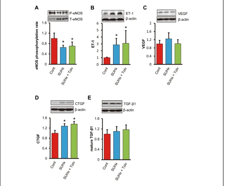
Figure S2. Western blotting analysis of (A) endothelial NOS phosphorylation ratio, (B) endothelin-1/β-actin ratio, (C) vascular endothelial growth factor/β-actin ratio, (D) collagen tissue growth factor/β-actin ratio and (E) mature transforming growth factor- β1/β-actin ratio. Cont, normal rats; SUHx, SU5416+hypoxia rats treated with a vehicle diet; SUHx+Tolv, SU5416+hypoxia rats treated with a tolvaptan diet. Values are presented as mean ± SD (n=6 in each group). *P<0.05 vs. Cont.