M
ol ec ul ar r ec ogni t i on by m
ul t i pl e m
et al
c oor di nat i on i ns i de w
avy- s t ac ked m
ac r oc yc l es
著者
N
akam
ur a Takas hi , Kaneko Yuya, N
i s hi bor i Ei j i ,
N
abes hi m
a Tat s uya
j our nal or
publ i c at i on t i t l e
N
at ur e c om
m
uni c at i ons
vol um
e
8
page r ange
129
year
2017- 07
権利
( C) The Aut hor ( s ) 2017
Thi s ar t i c l e i s l i c ens ed under a Cr eat i ve
Com
m
ons At t r i but i on 4. 0 I nt er nat i onal Li c ens e,
w
hi c h per m
i t s us e, s har i ng, adapt at i on,
di s t r i but i on and r epr oduc t i on i n any m
edi um
or
f or m
at , as l ong as you gi ve appr opr i at e c r edi t
t o t he or i gi nal aut hor ( s ) and t he s our c e,
pr ovi de a l i nk t o t he Cr eat i ve Com
m
ons
l i c ens e, and i ndi c at e i f c hanges w
er e m
ade.
The i m
ages or ot her t hi r d par t y m
at er i al i n
t hi s ar t i c l e ar e i nc l uded i n t he ar t i c l e’
s
Cr eat i ve Com
m
ons l i c ens e, unl es s i ndi c at ed
ot her w
i s e i n a c r edi t l i ne t o t he m
at er i al . I f
m
at er i al i s not i nc l uded i n t he ar t i c l e’
s
Cr eat i ve Com
m
ons l i c ens e and your i nt ended us e
i s not per m
i t t ed by s t at ut or y r egul at i on or
exc eeds t he per m
i t t ed us e, you w
i l l need t o
obt ai n per m
i s s i on di r ec t l y f r om
t he c opyr i ght
hol der . To vi ew
a c opy of t hi s l i c ens e, vi s i t
ht t p: / / c r eat i vec om
m
ons . or g/ l i c ens es / by/ 4. 0/ .
U
RL
ht t p: / / hdl . handl e. net / 2241/ 00150894
doi: 10.1038/s41467-017-00076-8
Cr eat i ve Commons : 表示
Molecular recognition by multiple metal
coordination inside wavy-stacked macrocycles
Takashi Nakamura
1, Yuya Kaneko
1, Eiji Nishibori
1& Tatsuya Nabeshima
1Most biological and synthetic receptors for small organic molecules employ a combination of relatively weak intermolecular interactions such as hydrogen bonds. A host compound that utilizes stronger yet reversible bonding in a synergistic manner could realize precise recognition, but the regulation and spatial arrangement of such reactive interaction moieties have been a challenge. Here, we show a multinuclear zinc complex synthesized from a macrocyclic ligand hexapap, which inwardly arranges labile metal coordination sites for external molecules. The metallomacrocycle forms a unique wavy-stacked structure upon binding a suitable length of dicarboxylic acids via multipoint coordination bonding. The saddle-shaped deformation and dimerization realize the differentiation of the interaction moieties, and change of guest-binding modes at specific metal coordination sites among the many present have been achieved utilizing acid/base as external stimuli.
DOI: 10.1038/s41467-017-00076-8 OPEN
1Graduate School of Pure and Applied Sciences and Tsukuba Research Center for Interdisciplinary Materials Science (TIMS), University of Tsukuba,
P
recise recognition of small molecules plays a vital role in nature. It serves as a basis for sophisticated functions such as signal transduction. Molecular recognition in biological systems1is usually realized by the combination of relatively weak intermolecular interactions, such as hydrogen bonds (typically 10–40 kJ/mol)2, aromatic–aromatic interactions (~5 kJ/mol)3, and van der Waals interactions (~1 kJ/mol)2. Many artificial synthetic receptors4–6 utilizing these interactions have already been developed, but they are still not optimized in terms of specificity. To achieve sophisticated recognition events, it is required to properly and three-dimensionally incorporate multiple inter-action moieties into the molecular-binding sites, but chemically synthetic receptors are no match against biological counterparts in that respect.In this context, utilization of stronger and more directional interactions in a synergistic manner could create a sophisticated artificial host. That is, a host that utilizes multiple coordination
bonds. Coordination bonds between metal atoms and Lewis bases are categorized to be stronger than hydrogen bonds, but weaker than typical covalent bonds (~500 kJ/mol, for a C–C bond)2. As an example using zinc, the binding enthalpy of the reaction [Zn(H2O)6]2+→[Zn(H2O)5]2++ (H2O) was calculated to be
~120 kJ/mol7. Although the coordination bonds are relatively strong, they are labile and reversible enough to be used for molecular recognition, which can be seen from the very fast exchange reaction of a water on the [Zn(H2O)6]2+with the
life-time on the order of 10−7s8. To utilize multiple coordination
bonds in the molecular recognition events9,10, labile coordination sites on the metal centers should be spatially arranged in the binding pocket. External small molecules bind to multiple metals in place of the exchangeable ligands.
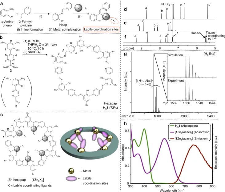
In this study, we create a rigid macrocyclic ligand hexapap H61,
and embed the metals in its cavity11–16 with their labile coordination sites directed toward the center of the pore.
g Simulation
Experiment
[1H n)Nan]+
(n
[H61Na]+
a.u. ) Intensity [ a.u. ] a a d d d b c b c ~ CHCl3
e f Hacac
e f ~ a
b c
e f
Emission intensity (
a.u.
)
Wavelength (nm) H61 (Absorption)
[1Zn6(acac)6] (Absorption)
[1Zn6(acac)6] (Emission)
(ii) Metal complexation
2-Formyl-pyridine
Zn-hexapap
acac– coordinating
to ZnII
a
+
(i) (ii)
(i) Imine formation
o-Amino-phenol
X = Labile coordinating ligands
b
2
3
[1Zn6Xn]
(1)p-TsOH,
THF/H2O = 3/1 (v/v)
(2) NaHCO3
60 °C, 10 h
Labile coordination sites
H61 (72%)
c coordination sites Labile Metal a b c d e f a b c d e f a b c d e f Hpap
Hexapap m/z 1532 1536 1540 1544
Intensity (
Intensity [
a.u.
]
1200 2000 2400
m/z 1600
d
e
f
(ppm) 9 8 7 6 5
h Absorbance 0 0.2 0.4 0.6
300 400 500 600 700 800 900
NH2 OH N O N OH
N M Xn N O N M Xn NH2 OH N tBu MeO OMe NH2 OH N O tBu N OH tBu N N OH tBu N N HO tBu N N HO tBu N N OH tBu N N OH tBu N N O tBu N N O tBu N N O tBu N N O tBu N N O tBu N N O tBu N Zn Zn Zn Zn Zn Zn Xn Xn Xn Xn Xn Xn 7
Fig. 1Synthesis and characterization of hexapap ligand and Zn-hexapap.aFormation of anN,N,O-type tridentate ligand Hpap and its metal complex. bSynthesis of hexapap H61from the bifunctional monomer2by a one-pot reaction.cChemical structure and schematic representation of a
metallomacrocycle, Zn-hexapap [1Zn6Xn], with inwardly arranged coordination sites.d–f1H NMR spectra (600 MHz, 298 K). Seeb,cfor the
assignment of NMR signals.d 2(CDCl3).eH61(CDCl3/CD3OD=10/1 (v/v)).f[1Zn6(acac)6] (CDCl3/CD3OD=10/1 (v/v)).gA MALDI TOF
mass spectrum of H61(positive, matrix: 2,5-dihydroxybenzoic acid). The simulated and observed isotope patterns of [H61Na]+are shown in the
inset.hAbsorbance spectra of H61(green) and [1Zn6(acac)6] (purple) and emission spectrum of [1Zn6(acac)6] (red,λex=546 nm)
(5μM, CHCl3/CH3OH=10/1 (v/v), 298 K,l=1.0 cm)
The complexation of H61 with ZnII produces a hexanuclear
complex, Zn-hexapap [1Zn6Xn] (X=exchangeable labile ligands).
Interestingly, the dicarboxylic acids with suitable chain lengths induce the formation of a uniquely-shaped wavy-stacked dimer of the Zn-hexapap via multiple coordination bonds between the carboxylate groups and Zn. Although the monomeric Zn-hexapap [1Zn6Xn] has six chemically equivalent metal
centers, the saddle-shaped deformation and dimerization of the macrocycles realize the differentiation of the Zn(pap) units. This desymmetrized dimeric macrocycle achieves the regulation and change of the guest-binding modes at specific metal coordination sites among the many available utilizing acid/base as external stimuli.
Results
Synthesis of hexapap ligand and Zn-hexapap complex. H61
possesses six inward Hpap (2-[(pyridin-2-ylmethylene)amino] phenol) chelate-binding units17,18(Fig.1a). Pap–is a
negatively-charged tridentate ligand. Upon binding of a metal, labile coordination sites not occupied by pap– are available for guest
binding. Meanwhile, the tridentate chelation of the metal is strong enough to prevent its removal by external guests.
Reversible imine bonds are often utilized for the construction of thermodynamically stable target products19–21, and most of them were constructed by mixing aldehyde and amine building blocks22–24. Here, H
61 was synthesized from a
bifunctional monomer 3 possessing both o-aminophenol and 2-formylpyridine moieties (Fig.1b). The compound2, a derivative of3, was designed whose formyl group was protected by dimethyl acetal to prevent spontaneous self-oligomerization25. 2 was prepared as an isolable cyclization precursor (1H nuclear magnetic resonance (NMR), Fig. 1d) (see Supplementary Figs.1–10for the synthesis of2and its synthetic intermediates). The angle between the formyl group and the amino group installed into3was about 120°. This geometrical feature realized the high-yield synthesis of the hexagonal macrocycle hexapap H61. That is, an aqueous acid-catalyzed reaction (p-toluene
sulfonic acid, tetrahydrofuran (THF)/H2O=3/1 (v/v)) facilitated
deprotection of the formyl group of2and imine-bond formation, and produced H61 in 72% yield. H61 was obtained as a yellow
precipitate in this reaction. The 1H NMR spectrum of the solid indicated the formation of H61as a single, pure product, and its
symmetric time-averaged structure on the NMR time scale (~msec) (Fig. 1e and Supplementary Fig. 11). The composition and purity of H61as a cyclic hexamer was further supported by
MALDI TOF mass (Fig.1g), infrared (IR), and elemental analysis (see Methods).
The reaction of H61and Zn(acac)2 (acac−=acetylacetonate)
led to the formation of Zn-hexapap [1Zn6(acac)6]. ZnIIcenters of
Zn-hexapap adopted a five-coordinate trigonal-bipyramidal geometry. Two labile coordination sites per Zn atom are available, which are directed upwardly inward and downwardly inward (Fig.1c). Changes in the chemical shifts of16–were observed in the 1H NMR spectrum upon the complexation of Zn, and [1Zn6(acac)6] retained its time-averaged sixfold symmetry
(Fig. 1f). The acac− ligands coordinating to the Zn from the
inside of the macrocycle were clearly discerned in the1H NMR spectrum. Complexation of Zn to pap− was further characterized
by the absorption and emission at the 546 nm and 762 nm, respectively (Fig. 1h). The absorption maximum (546 nm) of [1Zn6(acac)6] did not change at the concentrations of 1–100μM,
and 1H NMR signals of the complex were also not shifted depending on the concentrations (0.15–1.2 mM) (see Supple-mentary Figs.12and13). These results indicated that stacking of the macrocycles did not occur under these conditions.
Wavy-staked dimer of Zn-hexapap with dicarboxylic acids. Carboxylic acids were investigated as guest molecules in this study, for their ubiquity in nature and coordination ability of
N N O N N O N N O N N O N N O N N O t-Bu t-Bu t-Bu t-Bu t-Bu t-Bu X Zn Zn Xn Zn Zn Zn Xn Zn X N N O N N O N N O N N
N O N
N N O t-Bu t-Bu t-Bu t-Bu t-Bu t-Bu XZn Zn
XnZn
Zn ZnXn ZnX O O O O O O O O O
[1Zn6(acac)6]
m = 4, 5
a k b c d e f g h i
[ppm]10 9 8 7
j
CHCl3
a1,a4
~
a3,a6 a2,a5 d1,d4
d3
d6 b1
b4
b2 b5
d2 d5e3
e6 f3 f6 e1 e4 b3 b6 c1 c4 c3 c6 f2 f5 c2 c5 e2 e5 f1 f4
1.8 1.5 1.2
7.0 7.5 8.0 ~ g3 g6 CHCl3 g1 g4 g2 g5
d2,d5
b1,b4
f3,f6 c1,c4
c2,c5
e2,e5
m
Wavy-stacked dimer l
1, 2, 3. 4, 5, 6.
No guest 4b 4c 4d 4e 4f 4g 4h 4i
2
Hacac 12
[12Zn1242Xn]
H24
4a a b c d e f O tBu N N Zn X X
(CH2)m
O OH O HO OH O g 2
Upper ring parts parts Lower ring
Fig. 2Binding of dicarboxylic acids by Zn-hexapap and the formation of the wavy-stacked dimer.a–iInteraction of dicarboxylic acids H24a–H24iand
Zn-hexapap [1Zn6(acac)6].(1H NMR, 600 MHz, CDCl3/CD3OD=10/1
(v/v), 298 K, [1Zn6(acac)6]=2.5 mM).aMalonic acid H24a(m=1).m
indicates the number of methylene groups between the two carboxylic groups.bSuccinic acid H24b(m=2).cGlutaric acid H24c(m=3).dAdipic
acid H24d(m=4).ePimelic acid H24e(m=5). Seekfor assignment of the
signals.fSuberic acid H24f(m=6).gAzelaic acid H24g(m=7).hSebacic
acid H24h(m=8).iDodecanedioic acid H24i(m=10).j1H–1H ROESY
(rotating-frame Overhauser effect spectroscopy) NMR spectrum of the complex with two pimelates4e2−, [12Zn124e2Xn] (X=labile coordinating ligand) (600 MHz, CDCl3/CD3OD=10/1 (v/v), 323 K).Yellow circles
indicate ROE cross peaks between the top and bottom macrocycles. kChemical structure of [12Zn124e2Xn].Red arrowsindicate the pairs of
1H
–1H between which the ROE cross peaks were observed
(see Supplementary Fig.18). See also Fig.3d for the crystal structure of [12Zn124e2(H2O)4Cl8] colored in the same manner.lEmissions from
Zn-hexapap during UV irradiation (365 nm) upon binding of a series of dicarboxylic acids H24a–H24i(10μM, CHCl3/CH3OH=10/1 (v/v), 298 K).
their carboxylate groups to labile coordinating sites of metal centers. The recognition experiments of aliphatic dicarboxylic acids HOOC-(CH2)m-COOH (m=1–8, 10) by Zn-hexapap
[1Zn6(acac)6] are shown in Fig.2. A series of 1H NMR spectra
(Fig.2a–i) suggest that only adipic acid H24d(m=4) and pimelic
acid H24e (m=5) led to the formation of a single species. In
other words, a clear dependence of the molecular length was observed in this coordination-driven recognition event. The for-mation of a certain host–guest complex was also supported by the change in emission, where the samples in which H24dor H24e
was mixed with [1Zn6(acac)6] showed a stronger red emission
than the other dicarboxylic acids (Fig.2l, Supplementary Fig.14, and Supplementary Table 1). The 1H NMR spectrum of the host–guest complex with H24e suggested that its entire structure
was desymmetrized and six different pap− moieties were present in
the structure (Fig.2e). The ESI-TOF (electrospray ionization time-of-flight) mass spectrum of the sample with H24eindicated that the
dimer of Zn-hexapap [1Zn6Xn] was formed with two pimelate
4e2− molecules, that is, [12Zn124e2X
n] (see Supplementary Fig.15).
A single crystal suitable for X-ray diffraction analysis was obtained by the slow diffusion of acetone vapor into a 1,1,2, 2-tetrachloroethane/MeOH solution of [12Zn124e2Xn] (Fig.3and
Supplementary Fig. 16). Interestingly, it was found that the macrocyclic framework of hexapap16–uniquely warped, while it was tightly stacked around the total circumference of the macrocycle to form a dimeric structure (Fig. 3a, b). Each wavy-stacked dimer was not aligned on a concentric axis in the crystal, but found to be slip-stacked on a 41 helical axis (Fig. 3c).
As expected from the ESI mass measurement, two molecules of the pimelates 4e2− were captured inside the dimer of the
Zn-hexapap. 4e2− was recognized though multipoint
coordina-tion bonding with the metallomacrocycles. The two terminal
carboxylate groups of4e2− both bridged two Zn atoms. One of
the Zn atoms belonged to the top macrocycle, while the other to the bottom one (Fig. 3a, d). All the Zn centers adopted a fi ve-coordinate trigonal-bipyramidal geometry, but they can be categorized into three types in terms of the coordinating ligands at the inner exchangeable coordination sites (Fig. 3e). Thefirst type of Zn (depicted in red in Fig. 3d, e) was coordinated by a carboxylate oxygen atom of4e2− and a phenoxy oxygen atom of
the pap−. The coordination bond between the Zn (type1) and
phenoxy oxygen bridged the two Zn-hexapap macrocycles. The second type (depicted in blue) was bound by another carboxylate oxygen atom of 4e2− and a Cl−. The third type (depicted in
green) was bound by a Cl− and a water. Thus, Zn (type 3) was
free from the guest molecule 4e2− (Cl− probably derived from
the decomposition of 1,1,2,2-tetrachloroethane used as the crystallization solvent. We assumed that the slow generation of Cl− helped to grow single crystals with good qualities). From the
structural analysis described above, the following three main factors are considered to be the driving forces for the formation of the dimeric structure [12Zn124e2Xn]: (i) Inter-macrocycle
coordination bonds between the Zn (type 1) and phenoxy oxygen; (ii) Coordination of the carboxylate groups of4e2− bridging two
Zn atoms (types 1 and 2); and iii) π–π stacking between the hexapap aromatic frameworks.
The top and bottom macrocycles were in a different environment as the result of the binding of 4e2−. The overall
structure of [12Zn124e2(H2O)4Cl8] had a pseudo C2 symmetry
(Fig.3d), which is consistent with the1H NMR observation in the solution state. All the 1H NMR signals of [12Zn124e2Xn] were
successfully assigned based on the 1H-1H COSY and 1H-1H
ROESY measurements (Fig. 2e, j, Supplementary Figs. 17, 18). Several characteristic ROE crosspeaks confirmed that the wavy
a b c
d e f
Pimelate
H2O
Cl–
Type 2
Type 1
Type 3
Fig. 3Structure of [12Zn124e2(H2O)4Cl8] determined by X-ray crystallography. Solvents, hydrogens, andtBu groups are omitted for clarity. One disorder
pattern of4e2−is shown.a,bAn ellipsoidal model (30% probability).cPacking in the crystal (a stick model). Fora
–c, the atoms are colored according to
the elements: C,light green; N,blue; O,red; Zn,yellow; Cl,green.dA ball-and-stick model.eThree different coordination modes around the Zn centers. Ford ande, the atoms are colored to show the pseudoC2symmetry of the entire structure (see also Fig.2k). The Zn atoms are described in a space-filling model.f20 coordination bonds (magenta) that were not occupied by theN,N,O–chelating moieties of16–. Zn, yellow; non-metal atoms,light green
saddle-shaped structure was fixed on the NMR timescale and was essentially the same as the one determined by X-ray crystallography (Fig.2k).
The wavy shape was considered to be the result of the clipping by the intermacrocyclic coordination bonds. That is, the difference in lengths between the clipped parts caused the distortion. Looking at the right-half structure of [12Zn124e2X12]
in Fig.2k, the upper ring (light colors) has the part 3 between the clipped parts 2 and 1, but the clipped parts of the other ring (dense colors), parts 4 and 5, are adjacent to each other (see Fig.2k for the part numbers).
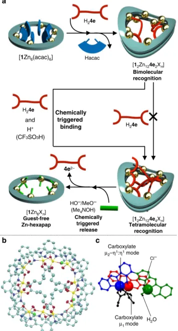
Regulation of guest binding at specific coordination sites. In the wavy-stacked dimer of the Zn-hexapap, four coordination bonds were used to connect the two stacked macrocycles,
while 20 coordination sites in total were unoccupied by theN,N, O-chelating moieties of the hexapap ligand and arranged inward (Fig.3f). Despite possessing many possible coordinating sites, the addition of more than two molar amounts of the pimelic acids H24eagainst the wavy-stacked dimer [12Zn12Xn] did not change
a binding mode, but the host–guest complex stably existed in a bimolecular recognition mode [12Zn124e2Xn] (see Supplementary
Fig. 19). Interestingly, however, an acid stimulus (CF3SO3H)
triggered further incorporation of two more 4e2−’s, and led
to a tetramolecular recognition mode [12Zn124e4Xn] (Fig. 4a,
Supplementary Fig.20). The conversion and the resulting com-plex were examined by 1H NMR (Supplementary Figs. 20–22),
ESI-TOF mass (Supplementary Fig. 23), and X-ray crystal-lographic analysis (vide infra). Three different [Zn(pap)Xn]
moieties were observed in the 1H NMR spectrum of
[12Zn124e4Xn], which suggested a change in the binding mode
from [12Zn124e2Xn] (C2 symmetry, six different [Zn(pap)Xn]
units) (Supplementary Fig. 20). The molecular structure of [12Zn124e4(H2O)4Cl4] was revealed by the X-ray crystallographic
analysis (Fig. 4b, c and Supplementary Fig. 24) (recrystallized from 1,1,2,2-tetrachloroethane/MeOH/acetone). The curvature of the wavy dimeric frameworks of the [12Zn12Xn] was slightly less
bent for the complex with four 4e2−’s than the one with two
4e2−’s, although the frameworks were basically the same for the
two complexes (see Supplementary Fig.25). The binding mode of the 4e2− in the structure of [12Zn124e4(H2O)4Cl4] showed an
interesting difference compared to the complex with two4e2−’s
(Fig.4c). One carboxylate group of4e2− bridged two Zn atoms in
aμ2–η1:η1 coordination mode, while the carboxylate group at the other end of 4e2− was bound to Zn in a monodentate
mode (μ1mode). In terms of the Zn centers, there are three types
of [Zn(pap)Xn] moieties as in the case with the complex with
two4e2−’s. The entire structure of [12Zn124e4(H2O)4Cl4] had an
S4symmetry, which is consistent with the1H NMR observation
in solution (see Supplementary Fig. 20).
The acid-triggered binding of H24e was explained by the
protonation and following release of HO− or MeO−
coordinat-ing to the [Zn(pap)] units. In solution, water and/or methanol molecules were bound to the labile coordination sites of [12Zn124e2Xn] (mainly water-bound complexes were observed
in ESI-TOF mass and X-ray measurements). It is considered that the water/methanol molecules at the inner labile coordination sites initially exist as deprotonated HO−/MeO− forms. The weak
carboxylic acid H24efailed to protonate those ligands, but only
the strong acid CF3SO3H was able to protonate them. This is
consistent with the fact that the pKavalues of H2O bound to a
Zn complex are in the range of 6–9, i.e., neutral pH9. This protonation weakened the coordination strength of the H2O/MeOH coordinating to Zn (type 2) in [12Zn124e2Xn],
resulting in the replacement of them with the additional pimelates 4e2− to produce [1
2Zn124e4Xn]. Furthermore, the strategy to use
acid/base stimuli to control the coordination strength can be applied to the release of guest molecules. That is, the addition of the Me4NOH base to the solution of [12Zn124e4Xn] released the
guest 4e2− and produced the guest-free Zn-hexapap [1Zn 6Xn]
(Fig.4a, Supplementary Fig.20). Here, HO− or MeO− worked as
relatively strong ligands under basic conditions, and dissociated the host–guest complex into [1Zn6Xn], whose inner coordinating
sites were occupied by HO−/MeO−. To summarize, the unique
property of the wavy-stacked macrocycles Zn-hexapap to express multiple modes of molecular recognition via coordination bonds was demonstrated.
Discussion
To summarize, we have designed and synthesized a hexapap ligand H61, and its Zn complex Zn-hexapap [1Zn6Xn] that
a
Bimolecular recognition
Tetramolecular recognition
b c
Carboxylate
µ2–η1:η1 mode
Cl–
Carboxylate
µ1 mode
H2O
[1Zn6(acac)6]
H24e
Chemically triggered
binding Hacac
[12Zn124e2Xn]
H+
Chemically triggered
release
4e2–
HO–/MeO–
(CF3SO3H)
[1Zn6Xn]
Guest-free Zn-hexapap
[12Zn124e4Xn]
H24e
and
H24e
(Me4NOH)
has the inner cavity in which labile coordination bonds are spatially arranged. Zn-hexapap recognized the dicarboxylic acid 4e2− though multiple coordination bonding to form the unique
wavy-stacked dimeric structure. Furthermore, the clear control and change of the binding modes of the guest was achieved, although the metallocyclic dimer [12Zn12Xn] possesses as many as
20 available labile coordination sites. Thus, hexapap is shown to be an artificial host molecule that achieves the binding and the control of small molecules via multiple coordination bonds in solution. Metal complexes of the hexapap are promising platforms for selective molecular sensors6as well as for allosteric catalysts26 that specifically interact with a target substrate via cooperative coordination, and such kinds of applications are now being investigated.
Methods
General. Unless otherwise noted, solvents and reagents were purchased from TCI Co., Ltd., Wako Pure Chemical Industries, Ltd., Kanto Chemical Co., Inc., Nacalai Tesque, Inc. or Sigma-Aldrich Co., and used without further purification. THF was purified by Nikko Hansen Ultimate Solvent System 3S-TCN 1.
Measurements were performed at 298 K unless otherwise noted.1H,13C, and
other 2D NMR spectra were recorded on a Bruker AVANCE III-600 (600 MHz) spectrometer or a Bruker AVANCE III-400 (400 MHz) spectrometer.
Tetramethylsilane was used as an internal standard (δ0.00 ppm) for1H and13C
NMR measurements when CDCl3or a mixed solvent with CDCl3was used as a
solvent. MALDI-TOF mass data were recorded on an AB SCIEX TOF/TOF 5800 system. ESI-TOF mass data were recorded on a Waters SYNAPT G2 HDMS system or an AB SCIEX TripleTOF 4600 system. Ultraviolet (UV)–Vis spectra were recorded on a JASCO V-670 spectrophotometer. Emission spectra were recorded on a JASCO FP-8600fluorescence spectrophotometer. Absolutefluorescence quantum yields were determined with a Hamamatsu Photonics absolute PL quantum yield measurement system C9920-02. Solvents used for measurements were air-saturated. IR spectra were recorded on a JASCO FT/IR-480Plus spectrometer. Elemental analysis was performed on a Yanaco MT-6 analyzer with tin boats purchased from Elementar. We appreciate Mr Ikuo Iida of University of Tsukuba for the elemental analysis.
Synthesis of hexapap H61. A solution of2(48.2 mg, 0.156 mmol, 1.0 eq.) and p-TsOH·H2O (6.0 mg, 0.03 mmol, 0.2 eq.) in a THF/H2O=3/1 mixed solvent
(4 mL) was stirred for 10 h at 60 °C under Ar atmosphere. The reaction mixture was neutralized with sat. NaHCO3aq. (5.0 mL), and the precipitation was collected
byfiltration. The solid was washed with H2O, CH3CN, and MeOH, and dried in
vacuo to give H61·6H2O as a yellow solid (29.6 mg, 18.26μmol, 72%).
mp:>280 °C;1H NMR (600 MHz, CDCl
3/CD3OD=10/1 (v/v)):δ9.12
(d,J=2.0 Hz, 6H), 8.91 (s, 6H), 8.32 (dd,J=8.1, 2.0 Hz, 6H), 8.14 (d,J=8.1 Hz, 6H), 7.56 (d,J=2.2 Hz, 6H), 7.46 (d,J=2.2 Hz, 6H), 1.44 (s, 54H); IR (Nujor): 3300 (m, OH), 1623 (m, C=N), 1578 (w), 1303 (m), 1260 (s), 1227 (m), 1202 (m), 1096 (w), 1021 (w), 955 (m), 861 (m), 838 (m), 738 (w), 636 (m) cm−1; UV/Vis
(CHCl3/CH3OH=10/1 (v/v)):λmax398 nm; MALDI TOF MS (m/z): [H61·Na+]
calcd. for C96H96N12O6Na, 1535.75; found, 1535.72; analysis (calcd., found for
C96H108N12O12(H61·6H2O)): C (71.09, 71.39), H (6.71, 6.57), N (10.36, 10.40).
It was difficult to obtain a good13C NMR spectrum due to low solubility of H 61. Complexation of hexapap H61 and Zn(acac)2. H61(1.00 mg, 0.66μmol, 1.0 eq.)
and Zn(acac)2(1.05 mg, 3.98μmol, 6.0 eq.) in a CDCl3/CD3OD=10/1 mixed
solvent (500μL) were mixed at room temperature. The complexation reaction was completed within 5 min and the formation of Zn-hexapap [1Zn6(acac)6]
was confirmed by1H NMR, UV–Vis absorption and emission measurements
(Fig.1f, h).
1H NMR (600 MHz, CDCl
3/CD3OD=10/1 (v/v)):δ9.31 (s, 6H), 8.68 (s, 6H),
7.95 (dd,J=7.9, 1.5 Hz, 6H), 7.68 (dd,J=7.9, 1.5 Hz, 6H), 7.57 (d,J=2.2 Hz, 6H), 7.32 (d,J=2.2 Hz, 6H), 1.38 (s, 54H); UV/Vis (CHCl3/CH3OH=10/1 (v/v)): 546
nm; emission (CHCl3/CH3OH=10:1 (v/v)): 762 nm (λex=546 nm); emission
quantum yield:ΦF=0.017 (λex=546 nm).
Binding experiments of dicarboxylic acids with Zn-hexapap. A representative procedure (pimelic acid H24e, Fig.2e): H61(1.00 mg, 0.66μmol, 1.0 eq.) and
Zn(acac)2(1.05 mg, 3.98μmol, 6.0 eq.) in a CDCl3/CD3OD=10/1 mixed
solvent (500μL) were mixed at room temperature.The formation of Zn-hexapap [1Zn6(acac)6] was checked by1H NMR. To the solution was added pimelic acid
H24e(2.0μmol, 3.0 eq. for H61) in a CDCl3/CD3OD=10/1 mixed solvent (3.0μL).
The reaction mixture was heated at 50 °C for 2 h. The resulting host–guest complexes were characterized by1H NMR, UV–Vis absorption and emission, and ESI-TOF-MS measurements.
X-ray crystallographic analysis of [12Zn124e2(H2O)4Cl8]. To the microtube
charged with H61(2.0 mg, 1.32μmol, 1.0 eq.) and 1,1,2,2-tetrachloroethane/
methanol=10/1 (v/v) (500μL) was added Zn(acac)2(2.09 mg, 7.96μmol, 6.0 eq.)
and pimelic acid H24e(0.64 mg, 3.96μmol, 3.0 eq.). The reaction mixture was
heated for 3 h at 50 °C. Single crystal of [12Zn124e2(H2O)4Cl8] suitable for X-ray
diffraction analysis was obtained by slow diffusion of acetone vapor into the solution.
The diffraction intensity data was measured at 100 K using MAR-CCD equipped at BL26B2 SPring-827. The wavelength of incident X-ray was 0.8 Å. The
collected diffraction images were processed by CrystalClear (Rigaku). The initial structure was solved using SIR9228and refined using SHELXL-201629. The
diffraction data up to 0.9 Å was used for the structure refinement.
Crystal data for [12Zn124e2(H2O)4Cl8]·1.5C2H2Cl4: C209H211Cl14N24O24Zn12, Fw=4723.99, purple prism, 0.10 × 0.07 × 0.05 mm3, tetragonal, space groupP4 1
(No. 76),a=30.029(3) Å,c=40.154(5) Å,V=36209(9) Å3,Z=4,T=100 K,
λ=0.800 Å,θmax=26.387°,R1=0.0981,wR2=0.2946 (after SQUEEZE30),
GOF=1.038.
Control of binding of a dicarboxylic acid by acid/base stimuli. Experimental procedure (Fig.4a, Supplementary Fig.20): Hexapap H61(1.96 mg, 1.29μmol, 1.0
eq.) was weighed in an NMR tube. To the tube were added Zn(acac)2(2.13 mg,
8.08μmol, 6 eq.) in a CDCl3/CD3OD=10/1 mixed solvent (550μL) and pimelic
acid H24e(2.6μmol, 2 eq. for H61) in a CDCl3/CD3OD=10/1 mixed solvent
(4.0μL). The reaction mixture was heated at 50 °C for 3.5 h to produce [12Zn124e2Xn]. To the solution was added CF3SO3H (4.0μmol, 3 eq. for H61) in a
CDCl3/CD3OD=10/1 mixed solvent (6.0μL). The conversion of the host–guest
complexes from [12Zn124e2Xn] to [12Zn124e4Xn] was characterized by1H NMR
and ESI-TOF-MS measurements. To the solution was added Me4NOH·5H2O
(18.1μmol, 14 eq. for H61) in CD3OD (28μL), which released the4e2−and
produced guest-free Zn-hexapap [1Zn6Xn].
X-ray crystallographic analysis of [12Zn124e4(H2O)4Cl4]·2C2H2Cl4. To a 20 mL
flask charged with H61(25 mg, 16.5μmol, 1.0 eq.) and CHCl3/CH3OH=10/1 (v/v)
(6 mL) was added Zn(acac)2(26.2 mg, 100μmol, 6.1 eq.) and pimelic acid H24e
(7.9 mg, 49μmol, 3.0 eq.). The mixture was stirred at 55 °C for 2 days. The solution wasfiltered through a membranefilter, and to thefiltrate was diffused the vapor of isopentane at 4 °C. The resultant precipitate was collected by
filtration and dried to yield a purple solid (31.9 mg). The obtained complex was dissolved in 1,1,2,2-tetrachloroethane/methanol=10/1 (v/v). Single crystal of [12Zn124e4(H2O)4Cl4]·2C2H2Cl4suitable for X-ray diffraction analysis was
obtained by slow diffusion of acetone vapor into the solution.
Single-crystal X-ray crystallographic measurements were performed using a Bruker APEX II ULTRA with MoKαradiation (graphite-monochromated,
λ=0.71073 Å) at 120 K. The collected diffraction images were processed by Bruker APEX2. The initial structure was solved using SHELXS-9731and refined using
SHELXL-201629. The diffraction data up to 0.9 Å was used for the structure
refinement.
Crystal data for [12Zn124e4(H2O)4Cl4]·2C2H2Cl4: C224H232Cl12N24O32Zn12, Fw=4982.42, purple block, 0.11 × 0.11 × 0.05 mm3, tetragonal, space groupI4
1/a
(No. 88),a=29.8662(12) Å,c=40.741(2) Å,V=36341(3) Å3,Z=4,T=120 K,
λ(MoKα)=0.71073 Å,θmax=23.282°,R1=0.1014,wR2=0.3590 (after SQUEEZE30), GOF=1.032.
Data availability. CCDC 1507879 and 1507880 contain the data for this paper. The data can be obtained free of charge from The Cambridge Crystallographic Data Centre viawww.ccdc.cam.ac.uk/getstructures. All the other data is available from the authors upon reasonable request.
Received: 8 November 2016 Accepted: 31 May 2017
References
1. Yin, H. & Hamilton, A. D. Strategies for targeting protein-protein interactions with synthetic agents.Angew. Chem. Int. Ed.44, 4130–4163 (2005). 2. Israelachvili, J. N.Intermolecular and Surface Forces, 3rd edn (Elsevier,
2011).
3. Burley, S. K. & Petsko, G. A. Aromatic-aromatic interaction: A mechanism of protein structure stabilization.Science229, 23–28 (1985).
4. Oshovsky, G. V., Reinhoudt, D. N. & Verboom, W. Supramolecular chemistry in water.Angew. Chem. Int. Ed.46, 2366–2393 (2007).
5. Yoshizawa, M., Klosterman, J. K. & Fujita, M. Functional molecularflasks: New properties and reactions within discrete, self-assembled hosts.Angew. Chem. Int. Ed.48, 3418–3438 (2009).
6. Hargrove, A. E., Nieto, S., Zhang, T., Sessler, J. L. & Anslyn, E. V. Artificial receptors for the recognition of phosphorylated molecules.Chem. Rev.111, 6603–6782 (2011).
7. Bock, C. W., Katz, A. K. & Glusker, J. P. Hydration of zinc ions: A comparison with magnesium and beryllium ions.J. Am. Chem. Soc.117, 3754–3765 (1995). 8. Helm, L. & Merbach, A. E. Water exchange on metal ions: experiments and
simulations.Coord. Chem. Rev.187, 151–181 (1999). 9. Kimura, E., Aoki, S., Koike, T. & Shiro, M. A
tris(Zn-1,4,7,10-tetraazacyclododecane) complex as a new receptor for phosphate dianions in aqueous solution.J. Am. Chem. Soc.119, 3068–3076 (1997).
10. Gan, Q., Ronson, T. K., Vosburg, D. A., Thoburn, J. D. & Nitschke, J. R. Cooperative loading and release behavior of a metal-organic receptor.J. Am. Chem. Soc.137, 1770–1773 (2015).
11. Clyde-Watson, Z.et al.Reversing the stereochemistry of a Diels–Alder reaction: use of metalloporphyrin oligomers to control transition state stability.New J. Chem.22, 493–502 (1998).
12. Bazzicalupi, C.et al.Molecular recognition of long dicarboxylate/dicarboxylic species via supramolecular/coordinative interactions with ditopic receptors. Crystal structure of {[Cu2L(H2O)2]⊃pimelate}(ClO4)2.Inorg. Chem.38,
620–621 (1999).
13. Fabbrizzi, L., Foti, F., Patroni, S., Pallavicini, P. & Taglietti, A. A sleeping host awoken by its guest: Recognition and sensing of imidazole-containing molecules based on double Cu2+translocation inside a polyaza macrocycle. Angew. Chem. Int. Ed.43, 5073–5077 (2004).
14. Hoffmann, M.et al.Enhancedπconjugation around a porphyrin[6] nanoring.
Angew. Chem. Int. Ed.47, 4993–4996 (2008).
15. Setsune, J., Kawama, M. & Nishinaka, T. Helical binuclear CoIIcomplexes of
pyriporphyrin analogue for sensing homochiral carboxylic acids.Tetrahedron Lett.52, 1773–1777 (2011).
16. Omoto, K., Tashiro, S., Kuritani, M. & Shionoya, M. Multipoint recognition of ditopic aromatic guest molecules via Ag-πinteractions within a dimetal macrocycle.J. Am. Chem. Soc.136, 17946–17949 (2014).
17. Muto, Y. Metal complexes with terdentate ligands. I. Synthesis of copper(II) and nickel(II) complexes having salicylaldehydesemicarbazone and related compounds as ligands.Bull. Chem. Soc. Jpn31, 1017–1020 (1958).
18. Asada, H., Hayashi, K., Negoro, S., Fujiwara, M. & Matsushita, T. Preparation and structures of trinuclear manganese(II) complexes withN
-2-pyridiylmethylidene-2-hydroxy-5-substituted-phenylamine.Inorg. Chem. Commun.6, 193–196 (2003).
19. Vigato, P. A., Peruzzo, V. & Tamburini, S. Acyclic and cyclic compartmental ligands: recent results and perspectives.Coord. Chem. Rev.256, 953–1114 (2012).
20. Nabeshima, T. Construction of cooperative and responsive supramolecular systems for molecular functional modulation.Bull. Chem. Soc. Jpn.83, 969–991 (2010).
21. Nabeshima, T. & Yamamura, M. Cooperative formation and functions of multimetal supramolecular systems.Pure Appl. Chem.85, 763–776 (2013). 22. Belowich, M. E. & Stoddart, J. F. Dynamic imine chemistry.Chem. Soc. Rev.41,
2003–2024 (2012).
23. Borisova, N. E., Reshetova, M. D. & Ustynyuk, Y. A. Metal-free methods in the synthesis of macrocyclic Schiff bases.Chem. Rev.107, 46–79 (2007). 24. Nakamura, T., Kimura, H., Okuhara, T., Yamamura, M. & Nabeshima, T.
A hierarchical self-assembly system built up from preorganized tripodal helical metal complexes.J. Am. Chem. Soc.138, 794–797 (2016).
25. Hartley, C. S. & Moore, J. S. Programmed dynamic covalent assembly of unsymmetrical macrocycles.J. Am. Chem. Soc.129, 11682–11683 (2007).
26. Lifschitz, A. M., Rosen, M. S., McGuirk, C. M. & Mirkin, C. A. Allosteric supramolecular coordination constructs.J. Am. Chem. Soc.137, 7252–7261 (2015). 27. Ueno, G.et al.RIKEN structural genomics beamlines at the SPring-8; high
throughput protein crystallography with automated beamline operation.
J. Struct. Funct. Genomics7, 15–22 (2006).
28. Altomare, A.et al.SIR92-a program for automatic solution of crystal structures by direct methods.J. Appl. Cryst.27, 435 (1994).
29. Sheldrick, G. M. Crystal structure refinement with SHELXL.Acta CrystC71, 3–8 (2015).
30. Spek, A. L. Structure validation in chemical crystallography.Acta CrystD65, 148–155 (2009).
31. Sheldrick, G. M. A short history of SHELX.Acta CrystA64, 112–122 (2008).
Acknowledgements
This research was supported by JSPS KAKENHI Grant Numbers JP15H00914, JP15H00723, JP26888003, JP17K14455, JP17H05351 (Coordination Asymmetry), TOBE MAKI Scholarship Foundation (16-JA-004), and Tokuyama Science Foundation. The synchrotron radiation experiments were performed at BL26B2 in SPring-8 with the approval of RIKEN (Proposal No. 20155498).
Author contributions
T. Nakamura and T. Nabeshima conceived the project. T. Nakamura and Y.K. designed and analyzed the experiments. Y.K. carried out the experimental work. E.N. performed X-ray measurement and analysis. T. Nakamura and T. Nabeshima prepared the manuscript, and all the authors contributed to the writing of the paper.
Additional information
Supplementary Informationaccompanies this paper at doi:10.1038/s41467-017-00076-8.
Competing interests:The authors declare no competingfinancial interests.
Reprints and permissioninformation is available online athttp://npg.nature.com/ reprintsandpermissions/
Publisher's note:Springer Nature remains neutral with regard to jurisdictional claims in published maps and institutional affiliations.
Open Access This article is licensed under a Creative Commons Attribution 4.0 International License, which permits use, sharing, adaptation, distribution and reproduction in any medium or format, as long as you give appropriate credit to the original author(s) and the source, provide a link to the Creative Commons license, and indicate if changes were made. The images or other third party material in this article are included in the article’s Creative Commons license, unless indicated otherwise in a credit line to the material. If material is not included in the article’s Creative Commons license and your intended use is not permitted by statutory regulation or exceeds the permitted use, you will need to obtain permission directly from the copyright holder. To view a copy of this license, visithttp://creativecommons.org/ licenses/by/4.0/.
![Fig. 3 Structure of [1 2 Zn 12 4e 2 (H 2 O) 4 Cl 8 ] determined by X-ray crystallography](https://thumb-ap.123doks.com/thumbv2/123deta/9855477.497962/5.892.158.735.73.487/fig-structure-zn-h-cl-determined-ray-crystallography.webp)