mice using anterograde axo‑astrocytic AAV transfer
Author Leonidas Georgiou Degree Conferral
Date
2020‑03‑31
Degree Doctor of Philosophy Degree Referral
Number
38005甲第48号 Copyright
Information
(C) 2020 The Author.
URL http://doi.org/10.15102/1394.00001528
Creative Commons Attribution 4.0 International(https://creativecommons.org/licenses/by/4.0/)
Thesis submitted for the degree
Doctor of Philosophy
Astrocyte calcium activity mapping in behaving mice using anterograde
axo-astrocytic AAV transfer
by
Leonidas Georgiou
Supervisor: Bernd Kuhn
March, 2020
Authorship
I, Leonidas Georgiou, declare that this thesis entitled Astrocyte calcium activity map- ping in behaving mice using anterograde axo-astrocytic AAV transfer and the data presented in it are original and my own work.
I confirm that:
• No part of this work has previously been submitted for a degree at this or any other university.
• References to the work of others have been clearly acknowledged. Quotations from the work of others have been clearly indicated, and attributed to them.
• In cases where others have contributed to part of this work, such contribution has been clearly acknowledged and distinguished from my own work.
• None of this work has been previously published elsewhere, with the exception of the following: (provide list of publications or presentations, or delete this part).
(If the work of any co-authors appears in this thesis, authorization such as a release or signed waiver from all affected co-authors must be obtained prior to publishing the thesis. If so, attach copies of this authorization to your initial and final submitted versions, as a separate document for retention by the Graduate School, and indicate on this page that such authorization has been obtained).
Date: March, 2020 Signature:
iii
Astrocyte calcium activity mapping in behaving mice using an- terograde axo-astrocytic AAV transfer
Astrocytes are considered active partners to neurons in information processing. Het- erogeneous, bidirectional interaction between neurons and astrocytes alludes to circuit specific communication. However, the role of astrocytes in information processing, primarily established through ex vivo experiments, has been challenged by a series of controversies that highlighted the importance of studying astrocytes under fully physi- ological conditions in behaving mice. Astrocytes extend highly ramified processes that cradle synapses. They form functionally independent microdomains where they exhibit a rich repertoire of localized calcium signals. How astrocyte Ca2+ microdomain signals relate to neuronal activity and behaviour in vivo is still unclear. My objective was to investigate circuit specific, single-astrocyte Ca2+ microdomain activity in mice during behavioural states and sensory stimuli. I found that adeno-associated viruses (AAVs) can transfer anterogradely along thalamocortical projections to transduce cortical as- trocytes and neurons. This axo-astrocytic AAV transfer enables the study of astrocytes and neurons embedded in specific neuronal circuits. Intersectional approaches, using anterograde axo-astrocytic AAV transfer in combination with membrane tagged genet- ically encoded calcium indicators (GECIs), enabled sparse, high contrast labelling of cortical astrocytes embedded in the somatosensory system of mice. Continuous imag- ing with two-photon microscopy of single astrocytes for about≈1 hour combined with automatic, unbiased extraction of Ca2+ activity revealed a rich repertoire of subsec- ond, µm scale, localized Ca2+ signals. The number, size and duration of astrocytic Ca2+ signals were modulated with locomotion but not with intermittent whisker-touch stimulation. Locomotion and whisker stimuli evoked rapid [Ca2+]i elevation in thalam- ocortical axon boutons, whose activity was not correlated with nearby astrocyte Ca2+
microdomain signalling. Astrocyte fine processes exhibit heterogeneous, non-random, Ca2+ signalling patterns giving rise to hotspots of higher activity that are stable over time. Hotspot patterns allude to subcellular specialization. Our study: a) provides a new toolkit for studying neuron-astrocyte interactions within brain circuits, b) ex- tends our understanding of astrocyte Ca2+ microdomain signalling and relationship to neuronal activity in behaving animals and c) suggests that there are astrocyte Ca2+
activity maps in the brain.
v
First, I would like to express my gratitude to my supervisor, Dr. Bernd Kuhn, who has supported me throughout the PhD as a mentor but also as a friend. I would like to thank you for the helpful discussions and feedback, continuous support whenever it was necessary, giving me the freedom to pursue my ideas, and letting me grow throughout the PhD. You were there anytime I had a problem (which was most of the time) and I never had to worry about access to equipment. Most importantly, I would like to thank you for being not just a good supervisor but also a great person. Science is a human endeavour in great need of people like you.
I would also like to thank Achilleas, who helped me with data processing and anal- ysis during his internship and beyond. Without your ongoing support and exemplary skills in programming this project wouldn’t have been completed on time. The fact that you continued supporting this project while doing your military service tells a great deal about your character, and it will be an honor to work with you again.
I would especially like to thank my lab members for supporting me and mentoring me. Thank you Sigita for teaching me how to perform surgeries when I just started.
It has been instrumental for this project. Kazuo, thank you for keeping us alive in the lab and making sure everything was available when it was needed. Every glass pipette I used was your own creation. Also thank you Chris, Claudia, Hiroko, Lina, Neil, Ray, Shinobu, Soumen and Tabbal for the support, conversations, feedback, fun and all the sweets over the years.
Bernd, Audrey, Claudia and Neil, thank you for taking the time to read over this thesis and give me feedback, especially considering the time constraints. Also, Audrey, I really enjoy our discussion about astrocytes. They have been very helpful.
To my close friends. You were perhaps the most important aspect of my experience in OIST. I will never forget the endless philosophical conversations of 224 from 2-4am with you. I will always treasure our treasure hunts in the jungles, adventures on the island and the beautiful ocean, cooking experiments and board games among many others. You gave life to PhD life. You know who you are. To all the amazing people I met at OIST, it was a great honor getting to know you and being part of this beautiful community.
A special thanks to my family. You have always been there for me, and without you I would never have reached this stage. Despite 8724 km you have always been in touch.
Finally I am grateful to the graduate school for all the support over the years, and to OIST for the generous funding, facilities, and welcoming us here. Of course this wouldn’t be possible without the initiative of the Japanese government and generous
vii
support of the Okinawan people. Through your support, I had the opportunity to complete this project and travel abroad to learn and present my work. In particular I especially appreciated the International Astrocyte School in Bertinoro, where I met amazing people and got introduced to the field.
1P single photon excitation microscopy 2P two photon excitation microscopy
Aldh1l1 aldehyde dehydrogenase 1 family, member L1 AM acetoxymethyl
AU Airy Unit AQP4 aquaporin 4
BTC brainstem trigeminal complex CaM Calmodulin
CI 95% confidence interval Cx Connexin
DREADD designer receptors exclusively activated by designer drug dsDNA double stranded DNA
EAAT excitatory amino acid transporter EE early endosome
ER endoplasmic reticulum GABA gamma aminobutyric acid
GAT GABA transporter GC genome copies
GECI Genetically encoded calcium indicator GFAP Glial fibrillary acidic protein
GLAST Glutamate aspartate transporter GLT1 glutamate transporter 1
GNR glia to neuron ratio
GPCR G-protein coupled receptor IHC immunohistochemistry
i.p intraperitoneal IP3 inositol triphosphate
IP3R Inositol trisphosphate receptor
Kir4.1 inward rectifying potassium channel 4.1 KCC K+/Cl− co-transporter
LE late endosome LC locus coeruleus LTD long-term depression
LTP long-term potentiation
MAPK mitogen activated protein kinase MCT monocarboxylate transporters
ix
MCU mitochondrial calcium uniporter mGluR metabotropic glutamate receptor
MVB multivesicular body N.A. numerical aperture
NA noradrenaline
NBM nucleus basalis of Maynert NCX Na+/Ca2+ exchanger NKA Na+/ K+ ATPase
NKCC Na+/ K+/Cl− co-transporter NLCX Na+/Li+/Ca2+ exchanger
NO nitric oxide
OCT optimal cutting temperature PAPs perisynaptic astrocytic processes
PI3K phosphoinositide 3-kinase
PIP2 phosphatidylinositol bisphosphate PLC phospholipase C
PMCA plasma membrane Ca2+ ATPase PMT photomultiplier tube
POm posterior medial nucleus PrV principal sensory nucleus PSF point spread function
PV parvalbumin RYR ryanodine receptor
S1 primary somatosensory cortex
SBF-SEM serial block-face scanning electron microscopy SD standard deviation
SERCA sarco-endoplasmic reticulum ATPase SNR signal to noise ratio
SpV spinal nucleus
SpVc spinal nucleus subnucleus caudalis SPVi spinal nucleus subnucleus interpolaris SpVo spinal nucleus subnucleus oralis SR101 sulforhodamine 101
ssDNA single stranded DNA SST somatostatin
TGN trans-golgi network TLL "Traffic Light Labelling"
TNFα tumor necrosis factor alpha
TRP transient receptor potential cation channel V1 primary visual cortex
VPM ventral posterior medial nucleus xcorr cross correlation
Declaration of Original and Sole Authorship iii
Abstract v
Acknowledgment vii
Abbreviations ix
Contents xiii
List of Figures xvii
Motivation 1
1 Introduction 3
1.1 What are astrocytes? . . . . 3
1.1.1 Historical perspective . . . . 3
1.1.2 Distribution of astrocytes in the brain . . . . 3
1.1.3 Morphology . . . . 5
1.1.4 Molecular characteristics . . . . 5
1.1.5 Astrocyte networks . . . . 7
1.2 Multifunctional role of astrocytes . . . . 8
1.2.1 Homeostasis . . . . 8
1.2.2 K+ homeostasis . . . . 8
1.2.3 Water homeostasis, aquaporins and the glymphatic system . . . 10
1.2.4 Neurotransmitter homeostasis . . . . 10
1.2.5 Metabolic support . . . . 11
1.2.6 Perspective . . . . 12
1.2.7 Control of synapse form and function . . . . 12
1.2.8 Reactive astrocytes and pathology . . . . 13
1.3 Astrocyte calcium signalling . . . . 14
1.3.1 Calcium: the ubiquitous second messenger . . . . 15
1.3.2 Generation of Ca2+ signals . . . . 17
1.3.3 Spatiotemporal organization of Ca2+ signals . . . . 17
1.3.4 Diverse types of astrocyte Ca2+ signals . . . . 19 xiii
1.4 Astrocyte calcium responses to neuronal activity . . . . 20
1.4.1 Early studies revealed reliable astrocytic Ca2+ responses to neu- ronal stimulation . . . . 20
1.4.2 Do astrocytes respond with transient elevation of [Ca2+]i to neu- ronal activity? . . . . 21
1.4.3 Astrocytes respond reliably to neuromodulators . . . . 26
1.4.4 Perspective . . . . 27
1.5 Astrocyte modulation of neuronal activity . . . . 27
1.5.1 Mechanisms of gliotransmitter release . . . . 29
1.5.2 Modulation of gliotransmitter release . . . . 29
1.5.3 Role of astrocyte [Ca2+]i elevation and gliotransmission in neu- ronal circuits . . . . 30
1.5.4 Controversies . . . . 33
1.6 Methods for investigating astrocyte calcium signals . . . . 34
1.6.1 General principles for optical measurements of Ca2+ signals . . . 34
1.6.2 Organic Ca2+ indicator dyes . . . . 36
1.6.3 Genetically encoded calcium indicators (GECI) . . . . 36
1.6.4 Fluorescent microscopy: focus on two photon microscopy . . . . 37
1.6.5 Molecular toolkit for probing astrocyte physiology . . . . 39
1.7 AAV biology . . . . 40
1.7.1 Cell specific gene expression using AAVs . . . . 42
1.7.2 AAVs exhibit anterograde and retrograde properties . . . . 44
1.8 The mouse somatosensory system . . . . 45
1.8.1 The whisker somatosensory circuit . . . . 45
1.8.2 Whisker touch increases neuronal activity in barrel cortex . . . 46
1.8.3 VPM drives cortical states . . . . 46
1.8.4 Locomotion modulates sensory responses . . . . 46
1.8.5 Astrocytes of the barrel cortex . . . . 47
2 Anterograde axo-astrocytic AAV transfer to astrocytes 49 2.1 Motivation . . . . 49
2.1.1 Hypothesis . . . . 49
2.2 Methods . . . . 49
2.2.1 Animals . . . . 49
2.2.2 Surgery . . . . 51
2.2.3 Adeno associated viruses . . . . 51
2.2.4 Slice preparation and immunohistochemistry . . . . 52
2.2.5 Confocal imaging and analysis . . . . 52
2.2.6 Whisker removal . . . . 54
2.2.7 Exosome inhibition . . . . 54
2.3 Results . . . . 55
2.3.1 AAV1 vectors injected in the VPM mediate astrocytic and neu- ronal labelling in S1 . . . . 55
2.3.2 AAV capsids are found in astrocytes and neurons in the cortex following VPM injection . . . . 57
2.3.3 Mechanisms of intercellular AAV transfer . . . . 60
2.4 Discussion . . . . 64
2.4.1 A small number of AAV particles is anterogradely transferred to transduce 2nd-order astrocytes and neurons . . . . 64
2.4.2 Axo-astrocytic AAV transfer offers a flexible toolkit for studying brain circuits . . . . 66
2.4.3 The mechanisms involved in anterograde AAV transfer remain unknown . . . . 68
2.4.4 Summary . . . . 69
2.5 Summary of findings . . . . 70
3 Single-astrocyte [Ca2+]i microdomain mapping in behaving mice 71 3.1 Motivation . . . . 71
3.1.1 Hypotheses . . . . 72
3.2 Methods . . . . 72
3.2.1 Animals . . . . 72
3.2.2 Surgery . . . . 72
3.2.3 Adeno-associated viruses . . . . 73
3.2.4 Two photon imaging in awake animals . . . . 73
3.2.5 Data analysis . . . . 74
3.3 Results . . . . 79
3.3.1 Single-astrocyte Ca2+ microdomain 2-photon imaging in awake mice . . . . 79
3.3.2 State dependent astrocyte Ca2+ activity . . . . 81
3.3.3 Astrocyte Ca2+ event changes during state transitions . . . . 84
3.3.4 Astrocyte Ca2+ signals are not random . . . . 86
3.3.5 Astrocytes display Ca2+ activity hotspots that are stable over time 88 3.3.6 Axon-astrocyte interaction . . . . 91
3.4 Discussion . . . . 94
3.4.1 Anterograde axo-astrocytic rAAV transfer can be used for func- tional studies in barrel cortex astrocytes . . . . 94
3.4.2 Unbiased investigation of astrocytic Ca2+ microdomains during behaviour . . . . 95
3.4.3 Diversity of astrocyte Ca2+ microdomain signals . . . . 96
3.4.4 Locomotion modulates astrocyte Ca2+ microdomain signals . . . 97
3.4.5 Whisker stimulation does not modulate microdomain Ca2+ sig- nals in L2/3 S1 astrocytes . . . . 98
3.4.6 Thalamocortical axon Ca2+ activity increases with whisker stim- ulation and locomotion . . . 100
3.4.7 Astrocyte Ca2+ microdomain activity is not random . . . 101
3.4.8 Astrocyte Ca2+ activity maps in the brain . . . 102
3.4.9 Summary of findings . . . 103
Conclusion 105
Bibliography 107
1.1 Heterogeneous distribution and identity of astrocytes in the brain . . . 4
1.2 Astrocyte morphology . . . . 6
1.3 Astrocytes are connected to each other via gap junctions . . . . 8
1.4 Astrocytes maintain homeostasis in the brain . . . . 9
1.5 Astrocyte regulate the energy supply of the brain . . . . 11
1.6 Astrocytes control synaptic form, function and elimination . . . . 13
1.7 Ca2+ sources and sinks . . . . 16
1.8 A diverse Ca2+ signalling toolkit allows versatile control of cellular pro- cesses . . . . 18
1.9 Localized Ca2+ microdomains in astrocyte processes . . . . 20
1.10 Astrocytes responding to neuronal stimulation with Ca2+ microdomain signals . . . . 24
1.11 Neuromodulators modulate astrocyte Ca2+ signalling . . . . 25
1.12 Current model of bidirectional communication between astrocytes and neurons . . . . 28
1.13 Functions of astrocyte Ca2+ elevation . . . . 31
1.14 Heterogeneity in astrocyte - neuron interactions . . . . 32
1.15 Multiscale spatiotemporal relationship between astrocytes and neurons 35 1.16 Optical imaging . . . . 38
1.17 Schematic of AAV transduction pathway . . . . 41
1.18 rAAV construct building strategy for cell specific gene delivery . . . . 43
1.19 Whisker somatosensory system of mice . . . . 47
2.1 Hypothetical schematic of trans-synaptic AAV transfer to astrocytes and neurons. . . . 50
2.2 Intercellular AAV transfer injection strategies. . . . 56
2.3 AAV capsids enter thalamic cells within 24 hours after injection . . . . 58
2.4 AAV capsids are found in cortical astrocytes and neurons 12 days after injection in the thalamus. . . . . 59
2.5 “Traffic light labelling” . . . . 61
2.6 Number of labelled 2nd order cells in S1 was not affected by the exosome inhibitor GW4869. . . . 63
2.7 Number of labelled 2nd order cells in S1 was not affected by contralateral whisker removal. . . . . 65
3.1 Experimental design . . . . 80 xvii
3.2 State dependent modulation of astrocyte membrane Ca2+ signal char-
acteristics . . . . 82
3.3 State dependent astrocyte Ca2+ event characteristics . . . . 83
3.4 Astrocyte Ca2+ signal changes during state transitions . . . . 85
3.5 Astrocyte calcium activity is not random . . . . 87
3.6 Astrocyte activity heatmaps are stable over time . . . . 89
3.7 Axon-astrocyte interactions . . . . 92
The human brain contains≈100 billion neurons, connected through trillions of synapses involved in information processing. A tantalizing modern theory is that glial cells, such as astrocytes, may be active partners to neurons in information processing through active mechanisms supplementing their established homeostatic roles. This is a rev- olutionary idea in our understanding of brain function. These traditionally thought star-shaped cells arborize into nanoscopic processes that are in contact with synapses and blood vessels giving them a characteristics spongiform, cloud-like appearance. Con- sidering their proximity to synapses, are astrocytes involved in synaptic information processing? Astrocytes possess receptors that can sense chemical signals in their envi- ronment, including neurotransmitters and neuromodulators. A large body of evidence, suggests bidirectional communication between neurons and astrocytes. Astrocytes can respond to neuronal activity with Ca2+ signals that can in-turn drive mechanisms that modulate neurons. Reported mechanisms include astrocytic release of chemical messen- gers, termed "gliotransmitters". However, fast, bidirectional communication between neurons and astrocytes under physiological conditions is controversial. On one hand the spatiotemporal dynamics (whole astrocyte, multiple seconds) of Ca2+ signals are not compatible with precise and fast signalling between synapses. The bidirectional communication between astrocytes and neurons was also questioned on the grounds of pathology, since most of the supporting evidence was accumulated fromex-vivo prepa- rations. Since the identity and functions of astrocytes identity changes with pathology, age and the conditions of their environment it is important to minimize their pertur- bation in experimental preparations if we want to derive conclusions about physiology.
In-vivo preparations in awake, adult animals provides a more physiological platform to test these ideas. Modern research, suggests that astrocytic function and Ca2+ sig- naling are heterogeneous. For example, improved labelling techniques revealed a rich repertoire of fast (hundreds of milliseconds) Ca2+ signals localized in thin astrocytic processes that were previously unsampled. In addition, a heterogeneous and dynamic identity of astrocytes within different neuronal circuits starts emerging where astrocytes - neuron interaction exhibit circuit specificity. Therefore, functional tools for probing neuron - astrocyte interactions within specific circuits under physiological conditions, along with high spatiotemporal extraction of the rich Ca2+ activity at astrocytic thin processes, are bound to revolutionize our understanding about the role of astrocytes in information processing in the brain.
My aim was to develop a system for studying the function of astrocytes embed within specific circuits, at high spatiotemporal resolution, in behaving mice. Then use this system to investigate how Ca2+ signals in astrocytic fine processes are modulated
1
by neuronal activity and characterize them.
Introduction
1.1 What are astrocytes?
1.1.1 Historical perspective
Astrocytes are neuroglial cells. Rudolf Virchow (1858) coined the term neuroglia, referring to the passive ‘connective tissue’ holding neurons together. About a decade later Camillo Golgi (1873) stained this glia ‘goo’ with the ‘black reaction’ to reveal its cellular attributes. The ‘star like’ subset of these glial cells was termed astrocytes by Michael von Lenhossék (1891). Cajal’s pioneering modifications to the Golgi stain with gold chloride demonstrated the morphology of astrocytes [107]. Interestingly, the gold chloride stain targets intermediate filaments [178], consisting primarily of (GFAP) today used as an astrocytic marker.
1.1.2 Distribution of astrocytes in the brain
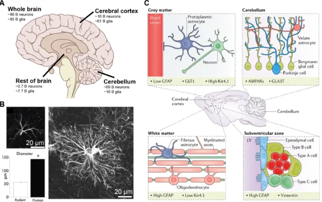
The human brain consists of≈1:1 glia to neuron ratio (GNR) [24]. The GNR ratio was a controversial topic in neuroscience for decades [5] [163]. It was thought that there were 10 times more glia than neurons until it was recently disproved by Herculano- Houzel’s group using the isotropic fractionator method [24] [26]. The GNR generally increases with brain size and varies between brain regions (Figure 1.1 A) and species [24] [380] [371]. The majority of glial cells in the human brain are oligodendrocytes (45-75%) followed by astrocytes (19-40%) and microglia (10%) [380]. Astrocyte tile the entire brain with little overlap to each other (Figure 1.2 A, B, C), thus occupying dis- tinct territorial domains [58]. They were classically divided based on their morphology and astrocyte enriched proteins into: fibrous astrocytes of the white matter, protoplas- mic astrocytes of the gray matter and specialized astrocytes. Specialized astrocytes include: radial glia, retinal Muller cells, Bergman cells, velate astrocytes, tanycytes, pituicytes, Gomori astrocytes, ependymocytes and surface associated astrocytes [373].
Some of these types of astrocytes are illustrated in Figure 1.1 C. Although this rough classification of astrocytes was helpful, defining the heterogeneity of astrocytes is still a work in progress. Astrocytes are defined by their morphology, molecular markers and function. Here, we will focus on protoplasmic astrocytes.
3
Figure 1.1: Heterogeneous distribution and identity of astrocytes in the brain. A) The glia to neuron ratio (GNR) varies between brain areas. Illustration of approximate number of neurons and glia in selected areas of the human brain. B) Human astrocytes (right) are ≈2.5 times bigger in diameter (bottom, left) compared to rodent astrocytes (top, left) as revealed by GFAP labelling. C) Astrocytes exhibit different morphology and molecular markers depending on their locations in the brain.
Protoplasmic astrocytes are found in the gray matter, have radial morphology and are in contact with synapses and blood vessels (top, left). Fibrous astrocytes are found in the white matter, have elongated morphology and are in contact with myelinated axons and oligodendrocytes (bottom, left). Bergman glia and velate astrocytes are found in the cerebellum. Bergman glia extend long apical processes who wrap the dendrites of Purkinje cells. Velate astrocytes are like protoplasmic astrocytes and are found particularly around granule neurons (top, right). The lateral ventricles of the subventricular zone are lined with astrocyte-like B cells(bottom, right). Different astrocyte types differentially express molecular markers including GFAP, GLT1, Kir4.1, AMPARs and vimentin. GFAP, Glutamate transporter 1 (GLT1), inward rectifying potassium channel 4.1( Kir4.1). Panel A was modified from wikimedia commons based on [24], panel B was modified from [254] and panel C was taken from [125].
1.1.3 Morphology
The name astrocyte was derived based on the star-like morphology observed when staining the GFAP enriched intermediate filaments [178]. However, more advanced visualization techniques have now revealed that astrocytes exhibit a bushy, spongiform morphology [299] [334] (Figure 1.2 A, B, C, D). Mouse protoplasmic astrocytes consist of a cell body emanating into 4-10 main branches that arborize into thin, nanoscopic processes often referred to as branchlets and leaflets that are devoid of GFAP. These astrocytes have a diameter of ≈40-60 µm occupying a volume of ≈6.6x10µm3 mostly composed of bracelets and leaflets (≈90-95% of volume). They also extend polarised specialized endfeet that wrap around blood vessels [299] [58] [257] [175]. The terri- tory of a single hippocampal astrocyte territory is estimated to encompass ≈140000 synapses [58] out of which ≈60% are juxtaposed to perisynaptic astrocytic processes (PAPs) [396] [370]. The anatomical and functional relationship between synapses and PAPs was consolidated into the concept of the “tripartite synapse” [14] or “astrocytic cradle” [372]. The intimate relationship between PAPs and synapses allows astrocytes to modulate a multitude of synaptic functions ranging from homeostasis to synap- tic plasticity and information processing [14] [372]. PAP coverage of synapses is a dynamic process [38] [280]. Synapses juxtaposed to PAPs tend to be larger [396], sug- gesting a positive correlation between synaptic strength and PAP coverage of synapses.
PAP motility was reported to be enhanced in response to synaptic activity, increasing synaptic coverage and stability [38]. For example, whisker stimulation in adult mice increases astrocyte coverage of synapses in the barrel cortex [116]. In contrast, a recent study [134] suggests that astrocyte processes might withdraw from mature synapses following long-term potentiation (LTP) induction (at least 30-40 min following LTP) thus enhancing glutamate spillover and cross talk between neighbouring synapses. The mechanism, conditions and heterogeneity of PAP mobility is bound to be very impor- tant for the functional relationship between astrocytes and neurons. The study of nanoscopic PAPs (Figure 1.2 E, F) is not amenable to diffraction limited optical meth- ods. While most studies so far relied on electron microscopy (often associated with fixation issues [182] and static images), super resolution microscopy techniques [147]
[309] [130] and expansion microscopy [389] have potential to “expand” the field. The size, diversity and morphological complexity of astrocytes varies across brain areas (i.e.
specialized astrocytes), within areas [194] and between species [253] [254]. For exam- ple, human astrocytes occupy a volume 15-20 times larger than rodents (Figure 1.1 B), possess more elaborate processes and have unique astrocyte subtypes [253] [254].
However, the comparative biology of astrocytes between species is not well established yet.
1.1.4 Molecular characteristics
There is still no systematic agreement on how to define astrocytes based on molecular markers. GFAP was the first molecular marker associated with astrocytes. However, GFAP is upregulated in pathology, not all healthy astrocytes express GFAP at con- centrations detectable by immunohistochemistry and GFAP is also expressed in cell types other than astrocytes. Thus GFAP expression alone is not sufficient or required
Figure 1.2: Astrocyte morphology. Astrocytes tile the brain with little overlap to each other shown by A) "Brainbow" expression in cortical astrocytes, B) Dye loading of hippocampal, dentate gyrus astrocytes and C) "Traffic light labelling" (tdTomato and eGFP) expression in cortical astrocytes. D) Fine astrocytic process architecture revealed by high resolution optical image of a Lucifer yellow-filled astrocyte. Colour scale represents fractal dimension. E) These nanoscopic processes exhibit high surface area to volume ratio as shown by serial block-face scanning electron microscopy (SBF- SEM) of perisomatic (top) and peripheral (bottom) volumes in Golgi-impregnated astrocytes. F) Astrocyte processes (pink) wrap around presynaptic (yellow) and post- synaptic (blue) synapses as shown by (SBF-SEM) in layer 6 and layer 2/3 of the somatosensory cortex. Panel A was taken from [202]; B was taken from [394]; C is from this thesis; D and E were taken from [334]; F was taken from [194].
for astrocyte identification [175] [234]. Other molecular markers commonly associated with astrocytes include: glutamate transporters GLT-1 and GLAST, connexins (Cx) Cx30 and Cx43, inward rectifying Kir4.1 channels, aldehyde dehydrogenase 1 family member L1 (Aldh1L1), glutamate synthetase, water channel aquaporin 4 (AQP4) and the calcium binding protein S100B [373] [175] [234] (Figure 1.1 C). The protein expres- sion profile of astrocytes shows variability within brain areas. For example, Cx30 and Cx43 are enriched within the barrels of the primary somatosensory cortex (S1) com- pared to septa [146] . Immunohistochemistry and transcriptome analysis revealed layer specific heterogeneity in S1 astrocytes [31] [194]. Also, transcriptomic analysis revealed molecular diversity of astrocytes between different areas [25] [59] [67]. The molecular characteristics of astrocytes is likely regulated by neuronal activity. For example, as- trocytes cultured with neurons change their expression of glutamate transporters [279], glutamine synthase [222], connexins [185], and neuropeptide receptors [230]. Prolonged whisker stimulation increases the expression of glutamate transporters in S1 astrocytes [116]. Disturbance of neuronal layers in S1 abolishes the layer specific heterogeneity of astrocytes [31] [194]. The molecular characteristics of astrocytes also change with age [350] [85] and pathology [175] [125]. Therefore, the molecular identity of astrocytes is heterogeneous and dynamic, changing in response to cues in their environment.
1.1.5 Astrocyte networks
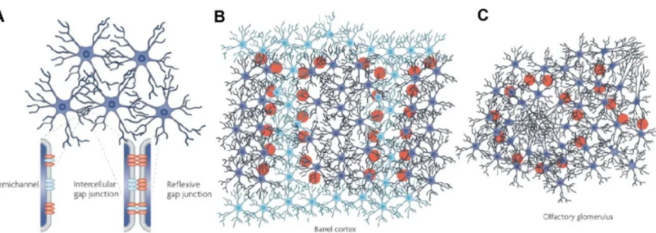
Astrocytes interact with all cell types in the central nervous system (CNS). They are connected to each other via gap junctions (Figure 1.3 A) composed primarily of con- nexins of subtype Cx43 and Cx30 [240]. Cx43 / Cx30 double knockout mice do not exhibit gap-junction mediated K+ [383] and metabolic [308] communication between astrocytes. Connexins assemble into hemichannels. Hemichannels which dock to each other form gap junctions [391] [56]. Unpaired hemichannels were reported to release gliotransmitters such as ATP [346], glutamate [408] and D-serine [227] in the extra- cellular fluid. Gap junctions (≈25%) also connect processes from the same astrocyte [302] (reflexive gap junctions, Figure 1.3 A), potentially coupling different subcellular compartments. Gap junctions allow rapid intercellular exchange of ions and small (≈1 kDa) molecules between astrocytes. This is critical for K+ buffering [383] and calcium wave propagation [105] [143] [114]. Astrocyte gap-junction networks are not random.
For example, Cx30 and Cx43 are focused towards the center of S1 [146] (Figure 1.3 B) and olfactory bulb [68] (Figure 1.3 C) functional units [117]. Gap junction commu- nication can be modulated by neurotransmitters and pathology [117]. Astrocytes are also known to communicate to each other through the release of gliotransmitters such as ATP [114].
Astrocytes communicate with microglia to mediate immune responses in the brain [199] [156] and synaptic pruning [315] [46]. They communicate with oligodendrocytes to modulate myelination [96] which could depend on neuronal activity [151]. Astrocytes cooperate with pericytes and neurons to control blood flow in the brain [229]. Finally, astrocytes communicate with neurons. While the interactions between astrocytes and neurons was considered to be homeostatic, a large amount of evidence suggests that astrocytes may be active partners to neurons in information processing in the brain [14] [16] [312] [88] [104]. In this thesis we will focus on astrocyte - neuron interactions.
Figure 1.3: Astrocytes are connected to each other via gap junctions. A) Gap junctions connect contacting astrocytes to each other. Gap junctions are com- posed of paired connexin (Cx) hemichannels. Same astrocyte process can connect with each other forming reflexive gap junctions. B) In the somatosensory cortex, gap junc- tion proteins (Cx30 & Cx43) are more highly expressed in barrel astrocytes (dark blue) compared to septal astrocytes (light blue). C) In the olfactory bulb, glomerular astro- cyte cell bodies are found in the periphery extending their processes to the center of the functional unit. Images taken from [117].
1.2 Multifunctional role of astrocytes
1.2.1 Homeostasis
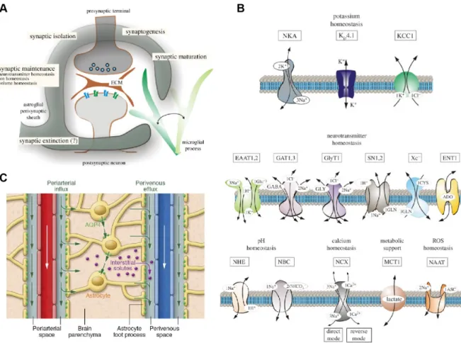
The most well accepted function of astrocytes is regulation of homeostasis. Astrocytes maintain ion homeostasis, regulate the water volume of the brain, maintain the pH, clear excess neurotransmitters (Figure 1.4) and provide metabolic support to neurons (Figure 1.5). While these functions are well accepted, they shouldn’t be overlooked, since they play critical roles in brain function and there is still a lot to be learned.
The spotlight of current research in astrocytes have been their emerging function as elements of information processing mediated by Ca2+ signalling. However, the home- ostatic functions of astrocytes are intimately linked to astrocyte Ca2+ signalling and can influence neuronal activity.
1.2.2 K+ homeostasis
When neurons fire action potentials, they release K+into the extracellular space which, if not cleared, can result in neuronal hyperexcitability. Astrocytes express a high con- centration of K+ channels and transporters that clear excess extracellular K+ through uptake and spatial buffering (Figure 1.4 B, top). These include Kir4.1 channels, Na+/ K+ ATPases (NKA), inward rectifying, Na+/K+/Cl− (NKCC) co-transporters and K+/Cl− co-transporters (KCC) [376] [35]. K+ uptake uses energy derived from the breakdown of ATP (i.e. NKA) or the electrochemical gradient through co-transporters (i.e. NKCC). Spatial buffering relies on passive transport of K+ ions from regions of high [K+] to regions of low [K+] through Kirchannels and the gap-junction coupled net-
Figure 1.4: Astrocytes maintain homeostasis in the brain. A) The "astro- cytic cradle" or "tripartite synapse" represents the functional and anatomical relation- ship between astrocytes and neurons at synapses. Astrocyte processes wrap around synapses and play important roles in the function, maturation, maintenance, isolation and elimination of synapses. B) Astrocytes express a plethora of proteins involved in the homeostasis of ions, neurotransmitters, metabolism, pH and reactive oxygen species summarized in this schematic. C) They also express aquaporin (AQP) chan- nels primarily located at their perivascular endfeet, allowing them to maintain water homeostasis and clear waste accumulated in the brain through the glymphatic system.
Panel A & B images taken from [372]; C taken from [206].
work of astrocytes [208] [35]. Disturbances to astrocyte mediated K+ homeostasis can lead to epilepsy [80] and a range of other disorders summarized in [35]. Interestingly, K+uptake could also be actively regulated through [Ca2+]idependent mechanisms. For example, [Ca2+]i elevation can increase the activity of Na+/Ca2+ exchangers (NCX), raising [Na+] that in turn activates Na+/ K+ ATPase mediated uptake of K+ and a decrease in extracellular [Na+]. This leads to neuronal hyperpolarization, reduction in excitatory synaptic activity and a decrease in synaptic failure [385]. Also computer simulations suggest that NCXs are sufficient for the induction of microdomain [Ca2+]i signals in PAPs [382]. These suggest that astrocytes may be able to modulate neuronal activity through passive and active, [Ca2+]i dependent, regulation of K+.
1.2.3 Water homeostasis, aquaporins and the glymphatic sys- tem
Astrocytes maintain water homeostasis and regulate extracellular volume in the brain by virtue of high expression of aquaporin (AQP) channels. AQP4 is the most preva- lent AQP in astrocytes, predominantly located at perivascular endfeet and, to a lesser extent, processes around excitatory synapses [239]. AQP4 channels were also linked to astrocyte [Ca2+]i elevations induced by osmotic stress [358]. AQP4 are also critical for the operation of the glymphatic system [150] (Figure 1.4 C). The glymphatic system clears cell waste accumulated in the brain. The system consists of a periarterial cere- brospinal fluid influx route and a perivenous interstitial fluid clearance path, coupled by convective flow facilitated by astrocytic AQP4 channels [155]. Interestingly, AQP4 isoforms (M23) regulate the motility of astrocyte process and glutamatergic synaptic transmission [75]. The position of astrocyte processes in relation to synapses is likely to have diverse effects on synaptic activity depending on the specific proteins expressed at the respective processes (i.e. transporters, channels and receptors).
1.2.4 Neurotransmitter homeostasis
Astrocytes rapidly remove neurotransmitters released by synapses in the extracellular fluid (Figure 1.4 B, middle), and constrain their spill over to nearby synapses [305]
(Figure 1.4 A). Astrocytes uptake the majority of glutamate released by synapses (>80%) through high affinity excitatory amino acid transporters (EAAT) 1 and 2, optimizing neuronal function and preventing excitotoxicity [213]. Considering that astrocytes can also release glutamate [214] astrocytes may maintain homeostasis by balancing the opposing functions of glutamate uptake and release [213]. Uptaken glu- tamate is converted to glutamine through the enzymatic action of glutamine synthetase that is expressed in astrocytes but not neurons [218], or used as energy by converting it to a-ketoglutarate used in ATP synthesis [323]. Glutamine transported to neu- rons is resynthesized to glutamate or gamma aminobutyric acid (GABA) and concen- trated into vesicles to be released as a neurotransmitters. This is referred to as the glutamate/GABA-glutamine cycle [28]. Similarly, astrocytes uptake GABA through GABA transporters (GAT) 1 and 3 where it is converted to glutamine or metabolized to succinic semialdehyde [106]. The number, position and distance of EAATs (and
Figure 1.5: Astrocyte regulate the energy supply of the brain. Current model on how astrocytes regulate energy supply. Astrocytes regulate the supply of nutrients by modulating the contractility of pericytes. Astrocyte [Ca2+]i elevation activates an enzymatic cascade that leads to prostaglandin release, relaxing pericytes and dilating blood vessels. Glucose can be taken up by neurons and astrocytes directly and used in glycolysis. In astrocytes glucose can be converted to glycogen for storage. Alternatively, astrocytes can produce lactate and shuttle it to neurons through monocarboxylate transporters (MCT) to use as energy source. For more detailed description of the model see [252]. Image taken from [252].
other transmitter transporters) relative to synapses is likely to be important in synap- tic modulation by controlling the spatiotemporal characteristics of neurotransmitter concentration availability at the synapse. For example, surface diffusion of glutamate transporters is modulated by synaptic activity and can influence synaptic transmis- sion [237]. Similarly transient receptor potential cation channel subfamily A member 1 (TRPA1) mediated [Ca2+]i elevation in astrocytes increase GAT-3 insertion in the membrane thus regulating GABAergic signaling [333]. Thus astrocyte [Ca2+]i elevation could lead to insertion of transporters near synapses to modulate synaptic activity.
1.2.5 Metabolic support
The human brain consumes ≈20% of the body’s energy, mainly to reverse ion fluxes involved in synaptic transmission [128]. Disruption in brain energy supply, quickly
leads to cell injury and death. Therefore, it is important to dynamically match energy supply to fluctuating demands. The response of increasing blood flow in response to higher neuronal activity is termed functional hyperaemia [21]. Astrocytes cover blood vessels with specialized endfeet [220] and enwrap synapses, the main energy consumers of the brain, with their fine processes [6] (Figure 1.5). They are thus in a privileged position to regulate functional hyperaemia. Early studies revealed that astrocyte [Ca2+]i elevation led to dynamic control of microcirculation, mediated by arachidonic acid derivatives [416] [236] [120] [353]. How astrocytes are involved in functional hyperaemia is still under intense investigation and debate, but it seems to involve the coordinated action between multiple cell types [252] (Figure 1.5). One important issue was that somatic Ca2+ signals in astrocytes triggered by neuronal activity were too slow to have a causative effect on neurovascular coupling. However, neurovascular coupling might be mediated by faster, inositol trisphosphate receptor (IP3R)-2 independent [Ca2+]i elevation. Astrocyte [Ca2+]i elevation (i.e. through ATP-gated P2X1 channels) can trigger an enzymatic cascade leading to the release of prostaglandin E2 acting on pericytes who can relax and dilate capillaries [229].
Diffused glucose then enters both neurons (via GLT3) and astrocytes (via GLT1) to be used for glycolysis [226]. In astrocytes glucose can be converted and stored as glycogen.
Pyruvate produced by glycolysis can be used by mitochondria for ATP production or be converted to lactate. According to the lactate shuttle hypothesis, lactate produced by astrocytes can be delivered to neurons (via monocarboxylate transporters) as an energy source [210] but the idea is still controversial [226]. Overall, astrocytes, along with other glia and neurons, play important roles in the regulation of energy supply in the brain.
The discovery of fast Ca2+ signals in astrocyte processes andin vivo preparations may help elucidate the functional role of astrocytes in functional hyperaemia [258]
1.2.6 Perspective
While Ca2+ mediated gliotransmission has gained enormous attention and popularity as a means for astrocytes to modulate synaptic activity, the homeostatic functions of astrocytes in regulating synaptic circuits shouldn’t be neglected. By mediating K+ uptake astrocytes control synaptic excitability. The motility of astrocyte processes and recruitment of channels, exchangers and pumps in PAPs could modulate neuronal activity by controlling the rate of transmitter and ion fluxes. Homeostatic processes can also be actively mediated by astrocytic Ca2+ signals and in turn can influence neuronal activity. It is thus possible that many of the effects attributed to the Ca2+
induced release of gliotransmitters may have been artifacts of astrocytes maintaining homeostasis. Considering that astrocytes are the principal homeostatic cells of the brain, would it be surprising if astrocyte Ca2+ signals often triggered by artificial neuronal stimuli, are responses of astrocytes trying to re-establish homeostasis?
1.2.7 Control of synapse form and function
Beyond their homeostatic function, astrocytes are required for the formation and func- tion of synapses. Seminal work by the Barres laboratory revealed that neurons grown in co-cultures with astrocytes had seven times more mature, functional synapses compared
Figure 1.6: Astrocytes control synaptic form, function and elimination. A) Astrocytes induce the formation of structural synapses by releasing thrombospondins (TSPs) and hevin. B) They can induce functional synapse formation through the release of glypicans and C) eliminate synapses directly through MEGF10 and MERTK mediated phagocytosis or indirectly by tagging synapses for elimination by microglia.
Image taken from [6].
to isolated neuron cultures [287] [365]. The role of astrocytes is synapse formation was demonstrated across species and types of synapse [6]. Astrocytes control the formation of synaptic structure, function and elimination by releasing different molecules. For example, releasing thrombospondins [72] and hevin [189] induces structural synapse formation (Figure 1.6 A). These synapses are presynaptically active but postsynap- tically silent. Releasing the hevin antagonist SPARC inhibits synapse formation and function [205] [4]. Astrocyte can enhance pre-synaptic function by releasing cholesterol [221] and postsynaptic function by regulating the number (tumor necrosis factor alpha (TNFα)) [343] and localization (Glypicans) [7] of AMPARs (Figure 1.6 B). Astrocytes also regulate synapse elimination directly through phagocytosis [74] and indirectly by tagging synapses for elimination by microglia [315] [46] (Figure 1.6 C). The hetero- geneity of astrocytes and the mechanisms of synaptogenic signal release in the control of synaptic development is currently an active frontier in the field. Interestingly, there is in vitro evidence suggesting that astrocyte synaptogenic factor release can be reg- ulated by ATP (thrombospondins) [362] and glutamate (SPARC, TNF-a) [159] [342].
Also, the effect on synaptogenic factors could have synapse specific roles [301]. Given that astrocytes are known to respond to neurotransmitters (i.e. ATP and glutamate) with [Ca2+]i elevations it will be interesting to determine if the release of synaptogenic factors is calcium dependent and to elucidate the mechanisms and conditions of their releasein vivo.
1.2.8 Reactive astrocytes and pathology
Astrocytes are involved in the coordinated multicellular response to brain trauma, dis- ease and inflammation. Astrocytes respond to pathological conditions and undergo a spectrum of morphological and functional changes; collectively referred to as reactive astrogliosis [338]. Reactive astrocytes were originally characterized by overexpression of GFAP and hypertrophy [36]. It is now becoming evident that astrogliosis is not a binary state, but rather a heterogeneous, graded continuum of context dependent changes in gene expression, morphology and function [9] [228]. Recent collaborative
work between the Barres and Steven laboratories [199] revealed two distinct types of re- active astrocytes termed A1 and A2. A1 astrocytes are induced by activated microglial secretion of Il-1α, TNF and C1q cytokines and are lethal to neurons and oligodendro- cytes. In contrast, A2 astrocytes upregulate neurotrophic genes and promote neuron survival. Considering the volatile nature of astrocyte identity, gene expression and function during pathological conditions it is worth asking whether experiments assess- ing physiological aspects of astrocytes in cultures, slices and maybe even in vivo are investigating a reactive state of astrocytes. Indeed, Takano et al. [354] showed that astrocytes in slice preparation exhibit observable reactive changes shortly (≈90 mins) after acute slice preparation. Another intriguing question is how different types of reactive astrocytes contribute to pathology and whether selective manipulation of as- trocytes using pharmacology or gene therapy can be used to treat disease. Astrocytes are involved in epilepsy, Alzheimer’s disease, Huntington’s disease, amyotrophic lat- eral sclerosis, CNS trauma, stroke, infections, cancer, psychiatric disorders, and others [338]. Reactive astrocytes also exhibit aberrant Ca2+ signalling generally character- ized by increased amplitude, frequency and duration [336]. Astrocytes rapidly respond with [Ca2+]i elevation to kainate induced epilepsy [138]. Reactive astrocytes display high frequency Ca2+ signals and Ca2+ waves close to amyloid plaques in Alzheimer’s disease model mice in vivo [90] [188]. The amplitude of Ca2+ signals was higher in the ischemic core compared to the penumbra region in acute stroke models [94]. Addition- ally, metabotropic glutamate receptors (mGluR5), often implicated in Ca2+ induced gliotransmission [260], while normally downregulated with age [350], are upregulated in reactive astrocytes [17] [366] [177]. Other receptors implicated in Ca2+ signalling are also upregulated in reactive astrocytes. For example, purinergic P2Y1 receptors [90] [187] [390] and TRP channels (TRPC4) [97] are upregulated during pathology.
Reactive astrocytes can in turn release gliotransmitters. For example, they can re- lease tonic GABA though bestrophin-1 channels or GABA transporter reversal [158]
[401] [73] and release neurotoxic levels of glutamate [375] [306]. Reactive astrocyte mGluR5 mediated [Ca2+]i signals may lead to thrombospondin 1 (TSP1) upregulation which contributes to synaptic remodelling and plasticity [177]. Therefore, the reactive state of astrocytes might be involved in Ca2+ mediated synaptic modulation by as- trocytes. Similarly, the sensitivity of astrocytes to neuronal stimulation might depend on pathology. For example, striatal astrocytes of wild type mice did not respond to cortical stimulation while Huntington’s disease model astrocytes responded robustly with Ca2+ signals [157]. Since every experimental preparation disturbs the system in some way, different reactive responses might be induced in astrocytes, depending on the specific experimental protocol. It is thus very important to maintain physiological con- ditions in experimental preparations assessing the functions of (healthy/non-reactive) astrocytes.
1.3 Astrocyte calcium signalling
Astrocytes, just like other glia, are electrically silent [190] as revealed by traditional electrophysiology techniques. Since astrocytes were not amenable to electrophysiolog- ical methods used to advance our understanding of neurons, the functional roles of


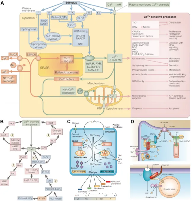
![Figure 1.7: Ca 2+ sources and sinks. The [Ca 2+ ] i of the cell is dynamically reg- reg-ulated through channels, pumps and transporters expressed in the plasma membrane and organelles of the cell](https://thumb-ap.123doks.com/thumbv2/123deta/6956899.2272986/35.892.154.724.189.763/figure-sources-dynamically-channels-transporters-expressed-membrane-organelles.webp)