Three-dimensional average face
‑
Introduction
Contemporary orthodontic treatment emphasizes i m p r o v i n g n o t o n l y t h e d e n t a l a n d s k e l e t a l relationships but also the soft tissue aspects of orthodontic problems1). Conventionally, the lateral appearance has mainly been evaluated for facial soft tissue morphology via lateral cephalometric r a d i o g r a p h s2 ). U s i n g l a t e r a l c e p h a l o m e t r i c radiographs, the dental and skeletal relationship were analyzed and compared with normative data to make an orthodontic diagnosis3). However, since cephalometric radiography involves the projection of an image onto the mid‑sagittal plane, it has been impossible to assess the three‑dimensional (3D) characteristics of the facial soft tissue.
Application of the three-dimensional average face in orthodontic practice
JOGAKI CHIHIRO, TOME WAKAKO, SASAKI MIE, SHIMIZU FUMI, KITAI NORIYUKI
Key words:3D, average face, upper central incisor labial inclination, anterior crossbite
Recently, new approaches have been developed to allow the facial soft tissue to be analyzed in 3D, but most of the current evaluation methods involve linear and angular measurements between numerous landmarks4, 5). Several studies have assessed the facial morphology using an average face model which has provided overall characteristics for facial growth6), ethnic characteristics7), and gender differences8, 9). However, a few reports concerning the evaluation of the soft tissue for the diagnosis and the facial eff ects of orthodontic treatment have been published10). Unfortunately, no study has yet compared the 3D average face before correcting a functional anterior crossbite with that immediately after the correction of the crossbite.
The purpose of the present study was to present
J. Gifu Dent. Soc.
Vol.46, No.1, 17 〜 21 June, 2019
ORIGINAL ARTICLES
between the individual landmarks were averaged and visualized to obtain an average face.
The average face of the patients in maxillary incisor protrusion and control groups. The average face of the patients in the functional crossbite group were created before and after the anterior crossbites were improved.
Results
The average face of the maxillary incisor protrusion group showed that the soft tissue below the nose and above the vermilion border placed anteriorly comparing to the control group (Figure 2). A comparison between the average face at pre‑
and post‑treatment showed that soft tissue below the nose and above the vermilion border moved anteriorly, and soft tissue in the submental region rotated backward with increased lower facial height over the period of observation (Figure 3).
Discussion
Compared with the normative average face, the entire upper lip was inclined to the labial side in the 3D average face of the maxillary incisor protrusion group. We believe that by creating a 3D average face, we can visualize the differences in the lip features in the maxillary incisor region from multiple directions and more clearly describe the characteristics of the facial soft tissue7, 11‑13).
The image of the 3D average face following correction of a functional anterior crossbite showed that the upper lip had moved forward while the lower lip had moved backward. The lateral image showed backward rotation of the mandibular soft tissue, resulting in an increase in the lower facial height. This visualizing method was able to show the overall characteristics of the facial changes before and after treatment. Of note, these changes may have included the effects of general growth.
However, the backward rotation of the mandibular soft tissue was considered to have been induced chiefly by treatment, as the treatment time was within six months, making it unlikely that the face had been altered signifi cantly by growth over such a short time period. The present method may also be useful for detecting changes in the facial morphology on correction of a functional posterior crossbite14) applying method of the 3D average face while
comparing (1) the normative average face with the average face of the patients with maxillary central incisor labial inclination and (2) the characteristics before and after the correction of a functional anterior crossbite.
Subjects
The subjects were 20 patients (maxillary incisor protrusion group; 9 males and 11 females; mean age, 9 years 10 months; age range, 7 years 5 months to 14 years 2 months) who exhibited Class I malocclusion with labial inclination of the upper central incisors, 15 patients (control group; 7 males and 8 females; mean age, 9 years 0 month; age range, 8 years 0 month to 14 years 4 months) who exhibited skeletal Class I malocclusion with standard inclination of the upper central incisors, and 25 patients (functional crossbite group; 7 males; 18 females; mean age: 8 years 6 months; age range: 6 years 10 months to 10 years 9 months) with an anterior crossbite and premature contact on at least 1 central incisor. The anterior crossbites were treated by labial movement of the upper incisors and completed within six months.
The subjects agreed to participate after receiving a full explanation of the studyʼs purpose and design.
The implementation of the study was approved by the Ethics Committee of the Asahi University Graduate School of Dentistry (No. 26003).
U t i l i z i n g a 3 D s u r f a c e ‑ i m a g i n g d e v i c e (3dMDcranial System; 3dMD, Atlanta, GA, USA), facial images were recorded for each subject.
The images were recorded with the subject in a seated and natural head position. The lips were closed, and the teeth were in slight contact in the intercuspal position. Three‑dimensional image data were transferred to a personal computer (HP Z210 SFF Workstation; Hewlett‑Packard Company, San Francisco, CA, USA) and analyzed using an image processing software program (3D‑Rugle Version 5.5;
Medic Engineering, Kyoto, Japan). An average face was created from the facial wire frame image using 25 landmarks for identifying the composite elements of the face (Figure 1). The positional data of the facial 25 points were normalized. The normalized distance
Three-dimensional average face
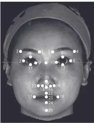
Figure 1. The 25 facial points for identifying the composing elements of the face to create the average face.(1, the inner end of the right eyebrow; 2, the inner end of the left eyebrow; 3, the outer end of the right eyebrow; 4, the outer end of the left eyebrow; 5, the most superior point of the right eye fi ssure; 6, the most superior point of the left eye fi ssure 7, the outer commissure of the right eye fi ssure; 8, the outer commissure of the left eye fissure; 9, the inner commissure of the right eye fi ssure; 10, the inner commissure of the left eye fi ssure;
11, the most inferior point of the right eye fi ssure; 12, the most inferior point of the left eye fissure; 13, the facial insection of the right alar base; 14, the facial insection of the left alar base; 15, the midpoint of the right nostril; 16, the midpoint of the left nostril; 17, the point located at right labial commissure; 18, the point located at left labial commissure; 19, the most anterior median point of the nasal root; 20, the most anterior median point of the nasal tip; 21, the deepest point on the nasolabial soft tissue contour between the columella crest and upper lip; 22, the median point of the vermilion line of the upper lip; 23, the median point of the horizontal labial fi ssure; 24, the median point of the vermilion line of the lower lip; 25, the median point of most anterior to the mandible).
Figure 2. Average face under the eyes created based on 3D facial images in the maxillary incisor protrusion and control groups (A, Maxillary nincisor protrusion group image; B, Control group image; left, frontal image; middle, three-quarter image; right, lateral image).
Figure 3. Average face under the eyes created based on 3D facial images before and after treatment (A, pre- treatment image; B, post-treatment image; left, frontal image; middle, three-quarter image; right, lateral image).
ୖ ᕥྑ
䝰䝜䜽䝻 ᇛᇉ༓ᐶ
ୖ ᕥྑ
䝰䝜䜽䝻 ᇛᇉ༓ᐶ
A B
ୖ ᕥྑ
䝰䝜䜽䝻 ᇛᇉ༓ᐶ
ୖ ᕥྑ
䝰䝜䜽䝻 ᇛᇉ༓ᐶ
ୖ ᕥྑ
䝰䝜䜽䝻 ᇛᇉ༓ᐶ
A B
of facial morphology surface changes in untreated children from 12 to 14 years of age.
. 2008; 134: 751‑760.
7 ) Gor T, Kau CH, English JD, Lee RP and Borbely P.
Three‑dimensional comparison of facial morphology in white populations in Budapest, Hungary, and
Houston, Texas. .
2010; 137: 424‑432.
8 ) Toma AM, Zhurov A, Playle R, Ong E and Richmond S. Reproducibility of facial soft tissue landmarks on 3D laser‑scanned facial images.
. 2009; 12: 33‑42.
9 ) Bugaighis I, Mattick CR, Tiddeman B and Hobson R. Three‑dimensional gender diff erences in facial form of children in the North East of England.
. 2013; 35: 295‑304.
10) Nanda V, Gutman B, Bar E, Alghamdi S, Tetradis S, Lusis AJ, Eskin E and Moon W. Quantitative analysis of 3‑dimensional facial soft tissue photographic images: technical methods and clinical application.
2015; 16: 21.
11) 井口 暁,中原リザ子.良好な側貌を有する日本人正 常咬合者の顔面形態の三次元的評価.Orthod Waves‑
Jpn Ed. 2009; 68: 124‑133.
12) Kau CH, Zhurov A, Richmond S, Cronin A, Savio C and Mallorie C. Facial templates: a new perspective in three dimensions. . 2006; 9:
10‑17.
13) Bugaighis I, Tiddeman B, Mattick CR and Hobson R.
3D comparison of average faces in subjects with oral clefts. . 2014; 36: 365‑372.
14) Primozic J, Richmond S, Kau CH, Zhurov A and Ovsenik M. Three‑dimensional evaluation of early crossbite correction: a longitudinal study.
. 2013; 35: 7‑13.
15) Foot R, Dalci O, Gonzales C, Tarraf NE and Darendeliler MA. The short‑term skeleto‑dental eff ects of a new spring for the intrusion of maxillary posterior teeth in open bite patients.
2014; 15: 56.
and in the intrusion of the posterior teeth when using skeletal anchorage devices in preadolescent patients15).
Using the visualizing method described in the present report, improved insight into the overall characteristics of facial morphology was gained without the need for radiation exposure. The present study provides useful information for the continuous and repeated evaluations of treatment outcomes in preadolescent children due to the lack of any need for radiation exposure.
Acknowledgements
The authors would like to thank Brian Quinn, (Japan Medical Communication) for the grammatical correction of the manuscript.
REFERENCES
1 ) Proffi t WR. Contemporary orthodontics 4th Edition.
St. Louis: Mosby Year Book Inc.; 2007: p.167‑233.
2 ) Hsu BS. Comparisons of the fi ve analytic reference lines of the horizontal lip position: their consistency
and sensitivity. .
1993; 104: 355‑360.
3 ) Ahsan A, Yamaki M, Hossain Z and Saito I. Craniofacial cephalometric analysis of Bangladeshi and Japanese adults with normal occlusion and balanced faces: A comparative study. 2013; 2: 7‑15.
4 ) Toma AM, Zhurov A, Playle R and Richmond S. A three‑dimensional look for facial diff erences between males and females in a British‑Caucasian sample aged 151/2 years old. . 2008; 11:
180‑185.
5 ) Berneburg M, Schubert C, von Einem C, Schaupp E, Moller M and Goz G. The reproducibility of landmarks on three‑dimensional images of 4‑to 6‑year‑old children. . 2010; 71: 256‑
264.
6 ) Kau CH and Richmond S. Three‑dimensional analysis
Three-dimensional average face
矯正歯科臨床における三次元平均顔の活用
城 垣 千 寛 留 和香子 佐々木 美 枝 清 水 ふ み 北 井 則 行
本研究の目的は,矯正歯科臨床において,三次元平均顔重ね合わせ法を活用することである.上顎中切 歯の唇側傾斜を伴う骨格性Ⅰ級患者(上顎切歯前突群),上顎中切歯の標準的な歯軸傾斜を伴う骨格性Ⅰ級 患者(対照群),少なくとも 1 歯以上の永久中切歯に早期接触を認める機能性反対咬合を伴う患者(機能性 反対咬合群)を被検者とした.機能性反対咬合群では,上顎中切歯の唇側移動を行うことによって前歯部 被蓋を 6 か月以内に改善した.各被検者に対して,非接触型三次元デジタルカメラ(3dMDcranial System, 3dMD, Atlanta, GA, USA)を用いて,顔面軟組織形態三次元画像を撮影した.この三次元顔面軟組織画像 を用いて,それぞれの被検者群の平均顔を,顔の構成要素を代表する軟組織上の 25 点に基づいたワイヤー フレーム画像から作成した.機能性反対咬合群では,被蓋改善前後の平均顔を作成した.
その結果,上顎切歯前突群では上唇全体が前突していることが示唆された.機能性反対咬合においては,
上顎中切歯唇側傾斜により被蓋改善後,上唇全体が前方へ移動し,下唇全体が後方へ移動し,オトガイが後 下方へ回転し,下顔面高が大きくなっていることが示された.
平均顔を用いた顔面軟組織形態の変化を評価する方法により,放射線被曝なしで,診断に有用な顔面全体 の特徴や治療による変化を表すことができた.放射線被曝を伴わないことから,若年者における短期間での 治療結果の評価に有用であると考えられる.
キーワード:三次元,平均顔,上顎中切歯の唇側傾斜,機能性反対咬合
朝日大学歯学部口腔構造機能発育学講座歯科矯正学分野
〒 501‑0296 瑞穂市穂積 1851
(平成 31 年 4 月 5 日受理)
岐 歯 学 誌
46 巻 1 号 17 〜 21 2019 年 6 月