Original paper (regular paper)
Comprehensive Analysis of Aerobic Benzene-Degrading Microorganisms
Using Modified and Conventional Stable-Isotope Probing Methods
Yuanbai Pan, Nobuhiko Nomura, Toshiaki Nakajima and Hiroo Uchiyama*
Graduate School of Life and Environmental Sciences, University of Tsukuba, Tsukuba, Ibaraki 305–8572, Japan
* TEL: +81–29–853–4927 FAX: +81–29–853–4927 * E-mail: [email protected] (Received; 2 November, 2013/Accepted; 24 November, 2013)
The contamination of groundwater and soil by benzene is a serious environmental problem worldwide, and cleaning polluted areas by bioremediation requires an understanding of the microbial community involved. To archive this purpose, benzene-assimilating degraders were classified in the bacterial community using conventional stable-isotope probing (SIP), whereas candidate co-metabolic degraders and non-degraders of benzene were classified with modified SIP (SIP-D) and 16S ribosomal RNA-targeted SIP. Conventional plating method was also accompanied. Consequently, bacteria belonging
Rhodo-coccus and Pseudomonas were mainly identified as benzene-assimilating degraders, one type of Pseudomonas was a
candi-date co-metabolic degrader, and Hydrogenophaga was a non-degrader, respectively. Some strains of the candicandi-date co- metabolic degraders and non-degraders of benzene that exhibited no benzene-assimilating capability displayed an active benzene-assimilating capability, which was confirmed with the culture-dependent method. Furthermore, functional gene analysis suggested that most benzene-assimilating degraders opened the benzene ring via a catechol ortho-cleavage pathway. We concluded that a comprehensive analysis using a combination of the two SIP methods would be effective for understanding the wide bacterial community in a chemically polluted environment.
Key words: bioremediation, benzene, stable-isotope probing, bacteria community, functional genes
1. Introduction
Petroleum pollution has become a serious environmental problem because it affects groundwater resources and causes soil deterioration. Benzene, the main mono-aromatic component in gasoline, is widely produced and used in many applications 2). However, its volatilization and carcinogenicity negatively influence human health 35). Bioremediation has recently been used as a low-cost and eco-friendly method to degrade benzene 16). The reaction-effective microorganism community must be defined and controlled in bioremedi-ation treatments. Head et al. 12) proposed a degradation network of oil in marine environments, consisting of an assimilating degrader, co-metabolic degrader 14), and non-degrader. A similar microbial community is also thought to exist and operate in soil environments. Many kinds of benzene-degrading bacteria have been described to date; sulfate-reducing 17), iron-reducing 1), nitrate-reducing 6), perchlorate-reducing 34), and methanogenic bacteria 9) as anaerobes, and various assimilating bacteria in aerobic envi-ronments 19,37). However, these previous studies mainly focused on assimilating degraders, and did not fully determine the bacterial community involved in benzene degradation. In this study, we identified an assimilating degrader, co-metabolic degrader, and non-degrader.
Culture-independent approaches have recently been devel-oped to observe microbial communities and their functions. Stable-Isotope Probing (SIP) is a particularly powerful method used to identify assimilating degraders. This method is based on the incorporation of stable isotopes (e.g., 13C) into cellular components 25). SIP has been used to examine the aerobic metabolism of methane 22), benzene 37), toluene 32), and many other chemical compounds. However, this conven-tional 13C-labeling method can only detect assimilating degraders 5). Therefore, we developed a modified SIP method to detect dissimilating bacteria (SIP-D), and this has been recommended for the biological dechlorination reaction 38). Here, tetrachloroethylene (PCE) dehalorespiring bacteria could gain energy via dehalorespiration in the presence of PCE, and grew with the assimilation of a 13C-labeled carbon source, which formed 13C-labeled DNA. However, dehalor-espiring bacteria could neither grow nor assimilate carbon in the absence of PCE. This physiological difference could be investigated with polymerase chain reaction-denaturing gradient gel electrophoresis (PCR-DGGE); differences between specific 13C-DNA bands in the presence or absence of a pollutant. In our previous study, we successfully identi-fied Dehalococcoides ethenogenes 195 as a dehalorespiring bacterium by SIP-D, subsequently applied this method to an artificially PCE-contaminated microcosm 38), and identified
the indigenous active dehalorespiring bacteria Dehalobacter sp. as a candidate dissimilative PCE-degrading bacteria.
The general objective of this study was to comprehen-sively analyze the bacterial community involved in degrading benzene under aerobic conditions. As shown in Fig. 1A, we performed SIP to investigate benzene-assimilating degraders using 13C
6-labeled benzene, and SIP-D to investigate candi-date co-metabolic benzene degraders using 13C
2-labeled acetate. Following SIP-D, to distinguish non-degraders of benzene that would also be identified with SIP-D, 16S ribo-somal (rRNA) gene-targeted SIP was performed. In this study of SIP-D, benzene, a pollutant that can be assimilated, was used in SIP-D instead of non-assimilative PCE, and the effectiveness of SIP-D for assimilative chemical pollutants was evaluated for the first time.
Accompanied by the culture-independent method, the conventional culture method was performed to investigate whether the benzene degraders identified by the culture-independent method were culturable or not (Fig. 1B). Furthermore, benzene degradation begins with the addi-tion of [OH-] to the benzene ring and proceeds via the catechol pathway under aerobic conditions 8). Thus, the key ring-opening functional genes, catechol 1,2-dioxygenase (C12O) 11) gene and catechol 2,3-dioxygenase (C23O) 21) gene, were quantified using real-time PCR (qPCR; Fig. 1C).
2. Materials and Methods
2.1 Benzene degradation assay and product measurement To assess benzene removal from soil by bacteria, we used benzene-contaminated soil (sandy loam) collected from a refinery site, in which benzene concentration was approxi-mately 500 μM. Benzene degradation test was performed with 5 g soil and 35 mL minimal medium containing (per liter) 0.5 g K2HPO4, 0.5 g KH2PO4, 2 g NaCl, 0.5 g (NH4)2SO4, 0.5 g MgSO4·7H2O, 0.1 g FeSO4·7H2O, and 100 mL tap water, pH 7.0, with 562.4 μM benzene as the sole carbon source in sealed glass bottles without degassing at 30°C in darkness without shaking. Negative slurry controls were prepared using the same conditions via autoclaving for 20 min at 120°C prior to the addition of benzene. Benzene was measured using a flame ionization detector on a gas chromatography system (GC-2010, Shimadzu, Japan) with 100 μL headspace gas from a bottle 26). The column was the fused-silica capillary column DB-1 (30 m long, 0.25-mm internal diameter, 0.25-mm film thickness; J & W Scientific Co., USA). The injector, oven, and detector temperatures were 270°C, 100°C, and 250°C, respectively. The split:splitless ratio was 1 : 15. Nitrogen (99.99% chemical purity, 0.55 mL/ min) was used as the carrier gas (detection limit of benzene, 3 μM). The liquid phase and total value of benzene in the bottle were calculated according to Henry’s law. The concen-tration of catechol, the intermediate product in aerobic benzene degradation, was measured with high-performance liquid chromatography (Waters Inc., USA) using a Symmetry C18 column (3.5 μm, 2.1×150 mm) with 30 mM NaH2PO4 and methanol (20 : 80, v/v) as the mobile phase (0.2 mL/ min) and a 239-nm fluorescence detector. All incubations were performed in triplicate.
2.2 DNA extraction, ultracentrifugation, and fractionation Slurry samples for culture-independent analysis were collected using a syringe at appropriate intervals and analyzed. The collected samples (5 mL) were centrifuged (10,000×g) into Lysing Matrix E tubes (MP Biomedicals, Santa Ana, CA, USA). After the samples were centrifuged and the liquid phase was removed, DNAs were extracted directly from each precipitation using a FASTDNA® Spin Kit for Soil (MP Biomedicals), loaded onto CsCl gradients in centrifuge tubes (2 mL, Quick-Seal®, Polyallomer, 11×32 mm; Beckman Coulter, Inc., USA) with CsCl gradient buffer containing 0.8 g CsCl, 775 μL TE (Tris ethylenediaminetetraacetic acid [EDTA]) and 25 μL ethidium bromide (10 mg/mL), and centrifuged at 354,100×g (20°C) for 20 h in a TLA 120.2 rotor (OptimaTM; Beckman Coulter). 13C-Labeled heavy DNA and unlabeled light DNA were detected with 365-nm ultraviolet (UV) after ultracentrifugation, and the separated fractions (100 μL) were directly collected using a 1-mL syringe. Ethidium bromide was removed from the samples using TE-saturated 3-methyl-1-butanol. DNAs were purified via ethanol precipitation, re-suspended in TE buffer, and Fig. 1. Details of the analytical methods used in this study.
Stable-isotope probing (SIP) and modified SIP for dissimilating bac-teria (SIP-D) are culture-independent methods (A) for identi-fying benzene-assimilating bacteria and co-metabolic bacteria, respectively. [13C] Benzene and [13C] acetate were used to label
DNA, respectively. The non-degraders of benzene were also identified in SIP-D. Therefore, RNA-based SIP was used to confirm candidate degraders in the presence of benzene using [13C] acetate. These bacteria were identified in PCR-DGGE
analysis. Degraders were also isolated from contaminated soil (B) for comparison with the results obtained by the culture-independent method. Functional gene analysis of the C12O and C23O genes using real-time PCR revealed their behavior during degradation (C).
stored at –30°C until use. 2.3 Stable-isotope probing (SIP)
SIP was performed to identify benzene-assimilating degraders in benzene-contaminated soil. Samples were treated with 562.4 μM 13C
6-labeled benzene (99% chemical purity; Cambridge Isotope Laboratories, Inc., USA) as a labeled carbon source at 30°C in darkness without shaking. Total DNA was extracted on day 2 (first degradation phase) and day 4 (second degradation phase) and fractionated via ultracentrifugation as described above.
2.4 Stable-isotope probing for dissimilation (SIP-D) To identify candidate co-metabolic benzene degraders, we treated the slurry samples described above with 562.4 μM 12C
6 benzene (99% chemical purity; Wako, Inc., Japan) in sealed glass bottles incubated at 30°C in darkness without shaking. Two time points (days 2 and 4) were selected for the addition of the labeling marker: 873.4 μM [13C
2] acetate (99% chemical purity; Cambridge Isotope Laboratories). As the most abundant intermediate of organic matter degra-dation in anoxic soil 13), [13C] acetate was used to identify the candidate co-metabolic benzene degraders involved in benzene degradation. On day 2 (first phase), [13C] acetate was added to make benzene-present (WITH-B) samples. Total DNA was extracted from these samples after 24 h and fractionated via ultracentrifugation as described above. To prepare day 4 DNA sample of WITH-B, another degrada-tion assay was conducted for 4 days and the same process was performed on day 4 (second phase) under the same conditions. Benzene-absent (WITHOUT-B) samples added with [13C] acetate were also made as controls for each phase. 2.5 RNA extraction, ultracentrifugation, and
fraction-ation
To distinguish the candidate degraders of benzene from the non-degraders, we extracted total RNA from WITH-B and WITHOUT-B samples after 18 h using Lysing Matrix E (MP Biomedicals) and an RNeasy mini kit (Qiagen, Germany) on day 4. The quantity of RNA was determined using a Nanodrop spectrophotometer (ND-1000; Thermo Scientific, Inc., USA) and fractionated via equilibrium density gradient centrifugation at 160,000×g (20°C) for 36 h in the same rotor described above with 2 g/mL cesium trifluoroac-etate, 375 μL diethylpyrocarbonate-treated water, and 75 μL deionized formamide as described elsewhere 15). Gradients were fractionated from the bottom into 200-μL fractions using a glass capillary liquid pump (0.2 mL min–1; Perista, ATTO Corporation, Tokyo, Japan) and RNAs were purified via 2-propyl alcohol precipitation. RNAs were reverse tran-scribed into complementary DNAs (cDNAs) with hexamer primers using a Superscript Kit III (Invitrogen) according to the manufacturer’s protocol.
2.6 PCR-denaturing gradient gel electrophoresis (PCR-DGGE)
PCR-DGGE analysis was performed with 13C-labeled DNAs and cDNAs as templates. The V3 regions of bacte-rial 16S rRNA genes were amplified using the PCR primers GC-357F and 518R (Table 1). Amplification was performed with the touchdown protocol, which comprised the following steps: 94°C for 4 min; 8 cycles of 94°C for 1 min, 65°C for 15 s, 72°C for 15 s, and 20 cycles of 94°C for 1 min; 55°C for 15 s, 72°C for 15 s, with a decrease of 0.5°C per cycle, and extension at 72°C for 3 min, as described elsewhere 20,27). DNA fragments of the expected size (0.23 kbp) were confirmed with electrophoresis on 2% (wt/vol) agarose gel (Agarose S; Nippon Gene, Japan) in Tris-acetate-EDTA buffer. Prod-ucts were confirmed and purified using an Ultraclean PCR clean-up DNA purification kit (Mo Bio Laboratories, Inc., Carlsbad, CA, USA). DGGE was performed with a DCode instrument (Bio-Rad Laboratories, Hercules, CA, USA) with a gel acrylamide concentration of 8% and denaturing gradient of 30%–60% run at 36 V for 18 h. The gel was subsequently stained with SYBR Gold (Molecular Probes, Eugene, OR) and imaged using a UV transilluminator (AE-691; ATTO Corporation, Japan). To verify the reproducibility of the method, we performed 3 independent PCR-DGGE analyses for each sample following a procedure described by Muyzer
et al. 23).
2.7 16S rRNA gene sequencing and phylogenetic analysis Unique DGGE bands were excised from the gel under UV illumination, placed in 50 μL TE buffer, and incubated over-night at 4°C. After centrifugation, the supernatant (0.5 μL) was used as the template DNA for 16S rRNA gene amplifica-tion with the primers 357F and 518R (Table 1). The product was subsequently purified and used for sequence analysis. The nucleotide sequences of the DGGE bands were determined with the 357F and 518R primers using a BigDye® Termi-nator v3.1 Cycle Sequencing Ready Reaction Kit (Applied Biosystems, Tokyo, Japan). The reaction and purification were performed according to the manufacturer’s protocol, and the products were analyzed on a 3130 Genetic Analyzer (Applied Biosystems). Sequence homology searches were conducted using the GenBank nucleotide sequence library
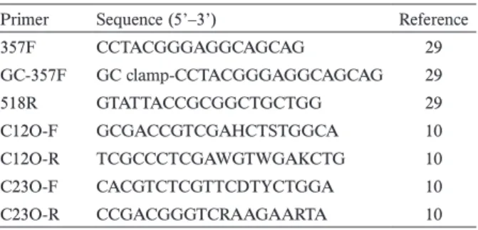
Table 1. Primers used in this study
Primer Sequence (5’–3’) Reference
357F CCTACGGGAGGCAGCAG 29 GC-357F GC clamp-CCTACGGGAGGCAGCAG 29 518R GTATTACCGCGGCTGCTGG 29 C12O-F GCGACCGTCGAHCTSTGGCA 10 C12O-R TCGCCCTCGAWGTWGAKCTG 10 C23O-F CACGTCTCGTTCDTYCTGGA 10 C23O-R CCGACGGGTCRAAGAARTA 10 GC clamp (5’-CGCCCGCCGCGCCCCGCGCCCGTCCCGCC-GCCCCCGCCCG-3’)
and Basic Local Alignment Search Tool program through the DNA Data Bank of Japan website (http://blast.ddbj. nig.ac.jp/ [accessed 31 October 2010]). Sequence data were analyzed with DNA Sequencing Analysis Software 4Peak (Version 1.7.2 for Mac). A phylogenetic tree was constructed with the evolutionary distance data by the neighbor-joining method 28) using CLUSTALX ver. 1.83 33).
2.8 Colony isolation and single-strain benzene degradation assay
Benzene-contaminated soil was suspended in distilled water and allowed to stand for 10 min without shaking for the culture-dependent method. The supernatant was spread onto 1.5% Luria-Bertani (LB) agar plates containing 1 μL/ ml of benzene, incubated at 30°C, and grown aerobically for 7 days. To isolate many types of benzene degraders (e.g. assimilating degraders, co-metabolic degraders), LB plate was used. Single colonies were isolated when colonies appeared. Genomic DNA was extracted using the alkali-sodium dodecyl sulfate method, and 16S rRNA gene V3 fragments were amplified using the described GC-357F and 518R primers. Bacteria cells were simultaneously harvested after 18 h incu-bation in LB and washed with buffer (50 mM Tris-HCl and 150 mM NaCl, pH 7.5). A benzene degradation assay was performed in sealed 5-mL vials with 5 mL minimal medium and a density of 107 cells/mL. Degradation was initiated by the addition of 562.4 μM benzene as the sole carbon source at 30°C without shaking. The concentration of benzene was determined using the described gas chromatography method. 2.9 Presence of C12O and C23O genes in enumeration
SIP fractions
To evaluate the behavior of the main functional genes, the C12O and C23O genes were quantified from separated 12C and 13C gradient fractions (2 μL per fraction as the template) at the 2 phases of degradation described above. The C12O gene was amplified using a protocol consisting of an initial denaturation (94°C, 10 min), 40 cycles of amplification (94°C, 1 min, 60°C, 30 s, 72°C, 20 s), acquisition (92°C), and a final extension (72°C, 5 min) with the C12O-F and C12O-R primers (Table 1). The C23O gene was amplified using the C23O-F and C23O-R primers (Table 1) with the following thermal conditions: initial denaturation (94°C, 10 min), 40 cycles of amplification (94°C, 1 min, 60°C, 30 s, 72°C, 5 s), and a final extension (72°C, 5 min). To detect a wide range of oil-degrading bacteria, we designed the degenerate primers with 35 sequences of the C12O gene and 137 sequences of the C23O gene, as described by Hara et al. 10). The V3 region of the bacterial 16S rRNA gene was also quantified using the primers 357F and 518R (Table 1) under the conditions of initial denaturation (94°C, 5 min), 40 cycles of amplifica-tion (94°C, 30 min, 55°C, 30 s, 72°C, 15 s), and a terminal extension (72°C, 5 min) using the LightCycler FastStart DNA MasterPLUS SYBER Green I kit (Roche Diagnostics, Mannheim, Germany). Melting curves were constructed from 65°C to 95°C with the recommended setting continuous
mode. LightCycler software version 3.5 (Roche Diagnostics) was used to generate the calibration curve and determine copy numbers. The relative abundance of functional genes was calculated by comparing the proportions of functional genes and 16S rRNA gene in each fraction.
3. Results
3.1 Details of the study strategy
We used 2 SIP methods to investigate benzene-degrading bacteria (Fig. 1A). SIP and SIP-D were performed to detect assimilation degraders and candidate co-metabolic degraders, respectively. However, as SIP-D was suggested to also detect the non-degraders of benzene, 16S rRNA-targeted SIP was performed using [13C] acetate to overcome this chal-lenge. In the presence of benzene, the degraders of benzene should incorporated more 13C carbon than that of the non-degraders, and higher levels of the 16S rRNA gene should be expressed, resulting in different proportions of expression. Thus, bacteria that expressed higher levels of the 16S rRNA gene in WITH-B samples (described in 2.4) were more likely to have responded by degrading benzene and could be classi-fied as candidate co-metabolic benzene degrader. The bands common to both the WITH-B and WITHOUT-B samples were classified into the candidate non-degrader of benzene.
A culture-dependent method was also used to verify the culturable strains that could be applied to bioaugmentation, and the V3 region of the 16S rRNA genes of the isolates were amplified and analyzed with PCR-DGGE (Fig. 1B).
Finally, to evaluate the key functional genes, we quantified the C12O and C23O genes using qPCR (Fig. 1C). The pres-ence of these genes was assessed in the 12C and 13C gradient fractions at 2 degradation phases as described in 2.3. 3.2 Occurrence of benzene degradation and preparation
of 13C-DNA
Benzene was measured by gas chromatography, and 562.4 μM benzene was degraded in 6 days during the experi-ments (Fig. 2). The phase from benzene addition to day 2 was defined as the first phase, and days 3 to 5 composed the second phase. To understand the diversity of the degraders at each phase, we selected these 2 phases for SIP and SIP-D analyses, respectively. Only a small amount of catechol appeared in the final stage of degradation during the entire process. No decrease in benzene levels occurred in the auto-claved control. [13C] DNAs were successfully separated from total DNA in both SIP and SIP-D using ultracentrifugation, and [13C] DNAs were directly collected under UV illumina-tion.
3.3 Detection of benzene-assimilating degraders
Benzene degradation was initiated on day 3 (Fig. 2), and several DNA bands appeared in the DGGE profile of the SIP sample from day 4 (Fig. 3, lane 2). DNA bands a–h revealed 13C carbon-incorporating bacteria as benzene-assimilating degraders. Non-labeled 12C samples were also analyzed and
the DNA band e was detected in the second phase (data not shown). Furthermore, bands a, b, g, and h in lane 2 had the same DNA sequences as those of bands a’, b’, g’, and h’ in lane 4, which suggested that these bacteria could assimilate both benzene and acetate.
3.4 Detection of candidate co-metabolic benzene de-grader and non-dede-grader of benzene
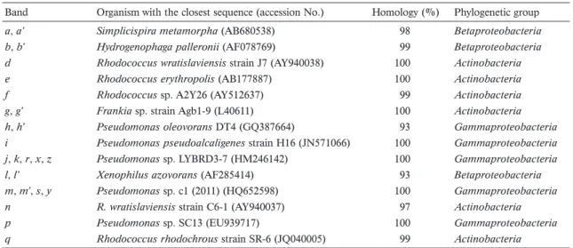
As shown in Figure 3, bands i–n in lane 4 were barely labeled with SIP (lanes 1 and 2). Compared to the WITHOUT-B sample in lane 6, the WITH-B sample in lane 4 showed several specific DNA bands, i, j, and n. These bands may represent candidate co-metabolic benzene degraders. Bands common to lane 4 (bands l, m) and lane 6 (bands l’ and m’) may represent the non-degraders of benzene, which assimi-lated acetate in the presence or absence of benzene. SIP-D may have been able to identify a non-degrader as a candidate co-metabolic degrader in this study. To eliminate this possi-bility, gene expression by 16S rRNA was monitored using SIP with [13C] acetate as follows. Fractionated [13C] RNA was reverse transcribed into cDNA, and the V3 region of the 16S rRNA gene was amplified and applied to DGGE. As shown in lane 7 in Figure 3, bacteria showing bands r and s, which existed in the 13C fraction, were suggested to be active in the presence of benzene. Moreover, as shown in Table 2, bands r and s had the same DNA sequences as those of bands j and
m, respectively. Thus, band j was concluded to be a candidate
co-metabolic benzene degrader, and bands i, m, and n were candidate non-degraders under this condition.
3.5 Detection of culturable benzene degraders using a culture-dependent method
The strains identified in the culture-independent method were culturable or non-culturable bacteria. This finding may be valuable when bioremediation is performed because culturable degraders may be feasible in bioaugmentation and non-culturable degraders in biostimulation. Therefore, the conventional colony isolation method was performed, and 12 colonies were observed on LB agar containing benzene after incubation for 7 days. Four of 12 colonies were selected based on different types of colony morphology (data not shown), and their benzene degradation capabilities were determined with the same concentration of 562.4 μM benzene as the sole carbon source (Table 3). The initial concentration of benzene was almost degraded by these strains, suggesting that these isolates exhibited benzene-assimilating capabilities in the pure cultivation condition. These isolates were detected using SIP-D (Fig. 3, lanes 8–11), and sequencing analysis showed that the DNA bands r and s in lane 7 were the same as z(x) and y (100% sequence similarity), respectively (Table 2). However, strain III (bands m, s, y) and IV (bands j, r, z) were not detected with SIP (lane 2). These results suggest that the isolated strains I, II, and IV were co-metabolic benzene degraders and strain III was a non-degrader in this benzene-contaminated soil.
Fig. 3. PCR-DGGE analysis for dependent and culture-independent methods. [13C] benzene was used to detect
assimi-lating bacteria with SIP (lanes 1 and 2). [13C] Acetate was
used to detect the other degraders with SIP-D (lanes 3–6). In lanes 1–7, PCR-amplified DNA obtained from the heavy DNA fraction was applied to the gel. [12C] or [13C] means the
label-ing carbon source, / means no additional; lanes 1, 3, and 5: day 2; lanes 2, 4, and 6: day 4; lane 7: reverse transcription-PCR products of the heavy fraction from [13C] acetate plus
the benzene-added sample; lanes 8–11: profile of the isolated benzene-degrading strains; M: marker.
Fig. 2. Benzene degradation. Added benzene (562.4 μM) was de-graded in benzene-contaminated soil (●) and negative auto-claved control (+; NC). Mixtures for the SIP method were collected on days 2 and 4, and total DNA was extracted. [13C]
acetate was added on days 2 or 4, and DNA was extracted after 24 h in each case for SIP-D. Catechol concentrations (■) were determined by HPLC. Error bars correspond to the stan-dard deviations calculated for 3 experimental replicates.
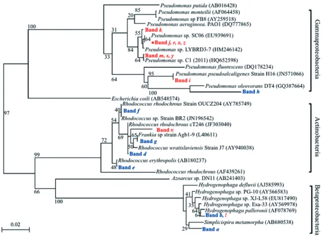
3.6 Phylogenetic analysis of detected bacteria
The phylogenetic tree showed that most SIP-identified bacterial strains were related to Actinobacteria and
Beta-proteobacteria in benzene-contaminated soil (Fig. 4). Rhodococcus, detected as bands d–f (Fig. 3, lane 2), has
been implicated in the degradation of benzene, toluene, and xylene 7,24). Several species of Hydrogenophaga (band
b), a genus of Comamonad bacteria, were formerly
classi-fied in the genus Pseudomonas 36), and band h was highly related to Pseudomonas oleovorans strain DT4, which has been described as a benzene degrader 39). In contrast, SIP-D-identified strains mainly belonged to Gammaproteobacteria. In lane 4 (Fig. 3), band i was closely related to
Pseudo-monas pseudoalcaligenes (100% sequence similarity), which
exhibited a highly enhanced benzene-degrading capability 31). Bands m and j were related to Pseudomonas sp. C1 and
Pseudomonas sp. strain LYBRD3-7 (100% sequence
simi-larity), respectively. This study is the first to show that the
Pseudomonas strain (band j) is a benzene degrader (marked
with red dots in Fig. 4). The most closely related strain to band n in lane 4 (Fig. 3) was a non-cultured bacterium,
Rhodococcus wratislaviensis (97% sequence similarity),
which has been reported to degrade a mixture of hydrocar-bons, gasoline, and diesel oil 3).
3.7 Quantification of C12O and C23O genes with qPCR Benzene was aerobically degraded via the catechol cleavage
pathway, and its key genes, the C12O and C23O genes, and the 16S rRNA gene were quantified using qPCR to determine which gene was functioning in benzene-ring opening (Fig. 5). The quantity of the 16S rRNA gene increased from the first to the second phase (from ~105 to ~106 copies per μL). In the SIP assay, most of the C12O gene was detected in the 13C fraction, suggesting that most benzene-assimilating bacteria harbor the C12O gene and metabolize benzene with it. And, the C23O gene was detected only in the 12C fraction of the second phase.
4. Discussion
Bacterial communities differ from site to site, and information regarding pollutant-degraders existing in the environment is important in bioremediation. Therefore, identifying functional bacteria is an essential component of effective bioremediation. To contribute our findings to the success of bioremediation, we considered the conditions of refinery sites and performed this study with approximately 500 μM of benzene, a minimal medium (without a carbon source), and mesophilic conditions.
Our results demonstrated the possibility of identifying the following bacteria. Xenophilus, a non-benzene-degrading bacterium, and the acetate-assimilating types of
Pseu-domonas were classified in the first degradation phase
of benzene because their DNA bands appeared in both samples (Fig. 3, lanes 3 and 5). In the second phase, the Table 2. Sequence analysis of the major DGGE bands in the 13C fractions
Band Organism with the closest sequence (accession No.) Homology (%) Phylogenetic group
a, a’ Simplicispira metamorpha (AB680538) 98 Betaproteobacteria
b, b’ Hydrogenophaga palleronii (AF078769) 99 Betaproteobacteria
d Rhodococcus wratislaviensis strain J7 (AY940038) 100 Actinobacteria
e Rhodococcus erythropolis (AB177887) 100 Actinobacteria
f Rhodococcus sp. A2Y26 (AY512637) 99 Actinobacteria
g, g’ Frankia sp. strain Agb1-9 (L40611) 100 Actinobacteria
h, h’ Pseudomonas oleovorans DT4 (GQ387664) 93 Gammaproteobacteria
i Pseudomonas pseudoalcaligenes strain H16 (JN571066) 100 Gammaproteobacteria
j, k, r, x, z Pseudomonas sp. LYBRD3-7 (HM246142) 100 Gammaproteobacteria
l, l’ Xenophilus azovorans (AF285414) 93 Betaproteobacteria
m, m’, s, y Pseudomonas sp. c1 (2011) (HQ652598) 100 Gammaproteobacteria
n R. wratislaviensis strain C6-1 (AY940037) 97 Actinobacteria
p Pseudomonas sp. SC13 (EU939717) 100 Gammaproteobacteria
q Rhodococcus rhodochrous strain SR-6 (JQ040005) 99 Actinobacteria
See Figure 3 for details of the DGGE analysis.
Table 3. Benzene degradation test for isolated strains
I II III IV NC
Decreased rate of benzene (%) 73.2 66.5 75.1 75.8 0.2
Residual absolute concentration (μM) 150.7±1.4 188.5±2.1 140.0±1.4 136.3±3.2 561.2±0.4 Benzene (562.4 μM) was added as the sole carbon source in minimal medium containing 107 cells/mL of each strain. The
concentra-tion was determined after 65 h using gas chromatography, and these samples were decreased to the detecconcentra-tion limit after 80 h. NC: au-toclaved control. Standard deviations were calculated for 3 experimental replicates.
benzene-assimilating bacteria belonging to Pseudomonas,
Rhodococcus, Frankia, and Simplicispira, were directly
identified by the SIP method (Fig. 3, lane 2). The candidate co-metabolic types of Pseudomonas, and the non-benzene-degrading types of Pseudomonas and Hydrogenophaga were mainly identified by the SIP-D method (Fig. 3, lanes 4 and 6). These results suggested that this combination of SIP methods may possible to compensate the comprehensive analysis in the microbe ecology field and delineate a comprehensive bacterial community that may contribute to biodegradation.
Benzene-assimilating degraders could be identified by the SIP method. Unfortunately, SIP-D covered a group of bacteria that assimilated both benzene and acetate in this study because bands were detected in both SIP (bands a, b, g, and h in lane 2) and SIP-D (bands a’, b’, g’, and h’ in lane 4) samples. 16S rRNA-targeted SIP (lane 7, Fig. 3) was used to address this question, and the results suggested that the most active bacteria in the second degradation phase belonged to
Pseudomonas sp. (bands j and m) and were considered to be
potential benzene degraders (Fig. 3).
These 2 strains displayed active benzene-assimilating capabilities, which were clarified using the culture-dependent method (Table 3). They could use acetate in benzene-contam-inated soil as shown in lane 4, Figure 3, and also the LB broth as their carbon source in the culture-dependent method.
Fig. 4. Phylogenetic relationships derived from 16S rRNA gene V3 region analysis (150 bp). SIP- and SIP-D-identified DNA bands are shown in blue and red, respectively. Red dots indicate candidate benzene co-metabolic degraders. Phylogenetic positions traced strains related to
Rhodococcus, Hydrogenophaga, or Pseudomonas strains based on the 16S rRNA gene V3 region sequence. Accession numbers of the
reference organisms are indicated in parentheses. Bootstrap values for branches are based on 1000 replicates. Bar=2% nucleotide substi-tution rate (Knuc).
Fig. 5. Quantification of the C12O and C23O gene proportions in degradation processes.
□, C12O gene; ■, C23O. Targeting assimilating bacteria de-tected by SIP. Quantitative analysis of the 12C- and 13C-DNA
fractions from SIP data. Evaluation of the functional genes of
12C and 13C gradient fractions from SIP data. (1st), first phase;
(2nd), second phase. The absolute values of all samples
exhibit-ed similar trends. Error bars correspond to the standard devia-tions calculated for 3 experimental replicates.
However, one bacterium (band j) showed co-metabolic degrading potential, while another (band m) exhibited no benzene-assimilating capability. For these strains could not be labeled by [13C] benzene in lane 2, Figure 3, they were classified as the candidate co-metabolic degrader (band j) and candidate non-degrader (band m) of benzene under this condition. These results suggested that a bacterium, even if it has an active benzene degradation capability, is not necessarily directly associated with degradation. And, an interaction between bacteria in the contaminated soil might inhibit the assimilating ability of this isolated strain. This important characteristic must be considered during bioremediation treatment. Furthermore, bands p and q in lane 6 were not detected in the WITH-B sample (Fig. 3, lane 4), suggesting that several acetate-assimilating bacteria, which may not have benzene-degrading capabilities, could be substituted in the WITH-B condition even in the presence of acetate.
Our findings also demonstrated the potential to trace uncultured bacteria (bands i, l, a’, b’, n, g’, and h’) by a comparison with a culture-independent method, which suggests that most degraders are rarely isolated under this condition, but could be detected with our combination of SIP methods. These results suggested that biostimulation (but not bioaugmentation) may be suitable at this benzene-contaminated site because many benzene degraders may be uncultured suggested by this conventional isolation method. An interesting challenge for the future will be to improve the SIP-D method by finding an appropriate labeling carbon source with which to target a group of bacteria, labeling the position, and optimizing the environmental pollutant concen-tration for a comprehensive analysis.
Previous studies have shown that some Pseudomonas could co-metabolize aromatic compounds coexistent with organic acids 4,18), and our study shows the possibility of iden-tifying this kind of co-metabolizing bacteria. The existence of electron acceptors or donors also has a large impact on carbon flux in ecosystems, and for the selection of labeling carbon source to optimize SIP-D.
The relationship observed between the C12O and C23O gene is similar to that described by a previous study that analyzed genes associated with aromatic compound degrada-tion 30). Although acetate was added, the bacteria (bands c,
d, e, and f ) continued to express the C12O gene because no
band shifting of these bacteria was observed between lanes 2 and 4 (Fig. 3). The functional gene quantification experi-ment detected the C12O gene in both the light and heavy DNA fractions (Fig. 5), suggesting that the most benzene assimilating-degraders were processing the C12O gene and used this pathway to open the benzene ring. Although the C23O gene was processed in the second phase of the 12C fraction, they may not use this pathway because the C23O gene was not detected in the 13C fraction.
In conclusion, understanding the structure of the degradation community including assimilating degraders, co-metabolic degraders, and non-degraders, is essential for
improving bioremediation. In this study, a comprehensive analysis was performed to classify the bacterial members related to benzene degradation under mesophilic aerobic conditions. Benzene-assimilating degraders were classified with SIP, whereas the candidate co-metabolic degraders and candidate non-degraders of benzene were classified using SIP-D and RNA-based SIP. Furthermore, the qPCR results showed that the assimilating degraders used ortho-cleavage for benzene degradation. This combination of SIP methods is on the start line to be able to perform comprehensive analysis of mixed culture roughly, and could be used to confirm the structure of the bacterial community in the presence of chemical pollutants such as benzene.
Acknowledgements
This work was supported by a Grant-in-Aid for Scientific Research (B) (no.21310050, to H.U.), CREST program of Japan Science and Technology Corporation (JST), and the Monbukagakusho Scholarship (no.114042, to Y.P.) of the Ministry of Education, Culture, Sports, Science and Tech-nology. Japan. We thank Eri Hara for help with the primer construction, and Shouhei Yamasaki for helpful technical advice.
References
1) Anderson, R.T., J.N. Rooney-Varge, C.V. Gaw, and D.R. Lovley. 1998. Anaerobic benzene oxidation in the Fe (III) reduction zone of petroleumcontaminated aquifers. Environ. Sci. Technol. 32: 1222–1229.
2) Antti Tossavainen. 1978. Styrene use and occupational expo-sure in the plastics industry. Scand. J. Work Env. Hea. 4: 7–13. 3) Auffret, M., D. Labbé, G. Thouand, C.W. Greer, and F. Fayolle-Guichard. 2009. Degradation of a mixture of hydrocarbons, gasoline, and diesel oil additives by Rhodococcus aetherivorans and Rhodococcus wratislaviensis. Appl. Environ. Microbiol. 75: 7774–7782.
4) Basu, A., S.K. Apte, and P.S. Phale. 2006. Preferential utiliza-tion of aromatic compounds over glucose by Pseudomonas
putida CSV86. Appl. Environ. Microbiol. 72: 2226–2230.
5) Boschker, H.T.S., S.C. Nold, P. Wellsbury, D. Bos, W.D. Graaf, R. Pel, R.J. Parkers, and T.E. Cappenberg. 1998. Direct linking of microbial populations to specific biogeochemical processes by 13C-labelling of biomarkers. Nature 392: 801–804.
6) Coates, J.D., R. Chakraborty, J.G. Lack, S.M. O’Connor, K.A. Cole, K.S. Bender, and L.A. Achenbach. 2001. Anaerobic ben-zene oxidation coupled to nitrate reduction in pure culture by two strains of Dechloromonas. Nature 411: 1039–1043. 7) Deeb, R.A. and L. Alvarez-Cohen. 1999. Temperature effects
and substrate interactions during the aerobic biotransformation of BTEX mixtures by toluene-enriched consortia and
Rhodo-coccus rhodochrous. Biotechnol. Bioeng. 62: 526–536.
8) Ding, B., S. Schmeling, and G. Fuchs. 2008. Anaerobic me-tabolism of catechol by the denitrifying bacterium Thauera
aromatica-a result of promiscuous enzymes and regulators. J.
Bacteriol. 190: 1620–1630.
9) Grbic-Galic, D. and T.M. Vogel. 1987. Transformation of toluene and benzene by mixed methanogenic cultures. Appl. Environ. Microbiol. 53: 254–260.
10) Hara, E., N. Nomura, T. Nakajima, and H. Uchiyama. 2013. Bioremediation field trial of oil-contaminated soil with food-waste compost. Journal of J.S.C.E. 1: 125–132.
11) Harwood, C.S. and R.E. Parales. 1996. The beta-ketoadipate pathway and the biology of self-identity. Annu. Rev. Microbiol. 50: 553–590.
12) Head, L.M., D.M. Jones, and W.F.M. Röling. 2006. Marine microorganisms make a meal of oil. Nature Reviews Microbiol. 4: 173–182.
13) Hori, T., M. Noll, Y. Igarashi, M.W. Friedrich, and R. Conrad. 2007. Identification of acetate-assimilating microorganisms under methanogenic conditions in anoxic rice field soil by comparative stable isotope probing of RNA. Appl. Environ. Microbiol. 73: 101–109.
14) Janke, D. and W. Fritsche. 2007. Nature and significance of microbial cometabolism of xenobiotics. J. Basic Microbiol. 25: 603–619.
15) Kasai, Y., Y. Takahata, M. Manefield, and K. Watanabe. 2006. RNA-based stable isotope probing and isolation of anaerobic benzene-degrading bacteria from gasoline-contaminated ground-water. Appl. Environ. Microbiol. 72: 3586–3592.
16) Kasai, Y., Y. Kodama, Y. Takahata, T. Hoaki, and K. Watanabe. 2007. Degradative capacities and bioaugmentation potential of an anaerobic benzene-degrading bacterium strain DN11. Environ. Sci. Technol. 41: 6222–6227.
17) Laban, N.A., D. Selesi, C. Jobelius, and R.U. Meckenstock. 2009. Anaerobic benzene degradation by gram-positive sulfate-reducing bacteria. FEMS Microbiol. Ecol. 68: 300–311. 18) Lerch, T.Z., M.F. Dignac, E. Barriuso, and A. Mariotti. 2011.
Effect of glucose on the fatty acid composition of Cupriavidus
necator JMP134 during 2,4-dichlorophenoxyacetic acid
deg-radation: implications for lipid-based stable isotope probing methods. Appl. Environ. Microbiol. 77: 7296–7306.
19) Liou, J.S-C., C.M. DeRito, and E.L. Madsen. 2008. Field-based and laboratory stable isotope probing surveys of the identities of both aerobic and anaerobic benzene–metabolizing microorgan-ism in freshwater sediment. Environ. Microbiol. 10: 1964–1977. 20) Lu, T., P.G. Stroot, and D.B. Oerther. 2009. Reverse transcrip-tion of 16S rRNA to monitor ribosome synthesizing bacterial populations in the environment. Appl. Environ. Microbiol. 75: 4589–4598.
21) Mesarch, M.B., C.H. Nakatsu, and L. Nies. 2000. Deelopment of catechol 2,3-dioxygenase-specific primers for monitoring bio-remediation by competitive quantitative PCR. Appl. Environ. Microbiol. 66: 678–683.
22) Morris, S.A., S. Radajewski, T.W. Willison, and J.C. Murrell. 2002. Identification of the functionally active methanotroph population in a peat soil microcosm by stable-isotope probing. Appl. Environ. Microbiol. 68: 1446–1453.
23) Muyzer, G., E.C. de Waal, and A.G. Uitterlineden. 1993. Profil-ing of complex microbial populations by denaturProfil-ing gradient gel electrophoresis analysis of polymerase chain reaction amplified genes coding for 16S rRNA. Appl. Environ. Microbiol. 59: 695–700.
24) Na, K.S., A. Kuroda, N. Takiguchi, T. Ikeda, H. Ohtake, and J. Kato. 2005. Isolation and characterization of benzene-tolerant
Rhodococcus opacus strains. J. Biosci. Bioeng. 99: 378–382.
25) Neufeld, J.D., J. Vohra, M.G. Dumont, T. Lueders, M. Manefield, M.W. Friedrich, and J.C. Murrell. 2007. DNA stable isotope probing. Nature Protocol. 2: 860–866.
26) Peng, J. and A. Wan. 1997. Measurement of henry’s constants of high-volatility organic compounds using a headspace autos-ampler. Environ. Sci. Technol. 31: 2998–3003.
27) Pol, S.S., P.K. Dhakephalkar, and R.S. Bharadwaj. 2009. Char-acterization of leptospires using V3 region of 16S rDNA by denaturing gradient gel electrophoresis: a case study. J. Med. Microbiol. 27: 354–357.
28) Saito, N. and M. Nei. 1987. The Neighbor-joining method: a new method for reconstructing phylogenetic trees. Mol. Biol. Evol. 4: 406–425.
29) Sanchez, O., J.M. Gasol, R. Massana, J. Mas, and C. Pedros-Alio, 2007. Comparsion of different denaturing gradient gel electrophoresis primer sets for the study of marine bacterio-plankton communities. Appl. Environ. Microbiol. 73: 5962– 5967.
30) Sei, K., D. Inoue, K. Wada, K. Mori, M. Ike, T. Kohno, and M. Fujita. 2004. Monitoring behaviour of catabolic genes and change of microbial community structures in seawater micro-cosms during aromatic compound degradation. Water Res. 38: 4405–4414.
31) Suenaga, H., M. Mitsuoka, Y. Ura, T. Watanabe, and K. Furukawa. 2001. Directed evolution of biphenyl dioxygenase: emergence of enhanced degradation capacity for benzene, toluene, and alkyl-benzenes. J. Bacteriol. 183: 5441–5444.
32) Sun, W.M. and A.M. Cupples. 2012. Diversity of five anaero-bic toluene-degrading microbial communities investigated using stable isotope probing. Appl. Environ. Microbiol. 78: 972–980. 33) Thompson, J.D., T.J. Gibson, F. Plewniak, F. Jeanmougin, and
D.G. Higgins. 1997. The CLUSTAL_X windows interface: flex-ible strategies for multiple sequence alignment aided by quality analysis tools. Nucleic Acids Res. 25: 4876–4882.
34) Weelink, S.A., N.C.G. Tan, H.T. Broeke, C.V.D. Kieboom, W.V. Doesburg, A.A.M. Langenhoff, J. Gerritse, H. Junca, and A.J.M. Stams. 2008. Isolation and characterization of Alicy-cliphilus denitrificans Strain BC, which grows on benzene with chlorate as the electron acceptor. Appl. Environ. Microbiol. 74: 6672–6681.
35) WHO. INTERNATIONAL AGENCY FOR RESEARCH ON CANCER, IARC Monographs on the Evaluation of Carcino-genic Risks to Humans, Overall Evaluations of CarcinoCarcino-genicity: An Updating of IARC Monographs, Volumes 1 to 42, Supple-ment 7.
36) Willems, A., J. Busse, M. Goor, B. Pot, E. Falsen, E. Jantzen, B. Hoste, M. Gillis, K. Kersters, G. Auling, and J.D. Ley. 1989.
Hy-drogenophaga, a new genus of hydrogen-oxidizing bacteria that
includes Hydrogenophaga flava comb. nov. (Formerly
monas flava), Hydrogenophaga palleronii (Formerly Pseudo-monas palleronii), Hydrogenophaga pseudoflava (Formerly Pseudomonas pseudoflava and “Pseudomonas carboxydo-flava”), and Hydrogenophaga taeniospiralis (Formerly Pseu-domonas taeniospiralis). Int. J. Syst. Evol. Micr. 39: 319–333.
37) Xie, S.G., W.M. Sun, C.L. Luo, and A.M. Cupples. 2011. Novel aerobic benzene degrading microorganisms identified in three soils by stable isotope probing. Biodegradation 22: 71–81. 38) Yamasaki, S., N. Nomura, T. Nakajima, and H. Uchiyama.
2012. Cultivation-independent identification of candidate de-halorespiring bacteria in tetrachloroethylene degradation. En-viron. Sci. Technol. 46: 7709–7716.
39) Zhou, Y.Y., D.Z. Chen, R.Y. Zhu, J.M. Chen. 2011. Substrate interactions during the biodegradation of BTEX and THF mix-tures by Pseudomonas oleovorans DT4. Bioresour. Technol. 102: 6644–6649.