Title
[原著]頭蓋顔面の血管性病変と腫瘍に対する血管内手術
Author(s)
百次, 仁; 六川, 二郎; 金城, 利彦; 崎原, 永辰; 外間, 晶子; 豊
見山, 直樹; 佐村, 博史
Citation
琉球医学会誌 = Ryukyu Medical Journal, 12(3): 235-243
Issue Date
1992
URL
http://hdl.handle.net/20.500.12001/3160
Ryukyu Med. J. , 12(2) ,235-243, 1992
頭蓋顔面の血管性病変と腫痔に対する血管内手術
百次 仁 六川 二郎 外聞 晶子 豊見山直樹 金城 利彦 崎原 永辰 佐村 博史 琉球大学医学部脳神経外科 (1991年4月24日 受付、 1991年7月9日 受理) 緒 言 血管内手術法とは、血管性病変や腫癌性病変 に対し、血管内から操作を加えるtechniqueで ある。近年、カテーテルや栓塞材料の発明、改 良が進んだことや、疾患に対する知識が深くなっ たことにより、血管内手術は格段の進歩を遂げ ている。私どもは1989年11月から1990年11月ま での一年間で9例の頭蓋顔面病変に対して血管 内手術を行ったので報告する。 症 例 9例をつぎの3群に大別した。 第1群:顔面血管鹿の3例。栄養動脈を選択 的に栓塞した。 第2群:頭頚部悪性腫癌の4例O抗癌剤およ びLAK細胞の選択的動注療法を行った。 第3群:海綿静脈洞内血管性病変の2例。海 綿静脈洞部巨大内頚動脈癌の1例に対して離脱 式バルーソカテーテルによる内頚動脈遮断を行っ た。海綿静脈洞部硬膜動静脈奇形の1例に対し て流入動脈の経動脈的栓塞術と海綿静脈洞の経 静脈的栓塞術を行った。 症例の年齢、性別、病名、局在、治療法およ び材料をTable lに示す。以下に代表例を呈示 する。 [症例1] 4歳女児。出生時より舌、下口唇、 咽頭に血管腫癌が存在し、成長にしたがいとく に舌の腫癌が増大し歯槽変形、構音障害、閉口 障害、乾燥、易出血性などの症状を呈するよう になった。術前の選択的左舌動脈撮影で舌の尖端部 から背部にかけてstainが描出された(Fig.1), 右舌動脈撮影もほとんど同様の所見であった。 このvascular stainは腫癌に比して明らかに小 さかった。両側の舌動脈を介して250-500^ m 大のpolyvinyl alcohol(PVA)をvascular stainが消失するのに必要最小量用いて超選択的に栓 塞した(Fig.1),血管腫癌は辺縁部から次第 に刺離し、舌の機能を温存し美容上の問題を生 ずることなく48日目に脱落した(Fig.2)t [症例5] 50歳男性。右大脳半球の広範な神 経惨芽鹿の患者である(Fig.3)ォ 腫蕩部分摘 出術、放射線療法と抗癌剤の全身投与に加え、 抗癌剤の選択的動注療法を行った.内頚動脈に 留置した7Fr.カテーテルを介してTrackerl8 カテーテルを末梢に進め、眼合併症を避けるた めに眼動脈起姶部より遠位で動注した。すなわち後 交通動脈起始部より近位の内頚動脈からMCNU l-(2-chloroethyl)-3-(metyl α -D-glucopyranos-6-yl)-l-nitrosourea)を50m甘注入した。つぎにTracker カテーテルをさらに末梢に進め、中大脳動脈水 平部で50 J注入した(Fig.4)0 CT上著効が認 められたが術後合併症は生じなかった。 [症例8] 47歳女性。頭痛、複視、右眼喰下 垂で発症した右海綿静脈洞部巨大内規動脈癌。 MRIで最大径25mmの腫癖が右海綿静脈洞内に 認められ、右内頚動脈撮影で内腔の最大径が22mm の巨大動脈癌が証明された(Fig.5),癌頚部 が不明瞭なため、癌の栓塞術に代わって、癖近 位側の内頚動脈遮断術を選択した Swan-Ganz カテーテルで右内頚動脈を約20分間遮断し、神
236 頭蓋顔面の血管性病変と腫癌に対する血管内手術
Table 1 Summary of endovascular surgica一 cases
Group/Case No. Age/Sex Diagnosis/Location Treatments/Materials
I Facia一 hemangiomas
① 4 F hemolymphangiomaCtongue, lower I ip,pharynx) ③ 19 F hemangiotnadt.maxi l lary bone)
③ 14 M hemangioma(Lt.cheek)
Q Head and neck malignant tumors
④ 67 Y metastatic bone tumor (RCC ,skull base) ⑧ 50 M gl ioblastoma (Rt. hemisphere,diffuse) ⑥ 34 M squamous cel l ca.(Lt.max川ary sinus) ⑦ 15 F astrocytoma nKRt.thalamus,
Lt.frontal lobe) ID Intracavernous vascular lesions
⑧ 47 F Rt.carotid cavernous giant aneurysm
44 F cavernous duraI AVM
TAE* 1
(PvA ,gel foam microfibrilar IAI col lagen, ethanoI ) *3 LAK cell*5 MCNU*6 CDDP,*7 MCN〕 ZR ICproximal occlusionby balooncatheter TAEoffeeders gelfoam,micro- fibrillarco-llagen TVE*100fCSm metalcoils abbreviations :
# 1 ; transarterial embolization *7 ; Cis-Diamminedichloroplatinum *2; polyvinyl alcohol *8; internal carotid artery *3 ; intraarterial infusion *9 ; arteriovenous malformation #4; renal cell carcinoma *10; transvenous embolkation *5;レmphokine activated killer cell *11 ; cavernous sinus
# 6 ; 1-(2-chloroethyl)-3-( methyl a -D-glucopyranos-6-yl)-1-nitrosourea
経症状が出現しないこと、 Stump pressureが84/ 60mmHgと良好なこと(Fig.6)、また同時に施 99n 行した TeHMA-PAO SPECTによる脳血流分 布で左右差が出現しないことを確認した後、離 脱式バルーソカテーテルを用いて遮断した。術 後の血管撮影で後交通動脈および前交通動脈を 介する側副血行はともに良好であることが確認 された(Fig.7),術後、新たな神経症状の出 現は認めなかった。 [症例9] 44歳女性。左眼病、左結膜充血、 眼球突出、耳鴨を主訴とする海綿静脈洞部硬膜 動静脈奇形(CdAVM) 血管撮影では左眼動 脈、両側外頚動脈の多数の枝が流入動脈であり、 左上眼静脈、左下錐体静脈洞、 Labb e静脈が 流出静脈であった(Fig8)< まず、左内上顎動 脈から経動脈的栓塞術を行った,7Fr.カテーテ
Fig. 1 (case 1 )Superselective 一ingual angbgraph
(lateral view).
Lt. :Preembolization. The stain of hemangioma is seen.
Rt. :Postembolizatbn. Though the stain dissapeared completly, the main trunk of lingual artery is st= patent.
Fig.2 (case DOutview of tb diseased tongue. Lt. :Preembolkatbn. Huge hemangbma of the tongue is seen.
Rt. :Postembolizatbn. T「袷 hemangioma dissapeared wkhout functional deficit.
ルを左外頚動脈に留置し、 Tracker 18カテーテ ルを内上顎動脈まで進め、 50-100ォmのGelfoam およびmicrofibrillar collargenを注入した。術 後の内上顎動脈撮影では海綿静脈洞は描出され 、なくなった。患者の耳鴨は消失し、他覚的にも 雑音は聴取されなくなったO さらに右外頚動脈
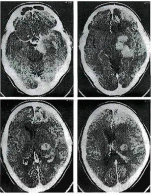
Fig.3 (case 5)Contrast enhanced CT scan shows multiple enhanced lesions at right
hemisphere. からの栓塞術を施行した。しかし、左内頚動脈 撮影で眼動脈を介してCdAVMが措出され、左 内上頚動脈からの硬膜枝の再開通も確認された ので、 3週間後、左下錐体静脈洞より経静脈的 塞栓術を施行した。左内頬静脈を直接穿刺し、 下錐体静脈洞とS状静脈洞との接合部に留置し た6Fr.カテーテルを介して、Trackerカテーテ ルを海綿静脈洞を越えて上限静脈まで進めた。 同部にHilalの金属コイルを3個、海綿静脈洞 後半部に5個、さらに下錐体静脈洞にも1個留 置した。術後の左内頚動脈撮影ではCdAVM, 上限静脈、下錐体静脈洞の描出は著明に減少し た(Fig.9)ォ 考 察 I.顔面血管厘に対する栓塞術について 血管腫には自然退縮する一群があり、これを 1)
Beltramello はimmatureまたはnewborn-he-頭蓋顔面の血管性病変と魔窟に対する血管内手術
F由.4 (case 5)Cerebral angiograph.
Upper Lt. :Selective internal carotid angiograph before intraarterial infusion
of MCNU(latera一 view).
Upper Rt. :Selective internal carotid angiograph before intraarteial infusion of MCNU(A-P view).
Lower Lt. '.Superselective intraarterial MCNU infusbn at internal carotid artery
between ophtha一mic artery and posterior
communicating artery.
Lower Rt. :Superselective intraartial MCNU infusion at horizontal portion of the middle cerebral artery.
The tip of Tracker catheter is shown by an arrow.
2)
mangioma、 Lasjaunias はinfantile hemangioma
と呼び、安易な外科的切除は慎むべきであると 述べている。すなわちこの群は6カ月以内に退 縮が始まり5-7歳までに完了するため、生命 の危険や機能障害を生じない限り治療を行わな い。 他方、自然退縮のない他の血管腫群では外科 的切除、放射線療法、栓塞術などの治療が必要 であるが、美容および機能上の問題や長期的な 副作用を考慮すると塞栓術が最も有利であると n* いわれる 。 血管睦の治療には塞栓材料のうち主として固
Fig. 5 (case 8 )Rt.internal carotid angtogram. Lt. :A-P view. Rt. :lateral view. A giant aneurysm is seen at cavernous portion. The neck of areurysm is indistinct.
F垣.6 (case 8 )Arterial blood pressure of the Rt. internal carotk】 artery during balbon occlusion test. An arrow indicates the beginning of balloon occlusion of the internal carotid artery. The pressure fall from 132/80 mmHg, which gives the evidence of good cross flow.
体材料が用いられる。組織障害性が弱いため (biocompatible)皮膚や粘膜の腫脹・壊死が 起こりにくいこと、操作が安全容易であること から、半永久的な栓塞材料であるpolyvinyl l-5) alcoholが有効と言われている ので症例1 に対してPVAを使用した。症例2、 3に対し ては皮膚に壊死が生じることを恐れてGelfoam, microfibrillar collagenを使用した。しかし、
F由.7(case 8)
Lt. : Lt. carotid angioraph (A-P view). Cross fbw via廿℃ anterior communicatト
ng artery remains well. Detachable balloons are pointed by white arrows.
Rt. : Lt. vertebra一 angiographOateral
view). Cross flow via the posterior communicating artery, pointed by black arrows is good. これらの症例ではすぐに再開通をきたし栓塞術 の効果が認められなかったので、血管厘の治療 においてPVAが有用であると考えられる。 Il.勤注化学療法について 選択的動注化学療法は1 )術前の腫痴体積の 縮小、 2)直達手術不能な症例の治療法、そし て3)術後の継続治療の1つとして有効である と考えられている。 頭頚部の悪性腫癖に対する動注化学療法は、 腫蕩内に高濃度の薬剤を移行させること、その さい薬剤の使用量を減じて腫癌周辺の健常脳や 全身の副作用を軽減することを目的として行な 6) われる。動脈内注入により腫痴内の抗癌剤濃 度は静脈内注入の約2倍に達し、腫蕩体積の縮小と 生存期間の延長が得られるという点で、有効性が高 7-9) いという報告がある 。またClaymanら が 指摘しているように内頚動脈から注入するさい 惹起する合併症(網膜障害、 encephalopathy) を避けるために、私どもは症例5および症例7 -・ -. -. I / . r ・ , . . . . . . I F由.8 (case 9)
Upper Lt. : Rt. ascending pharyngeal artery feeds cavernous dural artnoveno-us malformation ( CdAVM ).
Upper Rt. : Multiple branct℃s of the lt. internal maxillar artery feed CdAM. It drains into the 比. superior ophthalmic vein, inferior petrosal sinus and cortical Vems.
Lower Lt. : Recurrent meningeal branch of the rt. ophthalmic artery feeds C-dAVM.
Lower Rt. : After tramsareerial embolkat-ion. Shunt between the lt. externa一
artery and CdAVM remarkabかdisappear-ed. で限動脈起始部より遠位側で抗癌剤を注入した。 なお、抗癌剤の注入直前に血液脳血管関門を一 時的に開いて抗癌剤の腫癖への移行を高めるた め、 20%mannitol 200mlを約2分間で注入し 10) EB^^^^H?
頭蓋顔面の血管性病変と腫痔に対する血管内手術
F由.9 (case 9)Lt. interna一 carotid angiogram.
u. : Tracker18 catheter is cannulated
into the cavernous sinus via inferior petrosal sinus (arrows).
Rt. : After transvenous embohzation. Hilaf s coils are placed Into the superior ophthalmic vein, posterior portion of cavernous sinus and inferior petrosal sinus. Shunting fbw is markedly reduc-ed. lIl.海綿静脈洞内血管性病変に対する治療につ いて l.巨大内規動脈癖 海綿静脈洞部は巨大動脈癌の好発部位である。 クモ膜下出血を呈することは稀で海綿静脈洞症 候群として発症することが多い。同部が解剖学 的に複雑であることと出血のコントロールが困 難であることから以前は"thelast no man s ll) land と呼ばれていたが、近年直達手術の 1 1 . 1 2) 報告が増加している 。さらに血管内手 術法の進歩により、バルーソを用いた動脈癖の 閉塞や癌近位側内頚動脈の遮断も行われるよう 13、14) になった 。 本疾患の治療上の最大の問題点は内頚動脈の 血流遮断に対する患者の耐容能である。これを 確認するための確実な検査法はバルーソカテー テルを用いた内頚動脈balloon occlusion test
15.】6) である o 内頚動脈遮断中の神経症状の出 現の有無、 stump pressure,脳血流分布、脳波、 SEPなどの検査結果で総合判定する。とくに 99m ToHMA PAOを用いた脳血流分布は虚血に 16) 対して鋭敏であるので有用な検査である 。私 どもはこれらのテストの結果と血管撮影所見か ら前交通動脈および後交通動脈を介する側副血 行が良好であると判断したので、 bypass術を 併用せず離脱式バルーソカテーテルを用いて動 脈癖より近位の内頚動脈の遮断のみを行った。 このさいバルーソが早く外れたり(premature release)離脱する時にバルーソが移動したり、 膨脹させすぎて血管損傷をきたすことのないよ 17) うに注意した O 内頬動脈が遮断されると、 動脈癌は血栓化しその体積は縮小する。もし縮 小が著明でなくても周囲脳および脳神経への拍 動の伝播が消失し、神経症状は改善するといわ 18) れている 。しかし、症状発現から長期の経過 を経ると不可逆的変化が生ずるので、以上の検 査が済み次第すみやかな遮断が必要である。 2.硬膜動静脈奇形 海綿静脈洞部におけるこの疾患は症状が軽微 で進行性でない症例が多く、ときに何等かの誘 因で自然消失する例もあるので、症状や血管撮 影所見に基づいて治療の選択を行うことが必要 19、20) である 。 Barrowの分標に従うと私どもの症例はtypel)で 21) ある 。この型に対しては一定期間保存的に治 療するべきであるといわれるが、頭蓋内圧克進、 急激な眼球突出や視力低下、出血、一過性虚血 発作などの症状を呈する場合には積極的な治療 21,22) が必要でる 。すなわち1)用手的頚動脈 圧迫、 2)経動脈的栓塞術、 3)放射線療法、 4)バルーソによる内頚動脈遮断、 5)直達手 術などである。最近では上限静脈や下錐体静脈 洞から経静脈的にアプローチし、金属コイルや 絹糸、または液体栓塞物質などを用いて海綿静 23.24) 脈洞を閉塞する方法も行われている 。 私どもはまず外頚動脈からの塞栓術を行った が硬膜動静脈奇形の消失には至らなかった。そ こで下錐体静脈洞を介し、金属コイルを用いて 海綿静脈洞の塞栓術を行い、血管撮影上、硬膜 動静脈奇形はほとんど消失した。すなわち、ま ず経動脈的塞栓術によりflowを緩徐にし、つ
百次 いで経静脈的塞栓術を加えることが安全性、根 治性、難易度などの点から、最良の治療法であ ると考える。ただし、そのさいに静脈還流を海 綿静脈洞から脳裏静脈の方向へ変更して脳内出 血などの合併症を招くことのないように留意す :s.:s) べきである 。 血管内手術は小さな侵薬で大きな効果を得る ことができるため、今後ますます発展するであ ろう。 m 私どもがこの1年間に経験した9例の血管内 手術(顔面血管腫群- 3例、選択的動注化学療 法-4例、海綿静脈洞内血管性病変-2例)を まとめ、文献的考察を加えた。 文 献
1) Beltramello, A., Benati, A., Perini, S., Maschio, A. : lnterventional angiography in neuropediatrics. Child Nerv Syst 5:87-93,1989.
2) Lasjaunias, P., Berer鵜tein, A. : Surgical
neuroangiography, vol. 2 , Endovascular treatment of crardofadal lesions, Springer-Verlag. Berlin Heidelberg, 1987.
3) Latchaw, R.E., Gold, L.H.A. :
Polyvinyl foam embolization of vascular and neoplastic lesions of the head, neck, and spine. Radiology 131:669-679, 1979. 4) Forbes, G., Earnest, F., Jackson, I.
T., Marsh, W. R., Jack, C., Cross, S. A. : uIerapeutic embolization
angiogra-phy for extraaxial lesions in the head.
Mayo Clin Proc61:427-441, 1986. 5) Brawn, I.F.. Levy, S., Hoffman, J.
C. : The use of transarterial microemboli-zation in the management of hemangiomas of tl鳩perioral region. J Oral Maxillofac Surg 43:239-248, 1985.
6) Lee, Y. Y., Wallace, S., Dimery, I.,
仁 ほか 241
Goepfert , H. : Intraarterial chemotherapy of head and neck tumors. AJNR 7:343-348, 1986. 7)宮上 光祐、田副 誠、坪州 孝志:MC NUの悪性脳腫癌、正常脳組織内移行一静 注群と動注群との比較-.癌と化学療法17: 275-280 , 1990. 8) Greenberg, H. S., Ensminger, W. D., Chandler, W. F., Layton, P. B., Junck, L., Knake, J., Vine, A.K. :Intra-arterial BCNU chemotherapy for treatment of malignant glioma of the central nervous system. J Neurosurg 61:423-429, 1984.
9) dayman, D. A., Wolport, S.M., Heros, D. O. : Superselective arterial BCNU infusion in the treatment of patients with malignant gliomas. AJNR IO'. :767-771,
1989. 10)宮上 光祐、田副 誠、笠原 英司、坪川 孝志'蝣9L gliosarcoma実験脳腫癖に対す る20%マソニットール、 ACNU動注化学 療法、癌と化学療法16:2059-2065. 1989. ll)福島 孝徳・Cavernous Sinus血管病変に 対する直達手術:自験27例の総括. The Mt. Fuji Workshop on CVD Vol.6.:
169-178, 1988.
12)白馬 明、西村 周郎、白方 誠弥、塚本 政志:海綿静脈洞への直達手術19例の経験. Neuro Med Chir 22,295-308, 1982. 13) Higashida, R. T. , Halbach, V. V., Dowd.
C., Barnwell, S.L., Dormandy, B., Bell, J., Hieshima, G. B. : Endovascular detachable balloon embolization therapy of cavernous carorid artery aneLorysm: Result in 87 cases. J Neurosurg 72:857-863, 1990,
14) Fox, A. J., Vinuela, F., Pelz, D.M., Peerless, S. J., Ferguson, G. G., Drake, C. G., Debrun, G. :Use of
detachable balloons for proximal artery occlusion in the treatment of unclippable
242 頭蓋顔面の血管性病変と臆病に対する血管内手術
αrebral aneurysms. J Neu∫osurg 66U0-46, 1987. 15)三宅 一、岡田 雅博、中川義信、大林 正明、上田 伸、松本 圭蔵:主幹動脈血 流遮断に対する安全性の検討。第3回血管 内手術法研究会講演集1985, ppl67-173. 16)中川原譲二、武田利兵衛、井出 捗、瓢子 敏夫、宇佐見卓、高橋 州平、中村 順一、 末松 克美 Tc-HM-PAO SPECTによ るBallon Occlusion Test施行時の局所脳 循環動態の評価について、第4回血管内手 術法研究会講演集1987, pp 195-202. 17)滝 和郎、半田 肇:ノミル-ソテクニック.
脳外12:551-557, 1984.
18) Strother, C M., Eldevik, P., Kikuchi, Y., Graves, V., Partington, C., Merlis, A. : Thrombus formation and structure and the evolution of mass effect in intracranial ana.『ysms treated by balloon embolization : Emphasis on MR findings. AJNR 10:787-796, 1989.
19) Newton, T. H., Hoyt, W. F. :Dural
Arteriovenous shunts in the region of the nvernous sinus. Neuroradiology 1 : 71-81 ,
1970. 20)貫井 英明、柴崎 尚、宮城 修、玉田 潤平、金子 的実、佐々木秀夫、小松 俊 一、三塚 繁、豊田 収、篤塚 明能、堀 越 悟:特発性頚動脈海綿静脈洞唐の長期 追跡調査結果とそれに基づく治療方針. Neurol Med Chir 23:789-796, 1983. 21) Barrow, D. L., Sp∝tor, R. H., Braun,
I. F., Landman, J. A., Tindall, S. C., Tlndall, G. T. : Classification and treatm-ent of spontaneous carotid-cavernous sinus fistula. J Neurosurg 62:248-256, 1985. 22) Halbach, V. V., Hieshima, G. B.,
Higashida, R. T. , Reicher, M. : Carotid cavernous fistulae : indicatior迅for urgent treatment. AJNR 149:587-593, 1987. 23) Halbach, V. V., Higashida, G. B.,
Hardin, C. W. , Pribram, H. : nansvenous
embolization of dural fistulas involving the caernous sinus. AJNR 10:377-383, 1989. 24)高橋 明、菅原 孝行、吉本 高志、川上 喜代志:海綿静脈洞部硬膜動静脈シャソト の経静脈的栓塞術、脳卒中の外科18:349-354, 1990. 25)根来 真、寺島 圭一、王天 明、石口 恒男、佐久間貞行:硬膜動静脈奇形の病態 と治療法、第4回血管内手術法研究会講演 集1987, pp92-96.
26) Lasjaunias, P., Chiu, M., Brugge, K. T., Tolia, A., Hurth, M., Berenstein, M. : Neurological manifesations of
intracrar止al dural
百次 仁 ほか
Endovascular Surgery of Craniofacial Vascular Lesions and Tumors
Jin Momoji, Jiro Mukawa, Toshihiko kinjo, Eishin Sakihara, Akiko Hokama, Naoki Tomiyama, and Hirofumi Samura
Department of Neurosurgery, Faculty of Medicine, University of the Ryukyus
243
Key words ! endovascular surgery, hemangioma , intraarterial chemotherapy, intracavernous carotid aneurysm , cavernous dural arteriovenous malformation
Abstract
Nine patients were operated on endovascular surgery and grouped as follows.
Group 1 ( 3 cases of facial hemangioma):Feeding arteries of the hemangioma were embolized with microparticles (polyvinyl alcohol foam.Gel foam or microfibrillar collagen) or ethanol liquid.Endovascular surgery s巴ms to t治the best treatment leaving the mi山mal damage both in fumtional and cosmetic standpoints of view.
Group ll (4 cases of malignant tumor) ^Supersel∝live intraarterial infusion of LAK eells(lymphokine activated killer cells) ,MCNU,and CDDP was performed via the feeding arteries of tumors in four cases. Infusion should be performed as dose as possible to the tumor in order to give higher concentration of drugs into the tumor and minimize local and systemic side effects by exc器sive extratumoral path. Group 111(2 caces of intracavernous vascular lesion) : lntracavernous giant aneurysm was treated by proximal o∝iusion with detad旭ble balloons without making bypass. Balloon occlusion test is essential to make sure of the pn馬ence of si止fitient collaterals before the treatment. Cavernous dural arteriovenous malformation was successfully treated by staged embolization via transarterial route with gel foam and microfibrillar collagen, and transvenous route with metal coils.
R∝ent advances of microcatheter and gnbolic agents have made dramatic progress in endovascular surgery. Endvascular surgery is considered to be the measure of choice primarily in the treatment of craniofacial vascular lesions.