Original Article
J. St. Marianna Univ.Vol. 11, pp. 133–141, 20201 Department of Emergency and Critical Care Medicine, St. Marianna University Yokohama City Seibu Hospital 2 Department of Emergency Medicine, National Cheng Kung University Hospital, Tainan, Taiwan
3 Department of Emergency and Critical Care Medicine, St. Marianna University School of Medicine 4 Pulmonary, Critical Care, Allergy and Sleep Medicine, UCSF, San Francisco, CA, U.S.A.
CTX-M Group Distribution and Positivity of Extended-Spectrum
β-Lactamase (ESBL)-Producing Enterobacteriaceae in Urinary Tract
Infections in a Tertiary Metropolitan Hospital in Japan
Yuka Kitano
1, Hsiang-Chin Hsu
2, Hiroki Saito
1, Haruaki Wakatake
1,
Hideki Yoshida
3, Sari Umekawa
4, Ken Tsutsumi
1, Toru Yoshida
1,
Yoshihiro Masui
1, Yasuhiko Taira
3, and Shigeki Fujitani
3(Received for Publication: September 28, 2020)
Abstract
Urinary tract infection (UTI) is one of the most common clinical diagnoses managed by primary care physicians both in outpatient and inpatient care. The increasing ratio of extended-spectrum β-lactamase-produc‐ ing Enterobacteriaceae (ESBL-E) among UTI-causing pathogens is an epidemiological concern both in Japan and worldwide, leading to a higher empirical usage of broad-spectrum antibiotics. Commercially available gen‐ otypic diagnostic technologies for ESBL-E have been incorporated in clinical settings to facilitate earlier de-escalation to narrow-spectrum antibiotics. However, genotypic testing can detect only certain genotypes of the ESBL (CTX-M) group, and there are insufficient data regarding the ratio of other ESBL genotypes (TEM, SHV), especially in severe UTI cases. In this study, we collected consecutive ESBL-E isolates from UTI cases that fulfilled the systemic inflammatory response syndrome (SIRS) criteria from June 2012 to July 2017 in a tertiary metropolitan hospital in Japan and evaluated their genotypic distribution. Among 36 isolates, the num‐ ber of cases with genotypes CTX-M-1, CTX-M-2, CTX-M-9, SHV, and TEM were 12, 4, 21, 1, and 14, respec‐ tively. Notably, all isolates with SHV and/or TEM genotypes concomitantly had one of the CTX-M genotypes, and there were no ESBL-E isolates that harbored only SHV or TEM. Further research is warranted to investi‐ gate the utility of commercially available genotypic diagnostic technologies for ESBL-E in the clinical setting.
Key Words
Urinary tract infection (UTI), extended-spectrum β-lactamase producing enterobacteriaceae (ESBL-E), SIRS (systemic inflammatory response syndrome), sepsis, genotype distribution
Introduction
Urinary tract infection (UTI), including severe cases with pyelonephritis and/or bacteremia, is one of the most common diagnoses in primary care outpa‐ tient clinics, emergency departments, general wards, and critical care units1). An increasing number of
multidrug-resistant (MDR) isolates, especially ex‐ tended spectrum beta-lactamase producing Entero‐
bacteriaceae (ESBL-E) in recent years2) is a growing
global health concern. National and international re‐ search indicates that the proportion of ESBL-E among Enterobacteriaceae is 9.4-20% in Japan3,4).
Studies evaluating the clinical outcomes of patients with ESBL-E infection have shown a tendency to‐ ward higher mortality, longer hospital stay, greater hospital expenses, and reduced rates of clinical and microbiological responses5). However, prolonged use
of unnecessarily broad-spectrum antibiotics such as carbapenems, can lead to further increase in MDR isolates6,7). However, clinicians need to determine the
initial antibiotics empirically before the culture and sensitivity results are available. Until recently, the traditional method for ESBL-E identification was to use cultures and assessment of growth inhibition by antibiotical disc susceptibility tests as described in the Clinical and Laboratory Standards Institute (CLSI) standard methodology8). This method requires
several days to obtain antimicrobial resistance results, which hinders antimicrobial selection and early de-escalation.
To overcome this problem, rapid ESBL genome detection using polymerase chain reaction (PCR) technology has been developed9,10). The Verigene®
system is commercially available in Japan to detect ESBL-E from blood culture specimens11). Among
ESBL genotypes, the Verigene® system can detect
CTX-M-1, CTX-M-2, CTX-M-8, CTX-M-9, and CTX-M-25, but not the remaining ESBL genotypes, SHV and TEM11). Based on a comprehensive review
of several observational studies on ESBL-E genotype distribution in other countries12–14), CTX-M genotype
groups are the predominant genotype group that con‐ stitute ESBL-E. However, there are no sufficient pre‐ ceding reports regarding genotype distribution in clinical isolates from severe cases of adult UTI in Ja‐ pan to anticipate the current genotype distribution precisely. In Japan, the CTX-M group has been re‐ ported to constitute 90% of the ESBL genotype dis‐ tribution in adults15). This suggests that rapid PCR
methods can detect approximately 90% of ESBL-E with have a false negative ratio of 10%, which could be detrimental if the antibiotic is inappropriately de-escalated, especially in severe cases caused by ESBL-E. Birgy et al. have demonstrated differences in antibiograms with different genotypes and the im‐ portance of conducting epidemiological studies to monitor the bacterial evolution13).
Therefore, data regarding the distribution of ESBL genotypes in clinical isolates, especially in se‐ vere UTI cases, are essential in clinical decision mak‐ ing when utilizing PCR-based detection methods for ESBL-E. To this end, we conducted the present study to assess the genotypic distribution of ESBL-E and to evaluate the antimicrobial resistance patterns for each genotype in clinical isolates from severe UTI cases.
Material and Methods
The study population
This retrospective observational study was con‐ ducted from June 2012 to July 2017 at St. Marianna University School of Medicine, Yokohama City Seibu Hospital, Yokohama, Japan, which is a tertiary metropolitan hospital. The study protocol was ap‐ proved by the institutional review board of St. Ma‐ rianna University School of Medicine (Approval Number 2276). Basic information including patient characteristics, destination of care (outpatient vs. in‐ patient, etc.), underlying conditions, causative patho‐ gens, blood culture collection and positivity, empiri‐ cal initial antibiotics, definitive antibiotics, and clinical outcomes were obtained. This study was ini‐ tiated in 2012 when the SIRS criteria were the gold standard for categorizing severe and non-severe cases resulting in simple, objective, and practical categori‐ zation in a clinical setting.
Cases showing positive urine cultures (UCx) with ≥ 105 colony forming units (CFU)/mL were se‐
lected. The exclusion criteria included age younger than 18 years old, identical cases within 6 months, cases considered not to have active UTI based on the chart review {“considered not to have active UTI’’ was determined based on urine WBC < 10/high power field (HPF), chart review revealing the docu‐ mentation of the clinician’s assessment as urinary colonization and/or assessment of an infection source other than UTI (e.g., pneumonia, cellulitis, and surgi‐ cal site infection)}, cases with a “do not attempt to resuscitate (DNAR)” order or “comfort measures only,” and cases with missing data. ESBL-E bacterial isolates from UTI cases that fulfilled the systemic in‐ flammatory response syndrome (SIRS) criteria were collected16). Clinical outcomes were assessed as fol‐
lows. In the management of outpatient cases, the case was considered as ‘improved’ if it fulfilled the fol‐ lowing two criteria: 1) the chart did not indicate dete‐ rioration, return to the clinic with exacerbation of UTI, or inpatient management secondary to the dete‐ rioration of the current UTI episode, and 2) there was a record of outpatient follow-up at our facility after recovery from the UTI. The clinical outcome was considered as ‘unknown’ if the case did not show a follow-up visit to our facility. In the management of inpatient cases, the case was considered “improved” if the UTI symptoms, fever, and general condition in‐ cluding vital signs, had ameliorated. The case was considered “unchanged” if these factors did not amel‐
iorate as expected with antibiotic use. Microbiology and gene content analysis
ESBL-E was identified based on the CLSI de‐ scription utilizing discs with cefpodoxime, cefpodox‐ ime-clavulanic acid, ceftazidime, ceftazidime-clavu‐ lanic acid, cefotaxime, and cefotaxime-clavulanic acid8). The culture isolates were inoculated on
CHROMagar TM ESBL (Kanto Chemical Co., Inc.), which were evaluated for ESBL-E genotype utilizing the Cica Genius® ESBL detection kit 214). The ther‐
mal cycler PCR conditions were as described in the instruction manual of the Cica Genius® ESBL detec‐
tion kit 2 {(94°C 15 s, 63°C 15 s, 72°C 40 s) repeated for 30 cycles}. The PCR products were then electro‐ phoresed on a TAE buffered 2% agarose gel for ap‐ proximately 30 min under 100 V. Detection of the electrophoresed DNA bands was performed using 0.5 µg/mL ethidium bromide solution and an ultraviolet illuminator. Sensitivity to each antibiotic was deter‐ mined at the microbiology laboratory of Yokohama City Seibu Hospital, as described in CLSI8).
Statistical analysis
js-STAR version 9.8.4j was used for the statisti‐ cal analyses17). Categorical variables were evaluated
using Fisher’s exact test. A p-value < 0.05 was con‐ sidered statistically significant.
Results
In all, 36 isolates were obtained from ESBL-E UTI cases that fulfilled the SIRS criteria. The average age and standard deviation were 74.0 ± 13.4 years. Seventeen patients (47.2%) were male. The majority of cases (n = 35, 97.2%) were inpatients and 5 cases (13.9%) were in the intensive care unit (ICU). One case was managed as an outpatient. Overall, 20 cases were hospital-acquired infections, defined as UTI de‐ veloped on hospital day 4 or later (55.6%). The cases had a wide variety of underlying medical conditions. The majority (n = 33, 91.7%) of causative pathogens were Escherichia coli (E. coli), whereas were 1 (2.8%) and 2 (5.6%) isolates were Klebsiella pneumoniae (K. pneumoniae) and Proteus mirabilis (P. mirabilis), re‐ spectively (Table 1).
The initial antibiotics administered are summar‐ ized in Table 1. Among the 36 cases, 34 (94.4%) were treated with intravenous (IV) antibiotics. Piper‐ acillin-tazobactam (n = 8, 22.2%), ceftriaxone (n = 7, 19.4%), and meropenem (n = 7, 19.5%) were the three most frequently selected initial antibiotic regi‐
mens (Table 1). Twenty-four cases (66.7%) improved with the initial antibiotics and 9 cases (25.0%) im‐ proved after changing antibiotics. Three cases (8.3%) died, which all had severe comorbidities (transfusion dependent myelodysplastic syndrome, extreme of age (105 years old) with femoral neck fracture and acute kidney injury on chronic kidney disease with recent dialysis initiation, respectively) and UTI was one of the contributing factor but not the sole etiology of their death.
The PCR genotyping results are shown in Fig‐
ure 1 as gel electrophoresis images. Reaction mixture
1 (the left column) included the primer combinations to detect CTX-M-1, CTX-M-8, CTX-M-25, and CTX-M-64. Reaction mixture 2 (the right column) included the primer combinations to detect CTX-M-2, CTX-M-9, SHV, TEM, and GES-1.
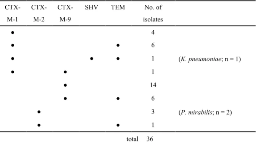
The genotyping results shown in Figure 1 are summarized in Table 2. The most detected genotypes were CTX-M-9 (n = 21), TEM (n = 14), and CTX-M-1 (n = 12). CTX-M-2 and SHV were detected in 4 and 1 isolates, respectively. M-8, M-25, CTX-M-64 (chimeric type), and GES-1 (ESBL type) were measured but were not detected among the 36 isolates included in our study. Among the 36 isolates, 22 har‐ bored only one ESBL genotype, whereas 14 isolates harbored two or three ESBL genotypes. The genotype combinations are summarized in Table 3. CTX-M-1/TEM, CTX-M-1/SHV/TEM, CTX-M-1/CTX-M-9, CTX-M-9/TEM, and CTX-M-2/TEM were found in 6, 1, 1, 6, and 1 isolate, respectively. Nota‐ bly, there were no isolates that harbored only SHV or TEM. Thus, all 36 isolates had one or more of the CTX-M group genotype(s).
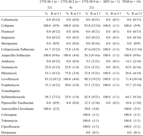
Table 4 describes the antibiotic-resistant isolates
of each ESBL-E genotype group. All available com‐ binations were analyzed. The rate of resistance to ampicillin sulbactam was significantly higher in the CTX-M-1 group than in the CTX-M-9 group and in the TEM group compared to the CTX-M-9 group (p = 0.0267 for both).
Discussion
This observational study investigated the geno‐ type distribution of ESBL-E isolated as causative pathogens in severe UTI cases positive for the SIRS criteria at a tertiary metropolitan medical facility in Japan. A substantial percentage of non-CTX-M geno‐ types were detected (15 PCR bands out of a total of 52 PCR bands) in the study; however, all isolates that harbored non-CTX-M genotypes (i.e., SHV and/or
Table 1. Patient Characteristics and Background in UTI Cases with SIRS Caused by ESBL-E Patient characteristics n=36 n % Male 17 47.2% Age (average ± S.D.) 74.0 ± 13.4 Destination Outpatient 1 2.8% Inpatient 35 97.2% Inpatient wards 18 50.0% HDU 12 33.3% ICU 5 13.9%
Acquisition of UTI of inpatient cases
Hospital Day 1–3 15 41.7%
Hospital Day 4 and later 20 55.6%
Department of admission for inpatient cases
Medical service 24 66.7%
Surgical service (excluding urology) 9 25.0%
Urology service 2 5.6% Underlying conditions Cardiac disease 7 19.4% Neurological disease 6 16.7% Pulmonary disease 7 19.4% Gastrointestinal disease 2 5.6%
Moderate-severe renal dysfunction† 13 36.1%
Orthopedic disease 3 8.3% Immune compromise 4 11.1% Malignancy 7 19.4% Urological disease 2 5.6% Pathogens ≥ 105 CFU/mL E. coli 33 91.7% K. pneumoniae 1 2.8% P. mirabilis 2 5.6% Blood culture Obtained 33 91.7% Positivity‡ 13/33 30.2% Initial antibiotics Oral antibiotics 2 5.6% Levofloxacin 2 5.6% IV antibiotics 34 94.4% Piperacillin-Tazobactam 8 22.2% Ceftriaxone (III-Cephalosporin) 7 19.4% Meropenem 7 19.4% Cefazolin (I-Cephalosporin) 3 8.3% Cefepime (IV-Cephalosporin) 3 8.3%
Ampicillin-Sulbactam 2 5.6% Cefmetazole 1 2.8% Levofloxacin 1 2.8% Imipenem/Cilastatin 1 2.8% Cefozopran 1 2.8% Clinical Outcomes
Improved with the initial antibiotics 24 66.7%
Improved after changing antibiotics 9 25.0%
Died 3 8.3%
UTI, urinary tract infection; SIRS, systemic inflammatory response syndrome; ESBL-E, extended-spectrum β-lactamase-producing Enterobacteriaceae; S.D., standard deviation; ICU, intensive care unit; HDU, high dependency unit; CFU, colony forming unit; E. coli,
Escherichia coli ; K. pneumoniae, Klebsiella pneumoniae; P. mirabilis, Proteus mirabilis ;
IV, intravenous; I-, III-, IV- cephalosporin, first, third, fourth generation cephalosporin. † EGFR < 44 mL/min·m2; ‡ All positive blood cultures showed pathogens identical to those
of the urine culture results (i.e., true bacteremia).
TEM) also harbored one of the CTX-M groups. This indicates that the genotype detection methods used to target the CTX-M group had 0 out of 36 false-nega‐ tive results and that clinicians could safely incorpo‐ rate the PCR genotyping results of ESBL-E in clini‐ cal decision-making. Currently, rapid genotype diagnosis of ESBL-E utilizing PCR is increasingly becoming the mainstream of early diagnosis for ESBL-E infection. In Japan, there are two commer‐ cially available modalities from Verigene® and Fil‐
mArray®. Both these systems detect only CTX-M
genotype groups. Our results showed that the modal‐ ity to detect CTX-M genotypes in cases with severe UTI is safe and reliable for antibiotic selection.
In a previous report from the United States, the rate of E. coli among pyelonephritis ranged from 70 to 80%18). A report from Japan on bacteremic UTI
demonstrated that the ratio of ESBL E. coli among all ESBL isolates was 81.8% (27/33)3. The proportion of
E. coli among ESBL isolates in the present study was 91%, which was higher than that in previous reports. Generally, E. coli tended to have a higher ratio (>90%) of CTX-M genotypes19) whereas K. pneumo‐
niae tended to have a higher ratio of concomitant multiple ESBL genotypes20), similar to our results.
The ratio of ESBL genotype distribution could differ from place to place. For instance, a lower ratio of CTX-M genotypes, 30% in K. pneumonia, has been reported in Iran14). A study from the United
States revealed that 91.7% (22/24) of ESBL E. coli harbored one or more CTX-M genotypes2). Another
report from Europe showed that 90.7% (107 /118) harbored one or more CTX-M genes21). Higher ratios
of CTX-M genotypes were also confirmed in our study.
Previous reports indicate that some genotypes may differ in resistance patterns19). The sample size in
the present study was not large enough to demon‐ strate statistical significance in most of the combina‐ tions, but the rate of resistance to ampicillin sulbac‐ tam was significantly higher in the CTX-M-1 group than in the CTX-M-9 group and in the TEM group compared to the CTX-M-9 group (p = 0.0267 for both). We hope that further investigation will provide more information.
Our study has several strengths and clinical im‐ plications. UTI with SIRS is a common clinical ob‐ servation1). In such cases, it is important to continue
effective antibiotics. When blood culture results are positive, PCR methods can be used for ESBL-E de‐ tection. However, a previous study from Japan re‐ ported that among ESBL-E isolates, 91.3% (95/104) were in the CTX-M group and 8.7% (9/104) were non-CTX-M group3). This suggests that application
of the PCR genotype detection method to detect only the CTX-M group may result in nearly 10% false negative results, which could be detrimental in severe cases of ESBL-E infection. Inappropriate de-escala‐ tion of antibiotics based solely on PCR ESBL-E de‐ tection methods may pose a risk to critically ill pa‐ tients. Simultaneously, prolonged use of broad-spectrum antibiotics, especially carbapenems, will
Figure. Gel electrophoresis results for genotype detection using PCR. Reaction mixture 1 (the left column) contains primer combinations to detect CTX-M-1, CTX-M-8, CTX-M-25, and CTX-M-64. Reaction mixture 2 (the right column) contains primer combinations to detect CTX-M-2, CTX-M-9, SHV, TEM, and GES-1. The number indicates each ESBL isolate. Only No. 36 has duplicate electrophoresis results. M, 100 bp DNA ladder; P, positive control; N, negative control, the numbers from 1 to 36, the serial number of each of the 36 clinical isolates.
lead to a higher incidence of carbapenem-resistant Enterobacteriaceae (CRE)22). CRE infection is associ‐
ated with higher mortality23). Therefore, early de-es‐
calation is ideal. In our study, ESBL-E clinical iso‐ lates were collected and evaluated. All isolates were found to harbor at least one of the CTX-M genotypes. These data thus provide supportive information to guide judicial antibiotic selection and de-escalation in clinical practice for primary care physicians based on ESBL-E PCR genotype detection methods.
The limitations of our study are as follows. First, microbiological distribution differs from place to place but this study was performed in a single urban
tertiary-care hospital. However, we excluded bacte‐ rial cultures from identical cases within six months and conducted this study over a period of 5 years. Thus, this study at least represents the standard distri‐ bution of ESBL-E genotypes in our region. Second, our study evaluated 36 cases of ESBL-E which is not a large enough number to lead definitive information. Third, our study evaluated genotype groups and does not include further genotypic differentiation using multi locus sequencing typing (MLST), pulsed-field gel electrophoresis (PFGE) or PCR-based ORF typ‐ ing (POT). Fourth, although our study aimed to eval‐ uate severe UTI case, one case was managed as an
Table 2. Positivity for Each of the ESBL Genotype Groups
No. of isolates positive for each of the following ESBL genotype groups
Species CTX-M-1 CTX-M-2 CTX-M-9 SHV TEM Total E. coli (n = 33) 11 2 21 13 47 K. pneumoniae (n = 1) 1 1 1 3 P. mirabilis (n = 2) 2 2 Total 12 4 21 1 14
CTX-M-8, CTX-M-25, CTX-M-64 (chimeric type), and GES-1 (ESBL type) were not detected among the 36 isolates tested.
Table 3. Number of Isolates Positive for ESBL Genotype Group Combinations
All the isolates were E. coli, except for those described as P. mirabilis or K. pneumoniae. Notably, there were no isolates positive only for SHV or TEM.
No. of isolates with the combination of genotype group(s) (n = 36) CTX-M-1 CTX-M-2 CTX-M-9 SHV TEM No. of isolates ● 4 ● ● 6 ● ● ● 1 (K. pneumoniae; n = 1) ● ● 1 ● 14 ● ● 6 ● 3 (P. mirabilis; n = 2) ● ● 1 total 36
outpatient and three cases did not have blood culture. This indicates the limitation of the usage of SIRS cri‐ teria to differentiate severe and non-severe cases. Therefore, further research collecting larger number of isolates from multicenter is warranted for further investigation of ESBL-E genotype distribution in se‐ vere UTI cases and safe de-escalation of antimicro‐ bial agents in the clinical setting.
In managing UTI cases caused by ESBL-E, rapid genome detection methods using PCR will be widely available in the near future. However, it is im‐ portant to know the false negative rates to utilize this novel technique for ESBL-E infection in clinical practice. All 36 ESBL-E isolates from severe UTI cases included in this study harbored one or more of the CTX-M genotypes, indicating that the false-nega‐
tive rate using ESBL genotype detection of CTX-M is quite low and reliably incorporated in the clinical practice in a specific region in Japan.
Conclusion
Clinicians may be able to safely de-escalate an‐ timicrobials in cases with urinary tract infection based on the ESBL-E genotyping modality.
Acknowledgements
We thank Yosuke Tanaka and Tatsuya Ono (De‐ partment of Clinical Laboratory of St. Marianna Uni‐ versity Yokohama City Seibu Hospital), Kenichi Na‐ kazono and Ayaka Katsu (St. Marianna University Yokohama City Seibu Hospital), Akiko Hosokawa (Department of Emergency and Critical Care Medi‐
Table 4. Number of Antibiotic Resistant Isolates in Each ESBL Genotype Group
There were statistically significant differences between CTX-M-1/CTX-M-9 and CTX-M-9/TEM in ampicillin sulbactam resistance rate (*, p value = 0.0267)
ESBL, extended-spectrum β-lactamase; R, resistant; I, intermediate. †R or I, the denominators are the number of isolates tested for antibiotic sensitivity. The numerators are the number of isolates with resistant or intermediate sensitivity results.
CTX-M-1 (n = 12) CTX-M-2 (n = 4) CTX-M-9 (n = 21) SHV (n = 1) TEM (n = 14) % R or I † % R or I † % R or I † % R or I † % R or I † Cefmetazole 0.0 (0/12) 0.0 (0/4) 0.0 (0/21) 0.0 (0/1) 0.0 (0/13) Cefepime 100.0 (9/9) 100.0 (4/4) 93.8 (15/16) 100.0 (1/1) 100.0 (9/9) Flomoxef 0.0 (0/12) 0.0 (0/4) 0.0 (0/21) 0.0 (0/1) 0.0 (0/13) Imipenem 0.0 (0/12) 0.0 (0/2) 0.0 (0/21) 0.0 (0/1) 0.0 (0/14) Meropenem 0.0 (0/9) 0.0 (0/4) 0.0 (0/16) 0.0 (0/1) 0.0 (0/9) Cefoperazone Sulbactam 41.7 (5/12) 75.0 (3/4) 47.6 (10/21) 100.0 (1/1) 78.6 (11/14) Ampicillin Sulbactam 100.0 (9/9)* 100.0 (4/4) 56.3 (9/16) 100.0 (1/1)* 100.0 (9/9)* Amikacin 0.0 (0/12) 0.0 (0/4) 9.5 (2/21) 0.0 (0/1) 14.3 (2/14) Gentamycin 25.0 (3/12) 25.0 (1/4) 23.8 (5/21) 0.0 (0/1) 42.9 (6/14) Minomycin 33.3 (4/12) 75.0 (3/4) 23.8 (5/21) 100.0 (1/1) 28.6 (4/14) Levofloxacin 83.3 (10/12) 100.0 (4/4) 90.5 (19/21) 100.0 (1/1) 71.4 (10/14) Fosphomycin 33.3 (4/12) 50.0 (2/4) 33.3 (7/21) 100.0 (1/1) 35.7 (5/14) Trimethoprim Sulfamethoxazole 58.3 (7/12) 25.0 (1/4) 42.9 (9/21) 100.0 (1/1) 64.3 (9/14) Piperacillin Tazobactam 0.0 (0/9) 0.0 (0/4) 12.5 (2/16) 0.0 (0/1) 10.0 (1/10) Amoxicillin Clavulanate 100.0 (2/2) 50.0 (3/6) 100.0 (5/5) Cefozopran 100.0 (1/1) 100.0 (1/1) Tobramycin 100.0 (1/1) 100.0 (1/1) Ciprofloxacin 100.0 (1/1) 100.0 (1/1) Doripenem 0.0 (0/1) 0.0 (0/1)
cine of St. Marianna University School of Medicine), and Keiko Nishida (St. Marianna University School of Medicine, Yokohama City Seibu Hospital) for pro‐ viding technical support. We would like to thank Ed‐ itage (www.editage.com) for English language edit‐ ing.
Conflicts of interest
The authors have nothing to disclose.
References
1) Belyayeva M, Jeong JM. Acute Pyelonephritis. http://www.ncbi.nlm.nih.gov/books/
NBK519537/. Retrieved on Jul 12, 2019. 2) Doi Y, Park YS, Rivera JI, et al. Community-as‐
sociated extended-spectrum β-lactamase-pro‐
ducing Escherichia coli infection in the United States. Clin Infect Dis 2013; 56: 641–648. 3) Mawatari M, Hayakawa K, Fujiya Y, et al. Bac‐
teraemic urinary tract infections in a tertiary hospital in Japan: the epidemiology of commun‐ ity-acquired infections and the role of non-car‐ bapenem therapy. BMC Res Notes 2017; 10: 336. doi:10.1186/s13104-017-2680-z.
4) CDDEP. Resistance map. https://resistance map.cddep.org/AntibioticResistance.php. Re‐ trieved on May 8, 2020.
5) Paterson DL, Ko W-C, Von Gottberg A, et al. Antibiotic therapy for Klebsiella pneumoniae bacteremia: implications of production of ex‐ tended-spectrum beta-lactamases. Clin Infect Dis 2004; 39: 31–37.
6) McLaughlin M, Advincula MR, Malczynski M, et al. Correlations of antibiotic use and carbape‐ nem resistance in Enterobacteriaceae. Antimi‐ crob Agents Chemother 2013; 57: 5131–5133. 7) Chang H-J, Hsu P-C, Yang C-C, et al. Risk fac‐
tors and outcomes of carbapenem-nonsuscepti‐ ble Escherichia coli bacteremia: A matched case–control study. J Microbiol Immunol Infect 2011; 44: 125–130.
8) Clinical and Laboratory Standards Institute (CLSI). Performance standards for antimicrobial susceptibility testing, 27th ed, Pennsylvania, Wayne, 2017.
9) Demirci M, Ünlü Ö, İstanbullu Tosun A. Detec‐ tion of O25b-ST131 clone, M-1 and CTX-M-15 genes via real-time PCR in Escherichia coli strains in patients with UTIs obtained from a university hospital in Istanbul. J Infect Public Health 2019; 12: 640–644.
10) Dallenne C, Da Costa A, Decré D, et al. Devel‐ opment of a set of multiplex PCR assays for the detection of genes encoding important beta-lac‐ tamases in Enterobacteriaceae. J Antimicrob Chemother 2010; 65: 490–495.
11) Luminex Japan. VERIGENE® Bloodstream In‐
fection Testing Panels. https://www.luminex corp.com/ja/bloodstream-infection-tests/. Re‐ trieved on May 8, 2020.
12) Tayh G, Al Laham N, Ben Yahia H, et al. Ex‐ tended-spectrum β-lactamases among Entero‐ bacteriaceae isolated from urinary tract infec‐ tions in Gaza Strip, Palestine. BioMed Res Int 2019; 2019: 1–11.
13) Birgy A, Madhi F, Jung C, et al. Diversity and trends in population structure of ESBL-produc‐ ing Enterobacteriaceae in febrile urinary tract in‐ fections in children in France from 2014 to 2017. J Antimicrob Chemother 2020; 75: 96– 105.
14) Yazdansetad S, Alkhudhairy MK, Najafpour R, et al. Preliminary survey of extended-spectrum β-lactamases (ESBLs) in nosocomial uropatho‐ gen Klebsiella pneumoniae in north-central Iran. Heliyon 2019; 5: e02349. doi:10.1016/j.heliyon. 2019.e02349.
15) Kishi K. Genotype destriution o ESBL produc‐ ing E. coli in Shimane Prefecture. Report of the Shimane Prefectural Institute of Public Health and Environmental Science. 2008; 50: 66–69. 16) Kitano Y, Wakatake H, Saito H, et al. Clinical
outcomes of urinary tract infection caused by extended spectrum beta‐lactamase producing Enterobacteriaceae: a retrospective observatio‐ nal study comparing patients with and without systemic inflammatory response syndrome. Acute Med Surg 2020; 7: e472. doi:10.1002/ ams2.472.
17) js-STAR. version 9.8.4j. http://www.kisnet.or.jp/ nappa/software/star/freq/2x2.htm#. Retrieved on May 10, 2020.
18) Czaja CA, Scholes D, Hooton TM, et al. Popula‐ tion-based epidemiologic analysis of acute pye‐ lonephritis. Clin Infect Dis 2007; 45: 273–280. 19) Nakane K, Kawamura K, Goto K, et al.
Long-term colonization by bla CTX-M-harboring Es‐ cherichia coli in healthy Japanese people en‐ gaged in food handling. Appl Environ Microbiol 2016; 82: 1818–1827.
20) Miyoshi S. Epidemiology of extended-spectrum β-lactamase-producing bacteria in Kagawa Uni‐ versity Hospital from 2006 to 2013. Igakukensa [In Japanese] 2014; 63: 714–718.
21) Doumith M, Dhanji H, Ellington MJ, et al. Characterization of plasmids encoding ex‐ tended-spectrum β-lactamases and their addic‐ tion systems circulating among Escherichia coli clinical isolates in the UK. J Antimicrob Che‐ mother 2012; 67: 878–885.
22) Marimuthu K, Ng OT, Cherng BPZ, et al. Ante‐ cedent carbapenem exposure as a risk factor for non-carbapenemase-producing carbapenem-resist‐ ant Enterobacteriaceae and carbapenemase-pro‐ ducing Enterobacteriaceae. Antimicrob Agents Chemother 2019; 63: e00845-19. doi:10.1128/ AAC.00845–19.
23) Xu L, Sun X, Ma X. Systematic review and meta-analysis of mortality of patients infected with carbapenem-resistant Klebsiella pneumo‐ niae. Ann Clin Microbiol Antimicrob 2017; 16: 18. doi:10.1186/s12941-017-0191-3.