Effects of dihydrotestosterone administration on the expression of reproductive and body weight regulatory factors in ovariectomized and estradiol-treated female rats
Authors
Takeshi Iwasa, Toshiya Matsuzaki, Kiyohito Yano, Yiliyasi Mayila and Minoru Irahara
Department of Obstetrics and Gynecology, Institute of Biomedical Sciences, Tokushima University Graduate School, 3-18-15 Kuramoto-Cho, Tokushima 770-8503, Japan
Corresponding author
Takeshi Iwasa
Department of Obstetrics and Gynecology, Institute of Biomedical Sciences, Tokushima University Graduate School, 3-18-15 Kuramoto-Cho, Tokushima 770-8503, Japan Phone number: +81−88−633−7177
E-mail: iwasa.takeshi@tokushima-u.ac.jp
Running title
Effects of DHT injection
Keywords
dihydrotestosterone, estradiol, kisspeptin, GnIH
This is an Accepted Manuscript of an article published by Taylor & Francis in Gynecological Endocrinology on 11/06/2017, available online: http://www.tandfonline.com/10.1080/09513590.2017.1337096.
Abstract
To clarify the direct effects of androgens, the changes in the hypothalamic levels of reproductive and appetite regulatory factors induced by chronic dihydrotestosterone (DHT) administration were evaluated in female rats. DHT treatment increased the BW and food intake of the ovariectomized rats, but not the estradiol (E2)-treated rats. DHT administration suppressed the expression of a hypothalamic anorexigenic factor. Although the kisspeptin (Kiss1) mRNA levels of the anterior hypothalamic block (the anteroventral periventricular nucleus, AVPV) were increased in the E2-treated rats, DHT administration did not affect the Kiss1 mRNA levels of the AVPV in the ovariectomized or E2-treated rats. Conversely, DHT administration reduced the Kiss1 mRNA levels of the posterior hypothalamic block (the arcuate nucleus, ARC) in the ovariectomized rats. Although the Kiss1 mRNA levels of the posterior hypothalamic block (ARC) were decreased in the E2-treated rats, DHT administration did not affect the Kiss1 mRNA levels of the ARC in these rats. Serum luteinizing hormone levels of these groups exhibited similar patterns to the Kiss1 mRNA levels of the ARC. These results showed that DHT affects the production of hypothalamic reproductive and appetite regulatory factors, and that these effects of DHT differ according to the estrogen milieu.
Introduction
Sex hormones influence reproductive function and body weight (BW) regulation in mammals and humans [1]. It has been well established that estrogen play pivotal roles in the regulation of gonadotropin-releasing hormone (GnRH)/luteinizing hormone (LH) secretion via positive and negative feedback effects on hypothalamic kisspeptin and its receptor Kiss1r, which is a positive regulator of GnRH [2]. Estrogen also affects the activity of hypothalamic RFamide-related peptides/gonadotropin inhibitory hormone (RFRP/GnIH) and its receptor, G protein-coupled receptor (GPR)147, which is a negative regulator of GnRH and LH [3,4]. It has been also well established that estrogen play pivotal roles in the regulation of appetite and BW through the modulation of hypothalamic orexigenic and anorexigenic factors [5-9].
In contrast to estrogen, the effects of androgens on reproductive function and BW regulation in females have not been fully established, although it has been reported that hyperandrogenemia is usually accompanied by visceral obesity and ovulatory disorders in humans [10]. Almost all of the basic studies that have investigated the effects of androgens on female physiological functions involved animal models of polycystic ovary syndrome (PCOS), which is a representative hyperandrogenism-related disorder [11]. Although the effects of androgens on reproductive and BW regulatory functions and the mechanisms underlying these effects have been partially clarified by the aforementioned studies [11], some other effects, particularly the effects of androgens on hypothalamic functions, have not been fully evaluated. Recently, we and other groups have investigated hypothalamic kisspeptin levels, as well as serum gonadotropin levels, in PCOS model
rodents; however, there were marked discrepancies between the results of these studies [12-14]. The protocols used to produce the PCOS model differed among the studies, and ovarian intact rodents were used; i.e., the rats were not only administered androgen, and androgen-induced changes in ovarian functions might affect hypothalamic functions, which could explain the discrepancies among the studies’ results. In addition, only a few studies have evaluated the effects of androgens on hypothalamic appetite regulatory factors in females [15], and there are no data about the effects of androgens on hypothalamic RFRP/GnIH in females.
To clarify the direct effects of androgens on reproductive and BW regulatory functions, we evaluated the effects of chronic dihydrotestosterone (DHT), which is a non-aromatizing androgen, on hypothalamic factors under ovariectomized (OVX) and stable estradiol (E2) milieu conditions.
Materials and Methods Animals
Wistar female rats were purchased from Charles River Laboratories Japan, Inc., (Kanagawa, Japan) and housed in a room under controlled light (12 h light, 12 h darkness; lights turned on at 0800 and turned off at 2000) and temperature (24°C) conditions. In total, 29 rats were used in this study. All animal experiments were conducted in accordance with the ethical standards of the institutional animal care and use committee of the University of Tokushima. All surgical procedures were carried out under sodium pentobarbital- (60-80 mg/kg, intraperitoneal, i.p.) or sevoflurane-induced anesthesia.
Effects of chronic DHT administration in ovariectomized (OVX) rats and OVX and estradiol-administered rats
At 15 weeks of age, the rats were randomly divided into the no treatment (control), DHT-administered (DHT), E2-administered (E2), and E2 and DHT-administered (E2+DHT) groups. All rats were OVX bilaterally and implanted with a silastic tube filled with crystalline steroid hormones under sodium pentobarbital-induced anesthesia. The parts of the tubes filled with DHT and E2 measured 30 mm and 3.0 mm in length, respectively [16,17]. In the control group, the rats were implanted with empty tubes. All of the tubes were removed and replaced with new ones 4 weeks later under sevoflurane-induced anesthesia. The rats were individually housed after surgery, and their BW and food intake (FI) were checked every 7 days. At 8 weeks after the OVX and implantation procedures, the rats were sacrificed by decapitation under sevoflurane-induced anesthesia. The weights of visceral fat (parametrial, perirenal, and mesenteric deposits) and subcutaneous fat (inguinal deposits) were measured, and the brain and blood were collected. Serum was separated by centrifugation and stored at -20°C, and tissues were stored at -80°C.
Hormone assays
Serum E2 levels were measured by a commercial laboratory (SRL, Tokyo, Japan) using an electrochemiluminescence immunoassay (ECLIA; Roche Diagnostics GmbH, Mannheim, Germany). Serum LH levels were measured using a radioimmunoassay (RIA) (rat LH [I-125] RIA kit, Institute of Isotopes Co., Ltd., Tokyo, Japan).
Whole hypothalamic explants were dissected from the frozen brains, as described previously [15]. Briefly, the target region of the brain was dissected out via an anterior coronal cut at the posterior border of the mammillary bodies, parasagittal cuts along the hypothalamic fissures, and a dorsal cut 2.5 mm from the ventral surface. Subsequently, the obtained brain tissue was divided into two blocks using coronal cuts at the posterior border of the optic chiasm. The anterior hypothalamic block contained the anteroventral periventricular nucleus (AVPV), and the posterior hypothalamic block contained the arcuate nucleus (ARC) [18]. Kisspeptin in the AVPV and ARC receives positive and negative feedback signals from estrogen, respectively [2]. Total RNA was isolated from the hypothalamic explants and visceral fat using a TRIzol® reagent kit (Invitrogen Co., Carlsbad, CA, USA) and an RNeasy® mini kit (Qiagen Gmbh, Hilden, Germany). cDNA was synthesized with oligo (deoxythymidine) primers at 50°C using the SuperScript III first-strand synthesis system for the real-time polymerase chain reaction (PCR; Invitrogen Co.). The PCR analysis was performed using the StepOnePlusTM real-time PCR system
(PE Applied Biosystems, Foster City, CA, USA) and FAST SYBR® green. The mRNA levels of neuropeptide Y (NPY), proopiomelanocortin (POMC), prepro-orexin (pporexin),
Kiss1 (the gene encoding kisspeptin), Kiss1r, RFRP/GnIH (the gene encoding the
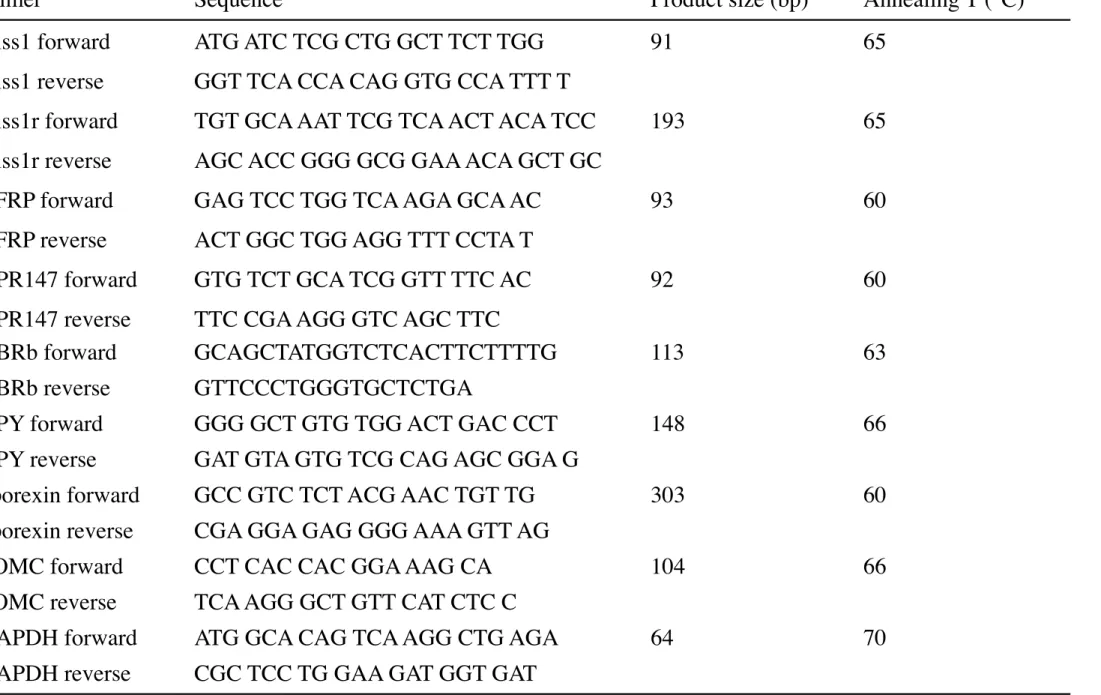
kisspeptin receptor), and GPR147 in the posterior hypothalamus, and the Kiss1 mRNA levels of the anterior hypothalamus were measured. The mRNA expression level of each molecule was normalized to that of GAPDH. Dissociation curve analysis was also performed for each gene at the end of the PCR. Each amplicon generated a single peak. The primer sequences, product sizes, and annealing temperatures are shown in Table 1.
The PCR conditions were as follows: initial denaturation and enzyme activation at 95°C for 20 s, followed by 45 cycles of denaturation at 95°C for 3 s, and annealing and extension for 30 s.
Statistical analyses
All data are presented as the mean ± standard deviation (SD). The statistical analyses were conducted using one-way or two-way analysis of variance (ANOVA) together with the Tukey-Kramer post-hoc test for comparisons among the groups.
Results
The E2-treated groups (E2, E2+DHT) exhibited significantly smaller BW changes than the non-E2-treated groups (control, DHT). The DHT group displayed greater BW changes than the control group, whereas the BW changes in the E2 group did not differ from those seen in the E2+DHT group (Figs. 1A and B). Similarly, the DHT group demonstrated greater cumulative FI compared with the control group, whereas the cumulative FI of the E2 group did not differ from that of the E2+DHT group (Figs. 1A and B). The weights of visceral fat, subcutaneous fat, total fat, and lean body mass recorded in the E2-treated groups (E2, E2+DHT) were significantly lighter than those observed in the non-E2-treated groups (control, DHT) (Figs. 2A-D). There were no significant differences in these parameters between the control and DHT groups or between the E2 and E2+DHT groups. The serum LH levels of the DHT, E2, and E2+DHT groups were significantly lower than that of the control group (Fig. 2E). The serum E2 levels of the E2-treated groups were significantly higher than those of the non-E2-treated
groups.
There were no significant differences in the hypothalamic NPY or pporexin mRNA levels among the examined groups (Figs. 3A and B), whereas the hypothalamic POMC mRNA level of the E2+DHT group was significantly lower than that of the control group (Fig. 3C). In the anterior hypothalamus, the Kiss1 mRNA levels of the E2-treated groups (E2, E2+DHT) were significantly higher than those of the non-E2-treated groups (control, DHT) (Fig. 3D). However, there were no significant differences in the anterior hypothalamic Kiss1 mRNA level between the control and DHT groups or between the E2 and E2+DHT groups. On the other hand, the posterior hypothalamic Kiss1 mRNA levels of the DHT, E2, and E2+DHT groups were significantly lower than that of the control group (Fig. 3E). The posterior hypothalamic Kiss1r mRNA level of the E2+DHT group was significantly lower than those of the other groups (Fig. 3F). The posterior hypothalamic RFRP and GPR147 mRNA levels of the E2+DHT group were lower than those of the control and DHT groups (Fig. 3H).
Discussion
Sex hormones play roles in reproductive function and BW regulation in mammals and humans [1]. It has been well established that the effects of estrogen on GnRH/LH are mainly mediated by hypothalamic kisspeptin and RFRP/GnIH [2-4] and that its effects on appetite are mediated by hypothalamic orexigenic and anorexigenic factors [1,5-9]. In contrast to estrogen, the effects of androgens on reproductive function and BW regulation, especially on hypothalamic functions, in females have not been fully established.
Although some studies have examined the effects of androgens on hypothalamic reproductive functions, the results were disputed since ovarian intact PCOS animal models were used [12-14]. Thus, to clarify the direct effects of androgens on reproductive functions, we evaluated the changes in the hypothalamic levels of reproductive factors induced by chronic DHT administration under OVX and stable E2 milieu conditions. Kisspeptin has been detected in both the AVPV and ARC, and it was reported that lower levels of estrogen reduce kisspeptin activity in the ARC and suppress GnRH/LH secretion, whereas higher levels of estrogen increase kisspeptin activity in the AVPV and induce surges in GnRH/LH production [2]. Thus, we separated the hypothalamus into anterior (AVPV) and posterior (ARC) blocks and evaluated the Kiss1 mRNA levels of these blocks. As a result, we found that although the Kiss1 mRNA levels of the anterior hypothalamic block (AVPV) were significantly higher in the E2-treated groups (E2 and E2+DHT groups) than in the non- E2-treated groups (control and DHT groups), they did not differ between the groups that were and were not administered DHT in the OVX or E2-treated rats. These results indicate that in the AVPV the Kiss1 gene is sensitive to E2, but not DHT. On the other hand, the Kiss1 mRNA levels in the posterior hypothalamic block (ARC) were significantly lower in the E2-treated groups than in the non-E2-treated groups. In addition, the Kiss1 mRNA levels of the posterior hypothalamic block (ARC) were significantly lower in the group treated with DHT than in the group that was not administered DHT among the OVX rats, but not the E2-treated rats. These results indicate that in the ARC the Kiss1 gene is sensitive to both E2 and DHT. As the serum LH levels of these groups exhibited similar patterns to their Kiss1 mRNA levels in the ARC; i.e.,
the levels of LH were decreased by both E2 and DHT, these steroids might have stronger effects on gonadotropin levels in the ARC than in the AVPV. Interestingly, although neither DHT nor E2 affected the hypothalamic mRNA levels of Kiss1r or RFRP/GnIH when administered alone, the co-administration of DHT and E2 suppressed the hypothalamic concentrations of these molecules. The administration of DHT reduced the hypothalamic mRNA levels of GPR147 in both the OVX and E2-treated rats. We could not evaluate the physiological roles of these alterations in this study; however, we speculate that these changes might represent a counter-regulatory mechanism that attenuates the effects of marked changes in kisspeptin activity.
In the present study, the BW gains observed in the OVX groups were significantly larger than those seen in the E2-treated groups. The BW gains and FI recorded in the group treated with DHT were significantly larger than those observed in the group that was not administered DHT in the OVX rats, whereas they did not differ in the E2-treated rats. On the other hand, the amounts of visceral and subcutaneous fat were not affected by DHT administration in either the OVX or E2-treated rats. These findings indicate that DHT itself does not affect the amount of body fat under OVX or stable E2 milieu conditions. It has been reported that both BW and fat weight were increased in DHT-treated PCOS models [11,19,20] and that they were also increased in testosterone-DHT-treated female rats [15]. As mentioned above, because androgens were administered to ovarian intact female animals and estrous cyclicity was disturbed in almost all of these animals in the aforementioned studies, some of the observed effects of androgens on BW and body fat might have been induced by disturbances in ovarian function; i.e., estrogen deficiency
or irregularities in estrogen production. In this study, the hypothalamic mRNA levels of orexigenic factors; i.e., NPY and pporexin, were not changed by the administration of DHT in the OVX or E2-treated rats. On the other hand, the hypothalamic mRNA level of
POMC, an anorexigenic factor, was reduced by the co-administration of DHT and E2. As
it has been reported that kisspeptin acts as a positive regulator of POMC expression in the hypothalamus, reductions in Kiss1 and Kiss1r expression might be related to the alterations in the POMC level seen in the DHT+E2 group [21,22]. However, such changes might not be directly involved in the regulation of BW or FI. Thus, we could not detect any hypothalamic mechanisms by which DHT increases FI under OVX conditions. Further studies are needed to clarify the mechanisms by which androgens affect BW and FI regulation.
In summary, this study showed that DHT affects BW, FI, and the expression of hypothalamic reproductive and appetite regulatory factors and that these effects of DHT differ according to the estrogen milieu. In addition, the effects of DHT on Kiss1 mRNA levels in the anterior (AVPV) and posterior (ARC) hypothalamus were markedly different. DHT might have stronger effects on gonadotropin levels in the ARC than in the AVPV.
Conflict of Interest
The authors declare that they have no conflict of interest.
[1] Hirschberg AL. Sex hormones, appetite and eating behavior in women. Maturitas 2012;71:248-256
[2] Putteeraj M, Soga T, Ubuka T, Parhar IS. A “timed” Kiss is essential for reproduction: lessons from mammalian studies. Front Endocrinol 2016;7:121
[3] Gibson EM, Humber SA, Jain S, Williams WP 3rd, Zhao S, Bentley GE, Tsutsui K, Kriegsfeld LJ. Alterations in RFamide-related expression are coordinated with the preovulatory luteinizing hormone surge. Endocrinology 2008;149:4958-4969
[4] Kriegsfeld LJ, Mei DF, Bentley GE, Ubuka T, Mason AO, Inoue K, Ukena K, Tsutsui K, Silver R. Identification and characterization of a gonadotropin-inhibitory system in the brain of mammals. Proc Natl Acad Sci USA 2006;103:2410-2415.
[5] Asarian L, Geary N. Sex differences in the physiology of eating. Am J Regul Integr Comp Physiol 2013;305:R1215-R1267
[6] Blaustein JD, Wade GN. Ovarian influences on the meal patterns of female rats. Physiol Behav 1976;17:201-208
[7] Butera PC, Beikirch RJ. Central implants of diluted estradiol: independent effects on ingestive and reproductive behaviors of ovariectomized rats. Brain Res. 1989;491:266– 273
[8] Palmer K, Gray JM. Central vs. peripheral effects of estrogen on food intake and lipoprotein lipase activity in ovariectomized rats. Physiol Behav 1986;37:187-189 [9] Santollo J, Yao D, Neal-Perry G, Etgen AM. Middle-aged female rats retain sensitivity to the anorexigenic effect of exogenous estradiol. Behav Brain Res 2012;15:159-164 [10] Gambineri A, Pelusi C, Genghini S, Morselli-Labate AM, Cacciari M, Pagotto U,
Pasquali R. Effect of flutamide and metformin administered alone or in combination in dieting obese women with polycystic ovary syndrome. Clin Endocrinol (Oxf) 2004;60:241-249
[11] Walters KA, Allan CM, Handelsman DJ. Rodent models for human polycystic ovary syndrome. Biol Reprod 2012;86:149
[12] Brown RE, Wilkinson DA, Imran SA, Caraty A, Wilkinson M. Hypothalamic kiss1 mRNA and kisspeptin immunoreactivity are reduced in a rat model of polycystic ovary syndrome (PCOS). Brain Res 2012;1467:1-9
[13] Matsuzaki T, Tungalagsuvd A, Iwasa T, Munkhzaya M, Yanagihara R, Tokui T, Yano K, Mayila Y, Kato T, Kuwahara A, Matsui S, Irahara M. Kisspeptin mRNA expression is increased in the posterior hypothalamus in the rat model of polycystic ovary syndrome. Endocr J 2017;64:7-14.
[14] Osuka S, Iwase A, Nakahara T, Kondo M, Saito A, Bayasula, Nakahara T, Takikawa S, Goto M, Kotani T, Kikkawa F. Kisspeptin in the hypothalamus of 2 rat models of polycystic ovary syndrome. Endocrinology 2017;158:367-377
[15] Iwasa T, Matsuzaki T, Tungalagsuvd A, Munkhzaya M, Yiliyasi M, Kato T, Kuwahara A, Irahara M. Effects of chronic testosterone administration on body weight and food intake differ among pre-pubertal, gonadal-intact, and ovariectomized female rats. Behav Brain Res 2016;309:35-43
[16] De Vries GJ, Wang Z, Bullock NA, Numan S. Sex Differences in the Effects of Testosterone and Its Metabolites on Vasopressin Messenger RNA Levels in the Bed Nucleus of the Stria Terminalis of Rats. J Neurosci 1994;14:1789-1794
[17] Le TY, Ashton AW, Mardini M, Stanton PG, Funder JW, Handelsman DJ, Mihailidou AS. Role of androgens in sex differences in cardiac damage during myocardial infarction. Endocrinology 2014;155:568-575
[18] Matsuzaki T, Iwasa T, Kinouchi R, Yoshida S, Murakami M, Gereltsetseg G, Yamamoto S, Kuwahara A, Yasui T, Irahara M. Fasting reduces the kiss1 mRNA levels in the caudal hypothalamus of gonadally intact adult female rats. Endocr J 2011;58:1003-1012
[19] Johansson J, Feng Y, Shao R, Lonn M, Billig H, Stener-Victorin E. Intense electroacupuncture normalizes insulin sensitivity, increases muscle GLUT4 content, and improves lipid profile in a rat model of polycystic ovary syndrome. Am J Physiol Endocrinol Metab 2010;299:E551-E559
[20] Manneras L, Cajander S, Holmang A, Seleskovic Z, Lystig T, Lonn M, Stener-Victorin E. A new rat model exhibiting both ovarian and metabolic characteristics of polycystic ovary syndrome. Endocrinology 2007;148:3781-3791.
[21] Fu LY, van den Pol AN. Kisspeptin directly excites anorexigenic proopiomelanocortin neurons but inhibits orexigenic neuropeptide Y cells by an indirect synaptic mechanism. J Neurosci 2010;30:10205-10219.
[22] Higo S, Iijima N, Ozawa H. Characterization of Kiss1r (GPR54)-expressing neurons in the arcuate nucleus of the female rat hypothalamus. J Neuroendocrinol 2017;29:10.1111
Figure legends Fig. 1
Weekly body weight (BW) changes (% of initial BW) (A, B) and cumulative food intake (FI; g/initial BW) (C, D) observed in the control, dihydrotestosterone (DHT)-treated, estradiol (E2)-treated, and DHT+E2-treated rats
Data are expressed as mean ± SEM values. Different letters (a-c) indicate significant differences (P <0.05).
Fig. 2
Visceral, subcutaneous, and total fat weight (A-C); lean body mass (D); and serum LH and serum estradiol levels (E, F) in the control, dihydrotestosterone (DHT)-treated, estradiol (E2)-treated, and DHT+E2-treated rats
Data are expressed as mean ± SEM values. Different letters (a-c) indicate significant differences (P <0.05).
Fig. 3
The mRNA levels of hypothalamic orexigenic and anorexigenic factors (A-C) and hypothalamic reproductive factors (D-H)
The mRNA expression levels of the control rats are expressed as 1.0. Data are expressed as mean ± SEM values. Different letters (a-c) indicate significant differences (P <0.05).
90 100 110 120 130 140 150 0wk 2wk 4wk 6wk 8wk control DHT E2 E2+DHT 1.0 2.0 3.0 4.0 5.0 control DHT E2 E2+DHT
ul
at
ive
F
I (g /
i
ni
ti
al
BW
)
BW
c
ha
nge
(% of i
ni
ti
al
BW
)
80 90 100 110 120 130 140 150 4wk 8wkBW
c
ha
nge
(% of i
ni
ti
al
BW
)
a b c c a b c c 1.0 1.5 2.0 2.5 3.0 3.5 4.0 4.5 a b c c a b a aul
at
ive
F
I (g /
i
ni
ti
al
BW
)
A
B
C
D
control DHT E2 E2+DHT control DHT E2 E2+DHTFig. 1
0 2 4 6 8 10 12 14 16 18 0 2 4 6 8 10 0 5 10 15 20 25 30
V
is
ce
ra
l fa
t (g)
S
ubc
ut
ane
ous
fa
t (g)
T
ot
al
fa
t (g)
a
a
b
b
a
a
b b
a
a
b b
100 200 300 400 500L
ea
n body m
as
s (g)
a
a
b b
50 100 150 200 250 300 350E
st
ra
di
ol
(
pg
/
m
L
)
2 4 6 8 10L
H
(
ng
/
m
L
)
a
b b b
control DHT E2 E2+DHTA
B
C
D
E
F
a a
b
b
Fig. 2
0.0 0.2 0.4 0.6 0.8 1.0 1.2