INTRODUCTION
The recent treatment of hematological malignancies reported that the event free survival (EFS) of children with acute lymphocytic leukemia (ALL) was approxi-mately 75% after reaching complete remission in more than 95%. The long-term survival for adults with ALL is only 20% after a complete remission rate of 80%. In childhood acute myeloid leukemia (AML), 85% reach complete remission, but EFS is only 35-50% after
remis-sion induction therapy. The EFS of adult patients with AML will not exceed 20% after about 85% of the patients reach complete remission (1-3). Despite the progress of the treatment for leukemia in recent decades, long term survival as shown in Table 1 remains unsatis-factory.
A major problem in the treatment of leukemia is caused by the development of drug resistance to chem-otherapeutic agents. It has been observed that a biphasic decline in the number of leukemia cells occurs during induction or re-induction chemotherapy after relapse (4). A schematic model is shown in Figure1, where the biphasic decline of leukemia cell number suggests that most leukemia cells are sensitive to treatment and are quickly killed, leaving behind a minor but substantial population of drug-resistant
REVIEW
Multidrug resistance in hematological malignancy
Masao Hirose
1, Eiji Hosoi
2, Shuichi Hamano
3and Ali Jalili
4 1Health Management Center, Naruto University of Education, Tokushima, Japan ; 2
Department of Morphological Laboratory Science, Major in Laboratory Science, School of Health, 3
Department of Laboratory of Medicine, and 4
Department of Medicine and Bioregulatory Sciences, The University of Tokushima School of Medicine, Tokushima, Japan
Abstract : The recent treatment of hematological malignancies appears to be unsatisfactory in child and adult patients with acute myeloid leukemia and adult patients with acute lymphocytic leukemia. A major problem in the treatment of leukemia is caused by the development of drug resistance to chemotherapeutic agents, which is already present at diagnosis or after chemotherapy as a minimal residual disease, their resistance having originated from genetic or epigenetic mutations during prior growth of the leukemia clone. It was suggested that the mechanisms of drug resistance consist of drug resistance proteins, which work as a drug efflux pump. These are the permeability- related glycoprotein (P- Gp), the multidrug-resistance associated protein (MRP), the lung resistance protein (LRP), and other MDR proteins such as the transporter associated with antigen processing (TAP), anthracyclin resistance associated protein (ARA), MRP 2-7, and breast cancer resistance protein (BCRP). In addition, anti-apoptosis mechanisms, alterations of tumor suppressor genes, altered immunogenicity, drug resistance mechanisms for individual drugs, and clinical risk factors such as white blood cell count, age, and other factors have been reported to act in drug resistance singly or in combinations.
Here we describe the update of research on the biology of MDR in the hematological malignancies and also discuss how to overcome MDR and adapt the updated treatment methods in the clinical medical field. J. Med. Invest. 50 : 126-135, 2003
Keywords : multidrug resistance, MDR, hematology, leukemia, lymphoma
Received for publication April 28, 2003 ; accepted June 2, 2003.
Address correspondence and reprint requests to Masao Hirose, M.D.,Ph.D., Health Management Center, Naruto University of Education, 748, Aza-Nakajima, Takashima, Naruto-cho, Naruto-shi, Tokushima 772-8502, Japan and Fax : +81-88-687-6107.
The Journal of Medical Investigation Vol. 50 2003
cells. A part of the fraction called minimal residual disease may eventually develop the drug-resistant subpopulations(4, 5). The most likely explanation for this phenomenon is that drug-resistant cells are already present at diagnosis, their resistance having originated from genetic or epigenetic mutations during prior growth of the leukemia clone. The patient whose white blood cell number is more may have more mutated cells or drug resistant subpopulations.
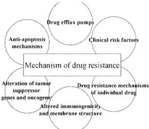
There are several factors that form drug resistance mechanisms against cytotoxic drugs in leukemia cells. It was suggested that mechanism of drug resistance consists of drug efflux pump, anti-apoptosis mechanism, alterations of tumor suppressor genes, altered immu-nogenicity, drug resistance mechanisms for individual
drugs, and clinical risk factors such as white blood cell counts, age, and others. Such defense mechanisms of leukemia cells to anti-cancer drugs develop singly or in combinations. These factors that can consist of drug resistance mechanisms are summarized in Figure 2.
CELLULAR ACTION SITES OF MECHANISMS
OF DRUG RESISTANCE.
The term multidrug resistance (MDR) describes the observation that tumor cell lines can become cross-resistant to another structurally unrelated chemo-therapeutic agent after exposure to a single cytotoxic drug. This is one of the major impediments in cancer treatment, which can be caused by alterations in drug transport, altered intracellular drug targets, altered apoptosis mechanisms, or altered metabolic mechanisms. As shown in Figure 3, three proteins that are related to the alteration of drug transport are found. One is the drug resistance protein, permeability-related glycoprotein (P-Gp)(6, 7), and the other factor is the multidrug-resistance associated protein (MRP)(8, 9). The third is the lung resistance protein (LRP) found in the cytoplasm of tumor cells showing MDR phenotype (10, 11). There are other MDR proteins such as the transporter associated with antigen processing (TAP) (12, 13), anthracyclin resistance associated protein (ARA)(14), MRP2-7 (15, 16), and breast cancer resistance protein (BCRP)(17,18), although their functions have not been fully elucidated.
CHARACTERISTICS OF THE
PERMEABILITY-RELATED GLYCOPROTEIN (P-GP).
As summarized in Table 2, P-Gp is encoded by the mdr-1 gene, localized at 7q21.1, a 170 kDa trans-membrane glycoprotein consisting of two domains.
Figure 1. Schematic model of cleavage of drug resistant cells.
Figure 3. Sites of action of some mechanisms of drug resistance. Table 1. Complete remission rate and event free survival rate
in childhood and adult leukemia by recent therapy. CR(%) EFS(%) Childhood Adult ALL AML ALL AML >95 85 80 85 75 35‐50 20 <20
Figure 2. Mechanisms of multidrug resistance.
Up-regulation of this protein results in a decreased intracellular concentration of anti-cancer drugs such as anthracyclins, epipodophylotoxins, and vinca-alkaloids. Themdr-1gene is differently expressed in a variety of normal tissues, particularly along the apical surface of secretory epithelium of the liver, pancreas, jejunum and colon, proximal tubular epithelium, and the glandular epithelium of the pregnant uterus, furthermore, it was also reported in the adrenal grand, placenta, capillary endothelium of the liver, testis and brain, in addition to hematopoietic precursors and lymphocytes (6, 19). Although the normal physiological function of P-Gp remains unknown, P-P-Gp exerts its action to reduce intracellular drug accumulation, which causes MDR as a result of the initial stage of the therapy or due to the chemotherapy after relapses. At the cellular level, the function of P-Gp has been extensively investigated in many types of cancer cells. In leukemia patients, cellular drug resistance profiles determined in vitro at the time of presentation showed a strong correlation with the outcome (18, 21, 22).
CHARACTERISTICS OF THE MULTIDRUG
RESISTANCE ASSOCIATED PROTEINS
(MRPs).
The characteristics of seven types of MRPs that have been found so far are shown in Table 3. MRP1 was identified in cell lines showing a typical MDR phenotype without elevated P-Gp. MRP is a 190 kDa protein and is encoded by the mrp gene, located at 16p 13.1(8). MRP possesses the characteristic structural motifs of P-Gp, and like P-Gp it is a member of the ABC-transporter superfamily. The amino acid homology between P-Gp and MRP1 is 15%. MRP1 has been detected in all human tissues and in all cell types from peripheral blood. Levels of MRP1 are low only
in erythrocytes and liver canaliculi (20). Although this is a transmembrane protein, anti-MRP antibodies stain mainly the intracellular epitopes. The physiological role of MRP1 in addition to another types of MRPs is unknown, but inside-out plasma membrane vesicles isolated from MRP1- overexpressing cells show an increased ATP-dependent transport of glutathione S-conjugates in addition to gluconate and sulphate conjugates(9). Evidence that intact cells require glutathione (GSH) for extrusion of several drugs by MRP1 has been obtained(9). Like P-Gp, MRP1 is in-volved in altered drug distribution within intracellular components in cytoplasm, leading to altered concen-trations of cytoplasmic drugs at their target sites (20).
CHARACTERISTICS OF LUNG RESISTANCE
PROTEIN (LRP).
LRP was initially identified in an anthracyclin-resistant, non-small cell type of lung cancer cell line which was characterized as an MDR-phenotype but which lacked P-Gp expression (10). As summarized in Table 4, the lrp gene was located at chromosome 16p13.2, proximal to the MRP gene on chromosome (21). LRP is a 110 kDa protein and is a member of vault protein family of ribonucleoproteins, where it is the major human vault protein, accounting for more than 70% of the mass of vault particles. LRP is expressed in normal hemopoietic cells and leukemia cells. LRP is not an ABC-transporter protein, although it is in-volved in transmembrane transporter of various sub-strates. The main function still needs to be identified, but the main target site of LRP may be intracellular and associated with the transport of drugs into and out of the nucleus since vaults are colocalized in the Table 2. The characteristics of P-Gp.
Gene, protein size & location : mdr-1, 170 kDa, 7q21.1 Drugs transported by P-gp : Anthracyclins, Epipodophylotoxins,
Taxanes, Vinca Alkaloids, Others Expressions in normal cells :
・apical surface of secretory epithelial cells of kidney, liver, pancreas, and intestine
・capillary endothelia in liver, testis, and brain ・CD34+
hemopoetic stem cells, NK cells, and CD8+
T cells Function :
・drug efflux pump ・ion channel
・increase of cytosol pH and alters membrane potential
Table 3. The characteristics of MRPs.
Gene, size & location : mrp1-7, 190 kDa, 16p13.1 Drugs transported Expressions MRP 1 : anthracyclins, vinca alkaloids all human tissues
normal hemopoietic cells, leukemia cells MRP 2 : vinca alkaloids, MTX, CDDP liver MRP 3 : eipodophylotoxins, vinca alkaloids, MTX MRP 4, 5 : thiopurine, anti-HIV drugs all tissues MRP 6, 7 : unknown
Function :
・altered drug distribution and sequestration of drug ・MRP1 transports drugs by binding GSH content,
glucuronide, and sulfate
・MRP6 is co-expressed with MRP1
M. Hirose et al. Multidrug resistance in hematological malignancy
nuclear membrane and vesicles. Like P-Gp and MRP, it has been suggested that LRP in normal tissues play a role in the detoxication process (20).
METHODS IN THE DETECTION OF MDR
PROTEINS.
To detect MDR expressions, there are several differ-ent methods to use such as in situ hybridization (ISH), PCR, RT-PCR, RNase protection, immunocytochemistry, flow cytometry, and functional assays combined with different types of inhibitors. These methods can detect the expressions of cellular or tissue DNA or mRNA , or proteins. On the other hand, functional assays using a test substance for each protein can measure retention or accumulation of anti-cancer drugs. The following detection methods, monoclonal antibodies, substrates for functional assays, and inhibitors are summarized in Table 5.
EXPRESSION OF MDR PROTEIN IN
HE-MATOLOGICAL MALIGNANCIES.
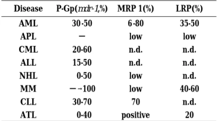
Several studies reported the frequency of the mdr-1 phenotype to be about 30 to 50% in AML patients with a higher percentage in older patients and in patients
whose leukemia relapsed after refractory chemotherapy (21-23). No P-Gp expression in acute promyelocytic leukemia has been found (24). In lymphoid malig-nancies such as non-Hodgkin’s lymphoma (NHL), multiple myeloma (MM), chronic lymphocytic leukemia (CLL), and adult T-cell leukemia/lymphoma (ATL), MDR-phenotypes have been reported. Detectable levels of P-Gp range widely from 0 to 50% in samples of NHL (25, 26). In de novo MM patients no elevated P-Gp expression has been found in the levels of either mRNA or protein stained with the anti-P-Gp antibodies. However, after exposure to vincristine, adriamycin, and dexamethazone (VAD) chemotherapy, the expression of mdr-1 reaches levels above detectable to75%, and reaches100% in refractory patients (27). However, because of various subtypes of the disease in NHL, CLL, and ATL, the expression already varies widely, making it difficult to decide on the significance of any MDR-phenotypes. Some of this validation can be accounted for by the threshold that was used to consider a sample positive for P-Gp. The expression level of the MDR protein in hematological malignancies is shown in Table 6.
Therefore, it is difficult to compare MDR expres-sions in cell lines and clinical samples from different studies. The difference in the results between studies is caused by the different detection methods such as immunocytochemistry, flow cytometry, RNase pro-tection assays, and quantitative PCR, use of different thresholds for positivity, use of different monoclonal antibodies, comparison of different expression levels among DNA/RNA/Protein, use of different internal controls, and differences in methods employed to purify leukemic blasts. In addition to these differences, clinical prognostic factors such as white blood cell number, age, cytogenetics, and gender in some types of malignancy might be significant. In addition, to explain the cause Table 4. The characteristics of LRPs.
Gene, size & location : lrp1-3, 110 kDa, 16p13.2
Drugs transported : anthracyclins, vinca alkaloids, CDDP, alkylating agents
Expressions : normal hemopoetic cells, leukemia cells Functions :
・transport of drugs into and out of the nucleus ・detoxication process
Table 5. Detection methods of MDR phenotypes. Detection methods
ISH, PCR, RT-PCR, RNase protection, Immunocytochemistry, Flow Cytometry, Functional Assay
Monoclonal antibodies P-Gp MRP LRP MRK16 MRPm6 LRP56 4E.3
UIC-2 JSB1
Substrates for Rho123 calcein functional assay DiO(C2)3 CF
Fluo-3
Inhibitors PSC833 PSC833 PAK104P Cy-A verapamyl CH-11 MoAb Verapamyl cremophor Dipyridamole MK-571 vanadate EL DNP-SG genistein GSSG
Table 6. Expression of MDR phenotypes in hematological malig-nancies.
Disease P-Gp(mdr-1,%) MRP 1(%) LRP(%)
AML 30 -50 6 -80 35-50
APL − low low
CML 20-60 n.d. n.d. ALL 15-50 n.d. n.d. NHL 0-50 low n.d. MM −→100 low 40-60 CLL 30-70 70 n.d. ATL 0-40 positive 20
− : negative, n.d. : not detected
of discrepancy between studies, some studies suggested the presence of a non-functional drug efflux pump among the MDR proteins and unknown proteins that can function as MDR phenotypes.
PROGNOSTICSIGNIFICANCEOFMDRPHENO-TYPES AT DIAGNOSIS IN HEMATOLOGICAL
MALIGNANCIES.
In AML, most of the study found that mdr-1over-expression at diagnosis is a strong impediment pre-dictor for complete remission and long-term survival, although there is a suggestion of a different “behavior” between adult and childhood AML (21, 22, 28). In childhood AML, P-Gp is not associated with a poor prognosis (28). This is controversial, but there is a correlation between clinical outcome of AML and MRP1 or LRP. Studies on LRP in AML emphasized the importance of the correlation between LRP-expression and anthracyclin accumulation and suggested that LRP-expression has a prognostic value at diagnosis (29, 30). However, there is an equal number of studies where a predictive value in the case of LRP-expression in de novo AML cannot be shown (31, 32). Co-expression of P-Gp and MRP has also been associated with poor outcome in AML (29, 33).
In ALL, mdr-1 expression is of minor importance for prediction of outcome, and MRP1expression at diagnosis is not associated with response and long term survival. The prognostic significance of LRP ex-pression in ALL is still controversial between positive and negative results (21).
There is a number of studies on the incidence of P-Gp expression in NHL. P-Gp expression has been correlated with drug sensitivity and clinical outcome in NHL. However, other studies did not find a correlation between response and P-Gp expression. Thus, it is
presently unclear whether MDR expression has a significant impact on the response to therapy in lymph-oma (25, 27).
In de novo MM patients P-Gp expression does not appear to occur, since myeloma cells at diagnosis neither express elevated levels of mdr-1 mRNA nor stain with the anti-P-Gp antibodies (34). MRP is not over-expressed in MM, but LRP is expressed in half of the MM patients, and is associated with a poor response to melphalan at conventional doses (35).
Most of these studies recognized the increased expression level in leukemia cells of relapsed or of the refractory stage compared with cells at diagnosis. The prognostic significance of MDR phenotypes at diagnosis in hematological malignancies is summarized in Table 7.
ROLEOFTHEAPOPTOSISCASCADETHROUGH
DEATH RECEPTORS IN HEMATOLOGICAL
MALIGNANCIES.
To bring about cell death through death receptors and apoptosis cascade proteins, there are two routes : either through mitochondrial or through non-mitochondrial paths, as summarized in Figure 4. As it is well known, cell death through the non-mitochondrial apoptosis cascade begins at receptor sites such as Fas (CD95/ Apo-1), TRAIL-R, and tumor necrosis factor receptor (TNF-R) on the cell membrane by binding the ligands of each receptor or by granzyme B. The receptor-ligand binding activates downstream caspases, leading to the subsequent cleavage of death substrates. These substrates can eventually cause DNA fragmentation. It is well known that bcl-2 and survivin (c-IAP 1/2) work as inhibitors for this pathway. P-Gp protects leukemia cells against caspase-dependent, but not caspase-independent cell death (36). Anti-P-Gp antibody induced Fas antigen in PBMC and accelerated apoptosis
Figure 4. Apoptosis cascade through death receptors. Table 7. Prognostic significance of MDR phenotypes in hematological
malignancies. Disease P-Gp(mdr-1) MRP 1 LRP AML strong(++) +(?) +(?) APL − − − CML − − n.d. ALL minor − n.d. NHL +(?) n.d. n.d. MM −→ + n.d. + CLL − − n.d. ATL n.d. +(?) +(?)
− : negative,+: positive, n.d. : not determined
M. Hirose et al. Multidrug resistance in hematological malignancy
through Fas-Fas-ligand interaction (37).
Alterations of apoptosis cascade genes associated with drug resistance have been found in studies using cell lines or fresh cells. Over-expression of bcl-2 with MAPK pathway links to drug resistance in AML (38). The con-stitutive expression level of Fas and bcl-2 is important, and the expression of these antigens is a predictive factor of the chemosensitivity in leukemia, especially in AML, though not in ALL (39, 40). Low or alteration of Fas expression leads to refractoriness in T-cell type ALL or normal T lymphocytes (41). Blocking of caspase 8 results in TRAIL resistance, and increased NF-κB can inhibit apoptosis in MM cells (42). Heat shock protein (HSP) 90 inhibits apoptosis in mononuclear phagocytic cells (43).
ALTERATIONS OF TUMOR SUPPRESSOR
GENES ASSOCIATED WITH DRUG RESISTANCE.
Among quite a few tumor suppressor genes, WT1 was found as a tumor suppressor gene in childhood Wilms’ tumor, which is located at chromosome 11 at band p13. The WT1 gene encodes a zinc finger tran-scription factor, which binds to GC-rich sequences and functions as a transcriptional activator or repressor for many genes. The WT1 protein is mainly expressed in cells or tissues of the genitourinary system. It is well known that its over-expression links to poor prognosis in leukemia. On the other hand, p53 was found at chromosome 17p13, which is activated or increased by injury in DNA or by stress, resulting in G1 arrest or apoptosis. P53 alteration including mutation was reported in various types of hematological malignancies such as CML, NHL, AML, and ALL (44, 45). No link has been found between the abnormality of the RB gene and drug resistance in hematological malignancy. Mdm2and ras genes are oncogenes. Mdm2 is ac-tivated by the induction of p53 and inactivates the functions of p53, which inhibits p53 induced G1 arrest and apoptosis. It was reported that mdm2 over-ex-pression often associates refractoriness in ALL (46, 47). The Ras gene causes immortalization or transformation of cells by associating with c-myc or p53. Thus, it was reported that a ras mutation leads to lowered mdr-1 in adult T-cell leukemia (48).In the future, a therapy that attacks the altered ex-pression of the apoptosis cascade, aimed at tumor suppressor genes, and in combination with conven-tional drugs may be promising.
REVERSAL OF DRUG RESISTANCE.
Two possible approaches to mdr-1 reversal by agents can be distinguished. The first option is the use of modulating agents that can restore drug accumulation by competing with cytostatic drugs for P-Gp binding sites. These agents include calcium channel blockers, some type of cardiovascular drugs, cyclosporin analogs, and anti-malarias. When initially trying to overcome MDR, we found that an anti-platelet drug, dipyridamole, could overcome MDR in leukemia cells (49, 50). Subse-quently, more substances have been tested and found to counteract MDR. We found that the activity of MRP1 can be blocked by a variety of chemical compounds, where the effect of P-Gp modulators on MRP1 was less than their effect on P-Gp over-expressing cells (51). Although the results of the few studies with P-Gp modifiers in the hematological malignancies are promising, the data are insufficient for recommending the routine use of such drugs to increase disease-free survival in leukemia. Most of these agents produced severe toxic effects at doses required to effectively block P-Gp function, and modulation of P-Gp in normal tissues can affect the pharmacokinetics and, thus, the toxicity of the associated chemotherapeutic agents (52). So far, clinical intervention studies with mdr-1 modifying agents have only been done with AML patients using a modified analog to cyclosporin D, PSC 833 (valspodar). Such third generation MDR modulators can be safely administered in combination with different chemotherapy regimens after a dose adjustment of cytotoxic drugs that are P-Gp substrates(53).In addition, some agents such as mdr-1-specific anti-sense oligonucleotides and protein kinase C inhibitors such as staurosporin have been demonstrated to be capable of down-regulating mdr-1expression (54, 55). Secondly, immunotherapy against a surface antigen of the membrane of leukemia cells may be promising (56). We confirmed the efficacy to be 5-fold higher the wild-type cell line of mouse human chimeric anti-CD 20 antibody on the VCR-resistant cell line of Daudi, an endemic Burkitt’s lymphoma cell line, in which the expression level of CD20 remained unchanged compared with those in the wild-type cells. In contrast, however, the anti-CD20 antibody was ineffective in a VCR-resistant BLTH, a non-endemic Burkitt’s lymphoma cell line, in which CD20 disappeared (57). Thus, it was suggested that the resistance to VCR in some tumor cell lines is associated with a modified antigen expres-sion of the target molecule and susceptibility to immu-notherapies.
Active oxygen radicals can damage the cell mem-The Journal of Medical Investigation Vol. 50 2003 131
branes by oxidizing their lipids. By such cytotoxic ac-tivity of oxygen radicals certain antineoplastic agents such as adriamycin, bleomycin, and etoposide exert their efficacy. Consequently, we found the cross-resistance in VCR-resistant cells with increased P-Gp and MRP expression to oxygen radicals which was produced by the hypoxanthin-xanthin oxidase reaction(58). Increased resistance to oxygen radicals may be caused by an altered membrane structure in VCR-resistant cells, being an impediment to treatment. These results may suggest a new mechanism of drug resistance in cells expressing P-Gp (59).
FUTURE DIRECTION OF THERAPY TO
OVERCOME MULTIDRUG RESISTANCE.
Clinical trials of the modulation of MDR have been limited by two major factors : the inability of achieving adequate blood levels of the modulator to reverse MDR in patients, and the presence of other resistance mech-anisms in addition to P-Gp. A third factor is that P-Gp modulators alter the pharmacokinetics of anti-cancer drugs by delaying their elimination : this can potentially increase toxicities if the dose of anticancer drugs is not appropriately reduced. However, because it was demonstrated that MDR modulators such as valspodar show substantial inhibition of P-Gp, reversal agents that only inhibit P-Gp in tumor cells and do not influence the pharmacokinetics of cytotoxic agents should be developed. Thus, to explore the potential of transporter-specific modulators in improving clinical outcome, more knowledge will be needed on the nature, substrate spe-cificity, inhibitory sensitivity, and expression of the efflux pump responsible for MDR in human cancer. The current therapy needs too high doses of anti-cancer agents to overcome drug resistance and often cause severe adverse effects, resulting in over-treatment. To avoid such over-treatment or under-treatment, the following is recommended ;
・identification of the type of drug resistance in each patient.
・evaluation of chemo-sensitivity of a patient’s normal cells or tissues in addition to those of malignant cells against anti-cancer drugs.
・determination of the treatment strategy such as dose of drugs and schedule of chemotherapy on the basis of these data.
REFERENCES
1. Pui CH, Campana D, Evans W : Childhood acute lymphoblastic leukemia-current status and future perspectives. Lancet Oncol 2(10) : 597-607, 2001. 2. Alcalai R, Ben -Yehuda D, Ronen I, Paltiel O : Ethnicity and prognosis in acute myeloid leukemia. Am J Hematol 72(2) : 127-134, 2003.
3. van den Heuvel-Eibrink MM, Sonneveld P, Pieters R : The prognostic significance of membrane trans-port-associated multidrug resistance (MDR) pro-teins in leukemia. Int J Clin Pharmcol Therap 38(2) : 94-110, 2000.
4. Brisco MJ, Sykes PJ, Dolman G, Hughes E, Neoh S-H, Peng L, Snell LE, Toogood IRG, Rice MS, Morley AA : Early resistance to therapy during induction in childhood acute lymphoblastic leukemia. Cancer Res 60 : 5092-5096, 2000. 5. Venditti A, Buccisano F, DelPoeta G, Maurillo
L, Tamburini A, Cox C, Battaglia A, Catalano G, Del Moro B, Cudillo L, Postorino M, Masi M, Amadori S : Level of minimal residual disease after consolidation therapy predicts outcome in acute myeloid leukemia. Blood 96(12) : 3948-3952, 2000.
6. Fojo AT, Ueda K, Slamon DJ, Poplack DG, Gottesman MM, Pastan I : Expression of a multi-drug resistance gene in human tumors and tissues. Proc Natl Acad Sci USA 84 : 265-269, 1987. 7. Noonan KE, Beck C, Holmayer TA, Chin JE,
Wunder JS, Andruis IL, Gazdar AF, Willman CL, Griffith B, Von Hoff DD, Roninson IB : Quantitative analysis of MDR-1(multidrug resistance) gene expression in human tumors by polymerase chain reaction. Proc Natl Acad Sci USA 87 : 7160-7164, 1990.
8. Cole SPC, Bhardwaj G, Gerlach JH, Mackie JE, Grant CE, Almquist KC, Stewart AJ, Kurz EU, Duncan AMV, Deeley RG : Overexpression of a transporter gene in a multidrug-resistant human lung cancer cell line. Science 258 : 1650-1654, 1992.
9. Versantvoort CHM, Broxterman HJ, Bagrij T, Scheper RJ, Twentyman PR : Regulationby glutathione of drug transport in multidrug-resistant human lung tumor cell lines overexpressing multidrug resistance-associated protein. Br J Cancer 72 : 82-89, 1995.
10. Scheffer GL, Wijingaard OLJ, Flens MJ, Izquierdo MA, Slovak ML, Pinedo HM, Meijer CJLM, Clevers HC, Scheper RJ : The drug resistance related protein LRP is the human major vault protein.
M. Hirose et al. Multidrug resistance in hematological malignancy
Nat Med 1 : 578 -582, 1995.
11. Wiemer E, Schoester M, Sonneveld P : Intracellular localization and dynamics of vault complex in non-P-gp multidrug resistant cell lines. Blood 92 : 676 a, 1998.
12. Neefjes JJ, Momburg F, Hammerling GJ : Selective and ATP-dependent translocation of peptides by the MHC-encoded transporter. Science 261: 769 -771, 1993.
13. Izquierdo MA, Neefjes JJ, Mathari AEL, Flens MJ, Scheffer GL, Scheper RJ : Overexpression of the ABC transporter TAP in multidrug-resistant human cancer cell lines. Br J Cancer 74 : 1961-1967, 1996.
14. Longhurst TJ, O’Neill GM, Harvie RM, Davey RA : The anthracyclin resistance-associated (ARA) gene, a novel gene associated with multidrug resistance in a human leukemia cell lines. Br J Cancer 74 : 331-1335, 1996.
15. Kool M, De Haas, Scheffer GL, Scheper RJ, Van Eijk MJT, Juijin JA, Baas F, Borst P : Analysis of expression of cMOAT(MRP2), MRP3, MRP4, and MRP5. Homologues of the multidrug resistance associated protein gene (MRP1), in human cancer cell lines. Cancer Res 57 : 3537-3547, 1997. 16. Borst P, Evers R, Kool M, Wijnholds J : A family
of drug transporters:the multidrug resistance-associated proteins. J Natl Cancer Inst 92(16) : 1295 -1302, 2000.
17. Chen YN, Mickley LA, Schwartz AM, Action EM, Hwang H, Fojo AT : Characterization of adriamycine resistant human breast cancer cells which display overexpression of a novel resistance-related membrane protein. J Biol Chem 265 : 10073-10080, 1990.
18. Doyle LA, Ross DD, Sridhara R, Fojo AT, Kaufman SH, Lee EJ, Schiffer CA : Expression of a 95 kDa membrane protein is associated with low daunorubicine accumulation in leukemic blasts. Br J Cancer 71 : 52-58, 1995.
19. Thiebaut F, Tsuruo T, Hamada H, Gottesman MM, Pastan I, Willingham MC : Cellular localization of the multidrug resistance gene product P-glycoprotein in normal human tissues. Proc Natl Acad USA 84 : 7735 -7738, 1987.
20. Simon SM, Schindler M : Cell biological mech-anisms of multidrug resistance in tumors. Proc Natl Acad USA 91 : 3497-3504, 1994.
21. van den Heuvel-Eibrink MM, van der Holt B, Boekhorst PAW, Pieters R, Lowenberg B, Sonnevelt P : MDR-1 is an independent prognostic factor for response and survival in de novo acute myeloid
leukemia. Br J Haematol 99 : 76 -83, 1997. 22. Sievers EL, Smith FO, Woods WG, Lee JW, Bleyer
WA, Willman CL, Bernstein ID : Cell surface ex-pression of the multidrug resistance P-glycoprotein (P-170) as detectable by monoclonal antibody MRK-16 pediatric in acute myeloid leukemia fails to define a poor prognostic group. Leukemia 9 : 2042-2048, 1995.
23. Covelli A : Modulation of multidrug resistance (MDR) in hematological malignancies. Annals of Oncology 10(suppl/6) : 53 -59, 1999.
24. Michieli M, Damiani D, Ermacora A, Geromin A, Michelutti A, Masolini P, Baccarani M : P-glycoprotein (PGP), lung resistance-related protein (LRP), and multidrug resistance-associated protein (MRP) expression in acute promyelocytic leukemia. Br J Haematol 108 : 703 -709, 2000.
25. Pileri SA, Sabatini E, Falini B, Tazzari PL, Gherlinzoni F, Michieli MG, Damiani D, Zucchini L, Gobbi M, Tsuruo T : Immunohisto-chemical detection of the multidrug transporter protein P170 in human normal tissues and malignant lymphomas. Histopathology 19 : 131-140, 1991. 26. Niehans GA, Jaszcz W, Brunetto V, Perri RT, Gajl-Peczalska K, Wick MR, Tsuruo T, Bloomfield CD : Immunohistochemical identification of P-glycoprotein in previously untreated, diffuse large cell and immunoblastic lymphomas. Cancer Res 52 : 3768 -3775, 1992.
27. Grogan TM, Spier CM, Salmon SE, Matzner M, Rybski J, Weinstein RS, Scheper RJ, Dalton WS : P-glycoprotein expression in human plasma cell myeloma :correlation with prior chemotherapy. Blood 81 : 490-495, 1993.
28. den Voer ML, Pieters R, Kazemier KM, Rottier MM, Zwaan CM, Kaspers GJ, Janka-Schaub G, Henze G, Creutzig U, Scheper RJ, Veerman AJ : Relationship between major vault protein/lung resistance protein, multidrug resistance-associated protein, P-glycoprotein expression, and drug resis-tance in childhood leukemia. Blood 91 : 2092-2098, 1998.
29. Hart SM, Ganeshuuru K, Scheper RJ, Prentice HG, Hoffbrand AV, Mehta AB : Expression of the human major vault protein (LRP) in acute myeloid leukemia. Exp Hematol 25 : 1227-1232, 1997.
30. Michieli M, Diamini D, Ermacora A, Raspadri D, Michelutti A, Grimaz S, Fanin R, Russo D, Lauria F, masolini P, Baccarani M : P-glycoprotein (P-gp) and lung resistance protein (LRP) expres-sion and function in leukemic blast cells. Br J The Journal of Medical Investigation Vol. 50 2003 133
Hematol 96 : 356-365, 1997.
31. Legrand O, Simon G, Zittoun R, Marie JP : Lung resistance protein (LRP) gene expression in adult acute myeloid leukemia : a critical evaluation by three techniques. Leukemia 12 : 1367-1374, 1998.
32. Pallis M, Turzansky J, Wheatley K, Langabeer S, Burmett AK, Russell NH : Use of standardized flow-cytometric determinants of multidrug re-sistance to analyse response to remission in-duction chemotherapy in patients with acute myeloblastic leukemia. Br J Hematol 104 : 258-265, 1999.
33. Borg AG, Burgess R, Green LM, Scheper RJ, Liu Yin JA : Overexpression of lung-resistance protein and increased P-glycoprotein function in acute myeloid leukemia cells predict a poor response to chemotherapy and reduced patient survival. Br J Haematol 103 : 1083 -1091, 1998. 34. Coenelissen J, Sonnevelt P, Schoester M, Raaijmakers HG, Nieuwenhuis HK, Dekker AW, Lokhorst HM: MDR1 expression and response to vincristine, doxorubicine and dexamethazone chemotherapy in multiple myeloma refractory to alkylating agents. J Clin Oncol 12 : 115-119, 1994.
35. Raaijmakers HG, Izquierdo MA, Lokhorst HM, de Leeuw C, Belien JA, Bloem AC, Dekker AW, Scheper RJ, Sonneveld P : Lung-resistance-related protein expression is a negative predictive factor for response to conventional low but not to inten-sified dose alkylating chemotherapy in multiple myeloma. Blood 91(3) : 1029 -1036, 1998. 36. Johnstone RW, Cretney E, Smyth MJ : P-glycoprotein
protects leukemia cells against caspase-depend-ent, but not caspase-independcaspase-depend-ent, cell death. Blood 93(3) : 1075-1085, 1999.
37. Gollapud S, Gupta S:Anti-P-glycoprotein antibody-induced apoptosis of activated peripheral blood lymphocytes : a possible role of P-glycoprotein in lymphocyte survival. J Clin Immunol 21(6) : 420 -430, 2001.
38. Milella M, Estrov Z, Kornblau SM, Carter BZ, Konopleva M, Tari A, Schober WD, Harris D, Leysath CE, Lopes-Berestein G, Huang Z, Andreeff M : Synergistic induction of apoptosis by simulta-neous disruption of the Bcl-2 and MEK/MAPK pathways inacute myelogenous leukemia. Blood 99(9) : 3461-3464, 2002.
39. Labroille G, Dumain P, Lacombe F, Belloc F: Flow cytometric evaluation of fas expression in relation to response and resistance to anthracyclines in leukemia cells. Cytometry 39(3):195 -202, 2000.
40. Wuchter C, Karawajew L, Ruppert V, Schrappe M, Harbott J, Ratei R, Dorken B, Ludwig W-D : Constitutive expression levels of CD95 and bcl-2 as well as CD95 function and spontaneous apoptosis in vitro do not predict the response to induction chemotherapy and relapse rate in childhood acute lymphoblastic leukemia. Brit J Haematol 110 : 154-160, 2000.
41. Beltinger C, Kurz E, Bohler T, Schrappe M, Ludwig WD, Debatin KM : CD95 (APO-1/Fas) mutations in childhood T-lineage acute lymphoblastic leukemia. Blood 91(10) : 3943 -3951, 1998.
42. Mitsiades N, Mitsiades CS, Poulaki V, Anderson KC, Treon SP: Intracellular regulation of tumor necrosis factor-related apoptosis-inducing ligand-induced apoptosis in human multiple myeloma cells. Blood 99(6) : 2162-2171, 2002.
43. Galea-Lauri J, Richardson AJ, Latchman DS, Katz DR : Increased heat shock protein 90(hsp 90) expression leads to increased apoptosis in the monoblastoid cell line U937 following induction with TNF-alpha and cycloheximide : a possible role in immunopathology. J Immunol 157(9) : 4109-4118, 1996.
44. Zheng A, Castren K, Saily M, Savolainen ER, Koistinen P, Vahakangas K : p53 status of newly established acute myeloid leukaemia cell lines. Brit J Cancer 79(3 -4) : 407-415, 1999.
45. Lam V, McPherson JP, Salmena L, Lees J, Chu W, Sexsmith E, Hedley DW, Freedman MH, Reed JC, Malkin D, Goldenberg GJ : p 53 gene status and chemosensitivity of childhood acute lymphoblastic leukemia cells to adriamycin. Leuk Res 23(10) : 871-880, 1999.
46. Cinti C, Claudio PP, Luca AD, Cuccurese M, Howard CM, D’ Esposito M, Paggi MG, Sala DL, Azzoni L, Halazonetis TD, Giordano A, Maraldi NM : A serine 37 mutation associated with two missense mutations at highly conserved regions of p53 affect pro-apoptotic genes expression in a T-lymphoblastoid drug resistant cell line. Oncogene 19(44) : 5098-5105, 2000.
47. Gu L, Findley HW, Zhou M : MDM2 induces NF-kappaB/p65 expression transcriptionally through Sp1-binding sites : a novel, p53-independent role of MDM 2 in doxorubicin resistance in acute lymphoblastic leukemia. Blood 99 (9) : 3367-3375, 2002.
48. Schaich M, Ritter M, Illmer T, Lisske P, Thiede C, Schakel U, Mohr B, Ehninger G, Neubauer A : Mutations in ras proto-oncogenes are associated with lower mdr-1gene expression in adult acute
M. Hirose et al. Multidrug resistance in hematological malignancy
myeloid leukemia. Brit J Haematol 112 : 300-307, 2001.
49. Hirose M, Takeda E, Ninomiya T, Kuroda Y, Miyao M : Synergistic inhibitory effects of dipyridamole and vincristine on the growth of human leukemia and lymphoma cells. Brit J Cancer 56 : 413 - 417, 1987.
50. Hirose M, Takeda E, Kuroda Y:Overcoming of vincristine resistance in HL- 60 human promyelocytic leukemia cell line by dipyridamole. Tokushima J Exp Med 40 : 27-33, 1993.
51. Hosoi E, Hirose M, Hamano S, Morimoto M, Kuroda Y : Effect of MDR antagonists on the cidal activity of vincristine for cells expressing MDR-1 is superior to those expressing MRP. Int J Oncol 13 : 343-348, 1998.
52. Fisher GA, Lum BL, Hausdorff J, Sikic BI : Pharma-cological considerations in the modulation of multidrug resistance. Eur J Cancer 32 A : 1082-1088, 1996.
53. Marie JP : Drug resistance in hematological malign-ancies. Curr Opin Oncol 13 : 463-469, 2001. 54. Revoltini L, Colombo MP, Supino R, Ballinari
D, Tsuruo T, Parmiani G:Modulation of multi-drug resistance by verapamil or mdr-1 anti-sense oligodeoxynucleotide does not change the high
susceptibility to lymphokine-activated killers in mdr-resistant human (LoVo) cell lines. Int J Cancer 46 : 727-732, 1990.
55. Chaudary PM, Roninson IB : Activation of the MDR-1(P-glycoprotein) gene expression by protein kinase C agonists. Oncol Res 4 : 281-290, 1992. 56. Linenberger ML, Hong T, Flowers D, Sievers
EL, Gooley TA, Bennett JM, Berger MS, Leopold LH, Appelbaum FR, Bernstein ID : Multidrug-resistance phenotype and clinical responses to gemtuzumab ozogamicin. Blood 98 : 988-994, 2001.
57. Hirose M, Hamano S, Tobinai K, Kuroda Y : Cytocidal activity of PBL, LAK, and IDEC-C2B8 and expression of HLA class1, ICAM-1, and CD 20 in vincristine-resistant hematologic cell lines. J Immunother 22 : 237-244, 1999.
58. Hirose M, Fukuzawa K, Kuroda Y : Vincristine resistant HL-60 cells show cross-resistance to hypoxanthine-xanthine oxidase. Cancer Lett 102: 217-221, 1996.
59. Hamano S, Hirose M, Hosoi E, Houchi H, Kuroda Y : Overcoming of cross-resistance to the cell-killing activity of oxygen radicals in VCR-resistant hematologic cell lines with cyclosporin-A. Anticancer Res 18 : 1543-1548, 1998.