Characteristics of Anti-Human
Leukocyte Antigen-DR Antibody and
Its Fc Modification to Avoid Toxicity
2008
学 位 論 文 の 要 旨
Characteristics of Anti-Human Leukocyte Antigen-DR Antibody
and Its Fc Modification to Avoid Toxicity
(HLA-DR 抗体の特性解析と Fc 変異体による毒性回避の研究)
氏 名 俵 知 紀 (1)B 細胞系の癌は、主に B リンパ腫及び B 細胞系白血病に分類され、その癌細胞は B 細胞の分化マーカー(CD20 等)を高発現している。その中でも、主要組織適合複合体(MHC) の一つであるヒト白血球抗原 DR(HLA-DR)は、古くから抗体医薬開発の標的分子として 臨床試験が実施されながら、現時点でも未だ成功例が無い抗原である。一方、CD20 抗体(リ ツキシマブ)の B 細胞系腫瘍における著効は有名で、リンパ腫及び白血病において抗体医 薬が効果を発揮しうることを裏付ける代表例となっている。しかし CD20 抗体においても、 不応例や再発例が存在し、さらには投与時の即時型毒性が問題となる場合もある。そこで、 HLA-DR 抗体を新たに作製し、その特性解析を行い、さらにその抗体の Fc 領域への変異導 入による毒性・薬効について検討した。 (2)医薬品の開発においては患者の遺伝子多型による効果のバラつきが障害となりうる が、HLA-DR 分子は最も多型を有する分子で、その数は 500 を超える。HLA-DR 抗体を医 薬品として応用する場合、膨大な多型による効果のばらつきを克服することが重要となる。 そこで、HLA-DR に対する完全ヒト抗体を、ヒト抗体産生マウスを用いて作製し、それ ら抗体の中から多くの HLA-DR 陽性細胞を認識する抗体 HD8 を選抜した。HD8 抗体の解析 の結果、検査した約 50 種の HLA-DR 陽性細胞すべてに強く反応することを確認した。しか し意外にも、合成ペプチドを用いた結合部位のマッピング解析で、HD8 抗体は多型に富む 領域を認識していることが判明した。さらに詳細に認識部位をペプチド解析した結果、HD8 抗体は、多型を有する領域の中でもほとんどの多型で保存されているアミノ酸を認識して いることが判明した。さらに、HD8 の認識部位のペプチドを各 MHC クラス II で作製し解 析した結果、HLA-DR の 99.2%、HLA-DP の 100%、HLA-DQ の 66%を認識しうることが 判明し、HD8 抗体の広い認識性が確認でき、多型克服のストラテジーが示された。傷害活性(ADCC)・補体依存性細胞傷害活性(CDC)を有すること、in vivo においてヒト B リンパ腫の担ガンマウスにおいて延命効果を示すことから、医薬品としての高い可能性が 示唆された。しかしながら、HD8 のヒト IgG1 及び IgG2 体をサルに投与した際、強いアナ フィラキシー様の毒性を示した。このアナフィラキシー様反応はインフュージョン反応と 呼ばれる免疫反応のひとつであり、抗体医薬品の投与の際に引き起こされる副作用として 知られている。この HD8 抗体のインフュージョン反応は、ADCC・CDC 能が強い IgG1 の みでなく、CDC のみが強い IgG2 においても発現したことから、CDC が原因であることが 予測できる。実際に、ラットを用いて新規に構築したインフュージョン反応モデルを用い たコブラ毒素による補体除去実験において、HD8-IgG1 抗体のインフュージョン反応が大幅 に軽減され、CDC が原因である可能性が強く示唆された。 そこで、毒性を回避し、且つ薬理作用を保持した抗体を作製するため、CDC を低減し ADCC を保持した抗体の作製を試みた。抗体の Fc 領域と Fc 受容体の立体構造等を基に Fc 領域の改変を行い、331 番目プロリンのセリン置換により CDC が低減されることが確認さ れた。さらには、322 番目リジンのアラニン置換の追加により、in vitro においては CDC 能 が全く検出されず、ADCC は若干の低減は認められたものの十分に保持されていた。ラッ トインフュージョン反応モデルにおいても、二変異抗体はそのインフュージョン毒性の発 現を大幅に軽減した。また、サルに投与した場合では、インフュージョン反応は全く発現 しなかった。薬効に関しては、マウスを用いたヒトリンパ腫担ガンモデルにおいて抗体投 与による延命効果が観察され、薬効の保持も確認できた。 (3)以上の結果、本研究により、Fc 領域に 2 変異を導入した HD8 抗体は抗腫瘍効果を保 持しつつ毒性を大幅に軽減し、医薬品としての高い可能性をもつことが明らかとなった。 また、HLA-DR 抗体のインフュージョン反応の主要な原因が CDC であることを明らかにし、 この 2 変異が CDC を原因とするインフュージョン反応の軽減法として有効であることを示 すことが出来た。
CONTENTS
Page
ABBREVIATIONS --- 1
GENERAL INTRODUCTION --- 4
CHAPTER I --- 14
A fully-human antibody exhibits pan-HLA-DR recognition and
high in vitro/vivo efficacy against HLA-DR-positive lymphomas
CHAPTER II --- 55
Complement activation plays a key role in antibody-induced
infusion toxicity in monkeys and rats
CONCLUSIONS --- 83
ACKNOWLEDGEMENTS --- 87
LIST OF PUBLICATIONS --- 89
Page
1
4
14
55
83
87
89
ABBREVIATIONS
Ab, antibody
Ag, antigen
ADCC, antibody-dependent cellular cytotoxicity
CDC, complement-dependent cytotoxicity
CVF, cobra venom factor
DDS, drug delivery system
DMEM, Dulbecco’s modified Eagle’s medium
DTT, dithiothreitol
EDTA, ethylene-diamine-tetra-acetic acid
ELISA, enzyme-linked immunosorbent assay
E/T ratio, effector to target ratio
Fab, antigen-binding fragment
FACS, fluorescence-activated cell sorter
Fc, constant region fragment
FCS, fetal calf serum
HLA, human leukocyte antigen
HLA-DR, human leukocyte antigen-DR
Ig, immunoglobulin
IMDM, Iscove’s modified Dulbecco’s medium
i.p., intraperitoneal
IU, international unit
i.v., intravenous
KD, affinity constant
kDa, kilodalton
mAb, monoclonal antibody
MFI, mean fluorescence intensity
mg, milligram
MHC, major histocompatibility complex
MLR, mixed leukocyte reaction
mRNA, messenger ribonucleic acid (RNA)
µg, microgram
µL, microliter
ND, not determined
NK cell, natural killer cell
p, probability
PAGE, polyacrylamide gel electrophoresis
PBMC, peripheral blood mononuclear cell
PBS, phosphate buffer saline
PE, phycoerythrin
RACE, rapid amplification of cDNA end
RT-PCR, reverse transcriptase polymerase chain reaction
s.c., subcutaneous
scid, sever combined immunodeficiency
SD, standard deviation
SDS, sodium dodecyl sulfate
SEM, standard error of the mean
TNF, tumor necrosis factor
TRAIL, tumor necrosis factor (TNF)-related apoptosis inducing ligand
v/v, volume to volume ratio (%)
Antibody for therapeutic use
Antibody can bind to a wide variety of potential targets and have been envisioned
as therapeutic targeting vehicles for a century (1, 2). The initial use of antibody as
therapeutic agents is polyclonal antisera, but in many cases they were proved unsuitable
because of their heterogeneity. Monoclonal antibodies (mAbs), which were first
demonstrated by Kohler and Milstein (3), have made us possible to create antibodies
with requisite pharmaceutical properties. MAb is capable of binding to a specific
antigen with remarkable specificity, which property can be refined so that mAbs and
their derivatives could be used in various therapies.
The first indication that mAbs might have significant therapeutic potential came in
1982 when an individual with lymphoma, Philip Karr, showed a complete response to a
brief treatment with a ‘tailor-made’ mouse anti-idiotype mAb (4). This notable success
elicited academic and commercial interest toward antibody researches and investments.
Nevertheless, the facts that many trials against cancer disclosed lack of efficacy, and a
lot of pharmaceutical problems of mAbs (too expensive to produce, needed specialist
expertise to administer, and often associated with considerable toxicity) waned the
enthusiasms of antibodies in the end of 1980s. Going through the early experiences
enabled to provide eight mAbs for clinical use which had been approved by US Food
and Drug Administration (FDA) in the late 1990s, and about a hundred of mAbs are
currently in are currently in clinical trials.
Structure and function of antibody
Antibodies are immunoglobulins (Ig) produced by B lymphocytes in response to
the antigenic stimuli (Figure A). Most immunoglobulins consist of two light and two
heavy chains that are composed of two different domains (Figure B). The Fab
domains serve as the antigen-binding sites, which are composed of variable regions of
heavy and light chains containing hypervariable regions. The Fc domains determine
the effector functions of antibodies, which depend on interactions with Fc receptors and
complement. Antibody-dependent cellular cytotoxicity (ADCC) is mediated by
neutrophils, mononuclear phagocytes, natural killer cells, some T cells and eosinophils,
which possess a variety of Fc receptors. Complement-dependent cytotoxicity (CDC)
is elicited by the cascade of complement components that exist in blood plasma. The
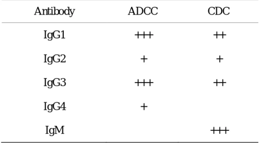
effector functions can be customized by selection of Ig subclasses (Table A) and
modification of the Fc domains. For example, human IgG1 is known to trigger both
IgG IgM IgD IgA1 IgE
(Small hexagons indicate sugar chains.)
Dimeric IgA
IgM (Janeway, Immunobiology Fifth edition, 2001)
Figure A. Human immunoglobulins
Figure B. Structure of immunoglobulin
Fc domain
Table A. Immune effector functions of human immunoglobulin isotypes Antibody ADCC CDC IgG1 IgG2 IgG3 IgG4 IgM +++ + +++ + − ++ + ++ − +++
Cancer Immunotherapy
MAbs and their derivatives possess a lot of characteristics that are useful for
cancer immunotherapy as follows;
(A) Antibodies with certain Fc domains possess ADCC and/or CDC that act as
anti-tumor agents through lysing target cells. ADCC is reported to play an important
role in tumoricidal activity in antibody therapies using anti-CD20 mAb and breast
cancers by anti-Her2/neu mAb (5, 6). The intensity of ADCC and/or CDC can be
modified by changing the Fc structures. Some recent researches revealed the critical
amino acids that affect ADCC and/or CDC by the tertiary structural analyses of Fc / Fc
receptor complex and the mutagenesis study of Fc region (7, 8). In addition, Ig-linked
sugar structure has also been reported to affect ADCC (9).
antibodies on their antigens themselves induce target cell death. For example, the cells,
expressing tumor necrosis factor (TNF)-related apoptosis inducing ligand (TRAIL)
receptor (TRAIL-R), induces programmed cell death (apoptosis) by its antibodies with
agonistic property that act like its ligand TRAIL (10).
(C) Drug delivery system (DDS) has been known to be effective for cancer
immunotherapy (11). For example, calicheamicin-conjugated anti-human CD33
antibody (Gemtuzumab Ozogamicin) acts on relapsed or refractory acute myeloid
leukemia cells by releasing toxins from internalized antibodies (12).
Radioisotope-labeled antibodies are proved to be effective on several cancers, for
example, anti-CD20 antibodies (131I-Tositumomab and 90Y-ibritumomab tiuxetan) on
rituximab-resistant B cell malignancies (13).
(D) Furthermore, many recombinant antibody-based molecules prepared by
antibody engineering techniques have been proposed and provided a wide range of
strategies to develop therapeutic antibody (14). For example, antibody-based small
molecules are reported to possess better kinetics of distribution and tissue penetration
The purpose of this study
In this dissertation, a new anti-human leukocyte antigen-DR (HLA-DR) antibody
with fully-human IgG structure is reported as a new possible therapeutic agent against B
cell malignancies. HLA-DR has been known to be highly expressed on B cell
lymphomas and leukemias and has been believed to be a good target of immunotherapy
for decades, but there has been no anti-HLA-DR antibody for clinical use until now (15).
Although the exact reasons of the failures of anti-HLA-DR antibodies are still unknown
yet, it seems to be possible to overcome the difficulties by combining a variety of
antibody engineering technologies.
In Chapter I, the new antibody, developed in the present research, is characterized
from the pharmacological viewpoints, especially its binding characteristics of the
recognition of polymorphic HLA-DR antigens. HLA-DR is known to possess 500
alleles and still increasing, and its polymorphism might have limited the patients
recognized by anti-HLA-DR antibodies. The newly developed anti-HLA-DR antibody
was revealed to have a wide binding property by immunoblot technique that could
recognize not only HLA-DR alleles but also other MHC class II alleles.
In Chapter II, the modification of the antibody by genetic engineering technique
story has started from the unexpected severe toxicities by the antibodies with human
IgG1 and IgG2 subtypes. From the hypothesis that the toxicities are caused by one
component that plays a part of effector functions of antibodies, a new structure of Fc
domain was created by amino acid substitutions. The newly designed antibody was
proved to show the significant reduction of the toxicity in the two models including
non-human primate studies.
In brief, a hopeful anti-HLA-DR antibody against B cell malignancies and a new
References
1 Himmelweit B (ed). The collected papers of Paul Ehrlich. Elmsford, NY, Pergamon
Press, 1975.
2 Pressman D, Day ED, Blau M. The use of paired labeling in the determination of
tumor-localizing antibodies. Cancer Res 1957; 17:845-50.
3 Kohler G, Milstein C. Continuous cultures of fused cells secreting antibody of
predefined specificity. Nature 1975; 256:495-7.
4 Miller RA. Treatment of B-cell lymphoma with monoclonal anti-idiotype antibody.
New Engl J Med 1982; 306:517-22.
5 Cartron, G., Dacheux L, Salles G, et al. Therapeutic activity of humanized
anti-CD20 monoclonal antibody and polymorphism in IgG Fc receptor Fc RIIIa gene.
Blood 2002; 99:754-58.
6 Carson WE, Parihar R, Lindemann MJ, et al. Interleukin-2 enhances the natural
killer cell response to Herceptin-coated Her2/neu-positive breast cancer cells. Eur J
Immunol 2001; 31:3016-25.
7 Lazar GA, Dang W, Karki S, et al. Engineered antibody Fc variants with enhanced
effector function. Proc Natl Acad Sci USA 2006; 103:4005-10.
on rituxan, a chimeric antibody with a human IgG1 Fc. J Immunol 2000;
164:4178-84.
9 Shields RL, Lai J, Keck R, et al. Lack of fucose on human IgG1 N-linked
oligosaccharide improves binding to human FcgRIII and antibody-dependent cellular
toxicity. J Biol Chem 2002; 277:26733–40.
10 Odoux C, Albers A, Amoscato AA, et al. TRAIL, FasL and a blocking anti-DR5
antibody augment paclitaxel-induced apoptosis in human non-small-cell lung cancer.
Int J Cancer 2002; 97:458-65.
11 Kreitman RJ. Immunotoxins for targeted cancer therapy. AAPS J 2006;
8:E532-551.
12 Voutsadakis IA. Gemtuzumab Ozogamicin (CMA-676, Mylotarg) for the treatment
of CD33+ acute myeloid leukemia. Anticancer Drugs 2002; 13:685-92.
13 Emmanouilides C. Radioimmunotherapy for non-Hodgkin's lymphoma. Semin
Oncol 2003; 30:531-44.
14 Holliger P, Hudson PJ. Engineered antibody fragments and the rise of single
domains. Nat Biotechnol 2005; 23:1126-36.
15 Dechant M, Bruenke J, Valerius T. HLA class II antibodies in the treatment of
CHAPTER I
A fully-human antibody exhibits pan-HLA-DR recognition
and high in vitro/vivo efficacy against HLA-DR-positive
Summary
HD8, a fully human monoclonal antibody specific for human leukocyte
antigen-DR (HLA-DR), was generated by using the transchromosome mouse that bears
the human immunoglobulin genes. HD8 could bind to all 13 tested HLA-DR-positive
cell lines and 35 B-cells from healthy donors. Epitope mapping revealed that while the
antibody recognizes the most polymorphic region of the HLA-DR-β chain, its critical
epitope residues are conserved in the major alleles. Indeed, HD8 could recognize 99.2%
of HLA-DRB alleles. Since its essential epitope residues are also largely conserved in
HLA-DP and HLA-DQ, HD8 could recognize 100% and 66% of the HLA-DP and
HLA-DQ alleles tested, respectively. HD8 exerted strong antibody-dependent cellular
cytotoxicity and complement-dependent cytotoxicity in vitro, and significantly extended
the life span of immunocompromised mice inoculated with non-Hodgkin lymphoma
cell lines. The HD8 antibody may be highly useful in HLA-DR-targeted immunotherapy
as it is likely to evoke similarly strong responses in individuals carrying different
Introduction
Many therapeutic antibodies that target B-cell malignancies are currently on the
market or are being tested in clinical trials (1-4). An example is the human-mouse
chimeric anti-CD20 antibody rituximab, which exerts significant anti-tumor effects with
limited toxicity in non-Hodgkin’s lymphoma and other B-cell malignancies (5, 6). The
clinical outcomes of rituximab treatment show clearly that antibody-based
immunotherapy is an effective and safe treatment for lymphoma. Rituximab is
believed to exert its anti-tumor effects by several mechanisms, including
antibody-dependent cellular cytotoxicity (ADCC), complement-dependent cytotoxicity
(CDC), and death signaling (7, 8). However, despite the clinical success of rituximab,
relapses and refractory tumors remain problematic (9). Consequently, several
strategies to improve the efficacy of this therapeutic approach have been devised. One
of these strategies has led to the radioisotope-conjugated anti-CD20 antibodies,
ibritumomab (Zevalin) (10) and tositumomab (Bexxar) (11,12). However, while both
antibodies augmented the response rate, they have failed to adequately prevent relapses
or control refractory tumors with CD20-negative cells (9). Several new anti-CD20
antibodies, which have recently been generated to exceed the efficacy of rituximab
(13-16), are all likely to suffer from the same limitations. Therefore, other approaches
These studies have led to an anti-CD22 antibody (17) and an anti-CD19 antibody (18).
An anti-CD52 antibody (alemtuzumab) has been shown to be successful, but only in
chronic lymphocytic leukemia (CLL), not in B-cell lymphoma (19).
Major histocompatibility complex (MHC) class II molecules are non-covalently
associated heterodimers of two transmembrane glycoproteins. Human leukocyte
antigen (HLA)-DR, one of three MHC class II molecules, is expressed on
antigen-presenting cells, including B lymphocytes, monocytes and dendritic cells, and
plays pivotal roles in antigen presentation and the induction of antigen-specific immune
responses. HLA-DR is also expressed on a wide variety of B-lineage lymphoma and
leukemia cells, which suggests that it may be a good target for antibody-based
immunotherapy (20-24). Many anti-HLA-DR antibodies have been generated, two of
which have been subjected to clinical trials, namely, humanized 1D10 (Hu1D10) and
mouse antibody Lym-1. However, these antibodies were only reactive in a proportion
of patients with HLA-DR-positive malignancies: Hu1D10 was reported to recognize the
tumors in only half of the HLA class II-positive hematological malignant patients tested
(25-27), while Lym-1 failed to recognize some HLA-DR-positive cell lines, 60% of
B-cell leukemia cells and 20% of the lymphoma patients (22, 28). These limitations
variable chain. Indeed, more than 500 different HLA-DRB alleles have been
identified to date (29). Thus, any immunotherapy targeting HLA-DR will be
confronted with the problem of target antigen polymorphisms.
In this chapter, we report the generation of HD8, a novel fully-human antibody
against the HLA-DR that reacts widely with human malignancies of B-cell lineage and
recognizes amino acid residues that are largely conserved in the vast majority of
HLA-DR, HLA-DP and HLA-DQ alleles. We also show here that HD8 has strong in
Materials and methods
Reagents
Phycoerythrin (PE)-conjugated anti-CD20 and fluoroscein-5-isothiocyanate
(FITC)-conjugated anti-HLA-DR (L243) antibodies were purchased from BD
Biosciences (San Jose, CA). The anti-CD20 human-mouse chimeric antibody
rituximab was purchased from Zenyaku Kogyo (Tokyo, Japan). Serum-derived human
IgG was purchased from SIGMA (MO).
Cell lines
The following 14 cell lines were obtained from the American Type Culture
Collection (ATCC): ARH77 (CRL-1621), Daudi (CCL-213), HS-Sultan (CRL-1484),
IM-9 (CCL-159), Jurkat (TIB-152), MC/CAR (CRL-8083), Namalwa (CRL-1432),
Ramos (CRL-1596), Raji (CCL-86), RL (CRL-2261), RPMI-1788 (CCL-156),
RPMI-8226 (CCL-155), SKW6.4 (TIB-215), WIL2/S (CRL-8885) and SP2/O-Ag14
(CRL-1581). Granta-519 was purchased from German Collection of Microorganisms
and Cell Cultures (ACC-342). HLA-DR-positive L929 cells were generated by
transfecting L929 cells (ATCC CCL-1) with the HLA-DR genes
Selection of the HD8-producing hybridoma
To obtain the HD8-producing hybridoma, KM mice (KM mouse) were immunized
with HLA-DR-transfected L929 cells. KM mice were generated by the
transchromosomal technique and produce fully human antibodies upon immunization
(30). The spleen cells from the immunized mice were fused with the mouse myeloma
cell line SP2/O-Ag14 and more than 100 hybridomas were screened for their ability to
bind to several HLA-DR-positive cell lines. One hybridoma that produces the
antibody HD8 (human IgG2, kappa) was selected as it produced a widely-reactive
anti-HLA-DR antibody.
Preparation of antibodies
The hybridomas producing the anti-HLA-DR mouse antibodies L243 (ATCC
HB-55) and Lym-1 (ATCC HB-8612) were transplanted into nude mice intraperitoneally.
The antibodies produced in the murine ascites were then purified by using protein A
(Amersham Biosciences, Piscataway, NJ). The hybridoma producing HD8 was
conditioned to D-MEM medium containing fetal calf serum (FCS) with low-IgG
concentrations (Hyclone, South Logan, UT) and the IgG was prepared from the
hybridoma-cultured media by using protein A.
hybridoma were cloned into pGEM-T Easy Vector (Promega, Madison, WI). The
antibody variable regions were genetically transferred into the N5KG1-Val Lark vector
(Biogen IDEC, Cambridge, MA), which has a puromycin resistance gene. The
antibodies were expressed in Chinese hamster ovary cells and purified from the culture
supernatant by using protein A. The HD8, L243, Lym-1 and serum-derived human
IgG antibodies were labeled with Alexa488 by using the Alexa Flour 488 antibody
labeling kit according to the manufacturer’s instructions (Invitrogen, Carlsbad, CA).
Reactivity of antibodies with various HLA-DR-positive cells, as determined by flow cytometry
Peripheral blood mononuclear cells (PBMCs) from healthy volunteers were
prepared by the density centrifugation separation method using Ficoll-Paque PLUS
(Amersham). The purified PBMCs and the cell lines (2×10⁵ cells each) were then
incubated for 10 minutes on ice with 1 mg/mL of purified human IgG from sera
(SIGMA) to block the Fc receptors. After centrifugation and resuspension in fresh
medium, the cells were incubated with fluorescence-labeled antibodies at concentrations
of 0.1, 1 or 10 µg/mL for 30 minutes on ice. After washing three times with staining
buffer (1% FCS, 1 mM EDTA in PBS), the cells were suspended in staining buffer
cytometry (FACS, BD Biosciences). The mean FL-1 fluorescence intensity of each
sample was calculated by the CellQuest software (BD Biosciences).
Reactivity of HD8 with HLA-DR protein, as determined by immunoblot analysis
HLA-DR protein was isolated from the membrane fraction of lymphoma cell line
SKW6.4 (ATCC TIB-215) by affinity chromatography using the originally derived
anti-HLA-DR chain mouse antibody. The affinity-purified protein was subjected to
electrophoresis in two 4%-20% gradient SDS-PAGE gels in non-reducing conditions.
One gel was subjected to silver staining, and the two major protein bands detected were
determined by N-terminal peptide sequencing to be the and chains of the HLA-DR
molecules. The other SDS-PAGE gel was transferred to a polyvinylidene fluoride
membrane (Hybond, Amersham), which was then incubated overnight at 4°C with 10%
blocking reagent (Block Ace: Dainippon Pharmaceutical, Japan) followed by washing
with TBS containing 0.1% Tween 20 (t-TBS). The membrane was then probed with 1
µg/mL HD8 for 1 h at room temperature, washed three times with t-TBS, and incubated
with horseradish peroxidase-conjugated rabbit anti-human antibody (Biosource, CA) for
1 h at room temperature. The membrane was subsequently washed and reacted with a
chemiluminescent reagent (ECL, Amersham) according to the manufacturer’s
measured by an image analyzer (LAS2000, FujiFilm, Japan).
Epitope analysis by peptide spot assays
A total of 94 13-mer peptides that span the entire extracellular domain of the
HLA-DR chain allele DRB1*15011 and overlap by 11 amino acid residues were
synthesized and placed on cellulose membranes (Jerini AG, Berlin, Germany) (31). To
precisely map the HD8 epitope, peptides in which each consecutive residue of the
DRB1*15011 sequence 61-WNSQKDILEQARA-73 was substituted with alanine were
synthesized. The HD8 epitope homologues in various HLA-DR, DP, and DQ alleles
were also synthesized. The sequence information of the various HLA-DR/DP/DQ
alleles was obtained from the ImMunoGeneTics HLA Database (European
Bioinformatics Institute, European Molecular Biology Laboratory) (29). The peptides
were engaged in immunoblot analysis with HD8. The chemiluminescent intensity on
each dot was then measured and analyzed by the appropriate software.
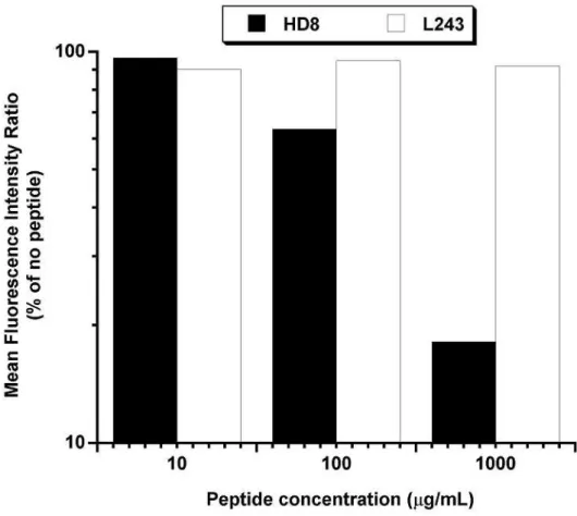
Epitope analysis by competition with epitope peptide
The HD8 epitope peptide (61-WNSQKDILEQARA-73 of DRB1*15011) was
prepared in PBS (-) solution. Raji cells (2×10⁵ cells) were incubated for 10 minutes on
After centrifugation and resuspension in staining buffer, the cells were incubated with 1
µg/mL Alexa488-labeled HD8 antibody in the presence of the peptide at final
concentrations ranging from 10 to 1000 µg/mL for 30 minutes on ice. After washing
three times with staining buffer, the cells were suspended in staining buffer containing
PI and analyzed by flow cytometry. The mean fluorescence intensity was calculated
by the CellQuest software.
Cytotoxicity assays
For antibody-dependent cellular cytotoxicity (ADCC) assay, PBMC effectors were
prepared from healthy volunteers by density centrifugation and suspended in complete
medium at a concentration of 4×10⁵ cells/mL (4× effector solution for ADCC). For
complement-dependent cytotoxicity (CDC) assay, human complement sera (SIGMA)
were diluted to 20% with complete medium (4× effector solution for CDC). To label
the target cells, 106 Raji cells were incubated with 100 µCi of sodium chromium
containing 51Cr (PerkinElmer Japan) in a total volume of 65 µL FCS-containing PBS (-).
After a 1-hour incubation at 37°C, the labeled target cells were washed three times to
remove free 51Cr and 2×10³ cells/50 µL were assayed in round-bottom microtiter plates
(BD Biosciences) with 100 µL of each antibody (2× antibody solution) at various
to each well. All samples were plated in triplicates. For the ADCC assay, the plates
were briefly centrifuged and incubated at 37°C for 4 hours. For the CDC assay, the
plates were incubated at 37°C for 2 hours. Tumor cell lysis was measured by
determining the amount of 51Cr released into the supernatant. Thus, after the
incubation, the plates were centrifuged and the supernatants from each well were
transferred into plates containing scintillator (Lumaplate-96, Packard Bioscience,
PerkinElmer). The plates were dried, and counted by a scintillation counter (Top
Count, Packard Bioscience, PerkinElmer). The percent specific lysis was determined
by the following equation: [(E - S)/(M - S)] × 100, where E is counts per minute for
sample, S is the average spontaneous release in the sample with target cells only, and M
is the average maximal release in the sample containing target cells lysed with 50 µL of
1% Triton X-100 detergent.
Mouse lymphoma-xenograft model
Female scid mice (C.B-17/Icr-scid/scid Jcl, CLEA Japan, Tokyo, Japan) and
NOD/scid mice (NOD/Shi-scid Jic, CLEA) were housed in autoclaved plastic cages,
and were maintained in an air-conditioned, specific pathogen-free animal room at a
and water ad libitum. All experiments in this study were approved by the Institutional
Animal Care and Use Committee in our laboratory.
For the intravenous lymphoma-xenografted model, 6- to 8-week-old scid and
NOD/scid mice were pretreated with 0.1 mg of anti-asialo GM-1 antisera (Wako Pure
Chemical Industries, Osaka, Japan) one day (day -1) before 5×10⁶ Raji lymphoma cells
(ATCC CCL-86) were inoculated intravenously (day 0). On day 5, various doses of
HD8 or the anti-CD20 chimeric antibody rituximab were injected intravenously, and the
survival of mice was monitored daily. The statistical significance of differences in the
Results
Binding spectrum of the novel pan-HLA-DR antibody HD8
The HD8 antibody against HLA-DR was generated by immunizing KM mice,
which produce fully human immunoglobulins (30), with HLA-DR-transfected L929
cells. Hybridomas derived from the splenocytes of these mice were screened for
reactivity with various lymphoma cell lines, and finally HD8 was selected by a wide
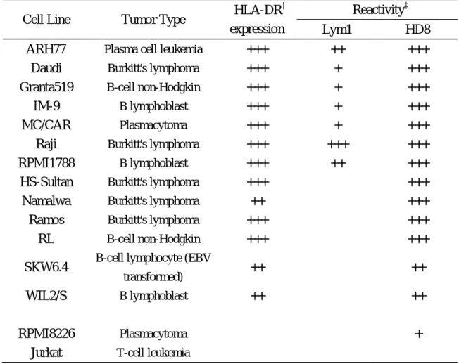
reactivity to HLA-DR-positive cells. The data for HD8 are shown in Table 1-1. The
HD8 antibody recognized all HLA-DR-positive 13 lines, mostly strongly (11/13, 85%).
In contrast, the chain-specific antibody Lym-1 was much less reactive, since it only
reacted with seven of 13 cell lines (only one was recognized strongly). Significantly,
HD8 recognized HLA-DR-negative plasmacytoma cell line RPMI-8226. As expected,
none of the antibodies recognized the HLA-DR-negative Jurkat cell line. Furthermore,
HD8 recognized B-cell population of the PBMCs from all 35 human volunteers that we
Table 1-1. Reactivity of HD8 with various lymphoma cell lines
Cell Line Tumor Type HLA-DR
†
expression
Reactivity‡
Lym1 HD8
ARH77 Plasma cell leukemia +++ ++ +++
Daudi Burkitt's lymphoma +++ + +++
Granta519 B-cell non-Hodgkin +++ + +++
IM-9 B lymphoblast +++ + +++
MC/CAR Plasmacytoma +++ + +++
Raji Burkitt's lymphoma +++ +++ +++
RPMI1788 B lymphoblast +++ ++ +++
HS-Sultan Burkitt's lymphoma +++ − +++
Namalwa Burkitt's lymphoma ++ − +++
Ramos Burkitt's lymphoma +++ − +++
RL B-cell non-Hodgkin +++ − +++
SKW6.4 B-cell lymphocyte (EBV
transformed) ++ − ++
WIL2/S B lymphoblast ++ − ++
RPMI8226 Plasmacytoma − − +
Jurkat T-cell leukemia − − −
Cells were stained with Alexa488-labeled antibodies HD8 and Lym-1 after blocking
with non-specific human IgG, and then the mean fluorescence intensity of FL-1
generated by the PI-negative live cells was measured by flow cytometry.
Alexa488-labeled non-specific human IgG was used as a negative control.
†
Expression level of HLA-DR on each cell line was confirmed by staining with
Alexa488-labeled L243. ‡The relative intensity indices for each antibody are defined
as follows: ‘−’, no staining; ‘+’, weak staining; ‘++’, intermediate staining; ‘+++',
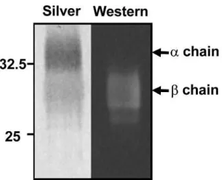
Epitope analysis of HD8
To determine whether HD8 binds to the or chain of HLA-DR, the purified
HLA-DR protein from the HLA-DR-positive B-lymphoblastoid cell line SKW6.4 was
subjected to immunoblot analysis using the HD8 antibody. SDS-PAGE of the protein
in non-reducing conditions allowed each HLA-DR protein chain to be detected
separately. HD8 recognized the lower band of HLA-DR protein (Figure 1-1a), which
was confirmed by N-terminal peptide sequencing to be the HLA-DR chain.
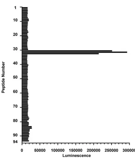
To identify the HLA-DRB epitope that is recognized by HD8, we synthesized 94
13-mer peptides that span the entire extracellular domain (199 amino acids) of the
HLA-DR chain allele DRB1*15011 (AAK51524.1, GenBank) and overlap by 11
amino acids. The peptides were blotted onto cellulose membranes and the ability of
HD8 to bind to each was tested. HD8 reacted strongly with peptide numbers 31-33,
which span amino acids 61-73, 63-75 and 65-77, respectively (Figure 1-1b). We then
confirmed that these peptides represent the HD8-binding epitope by determining
whether free #31 peptide (which has the sequence 61-WNSQKDILEQARA-73) could
compete with HD8 for binding to the HLA-DR-expressing Raji cell line. Indeed, the
#31 peptide significantly inhibited the binding of HD8 to Raji cells in contrast to no
is located on the helix of the HLA-DR chain, which is part of the HLA-DR groove
bound by peptide antigen (32, 33). Analysis of the IMGT/HLA database, which
contains the sequences of all known HLA-DRB alleles (29), revealed that while the
HD8 epitope contains highly polymorphic sequences, it also bears several amino acid
residues that are largely conserved in HLA-DRB alleles.
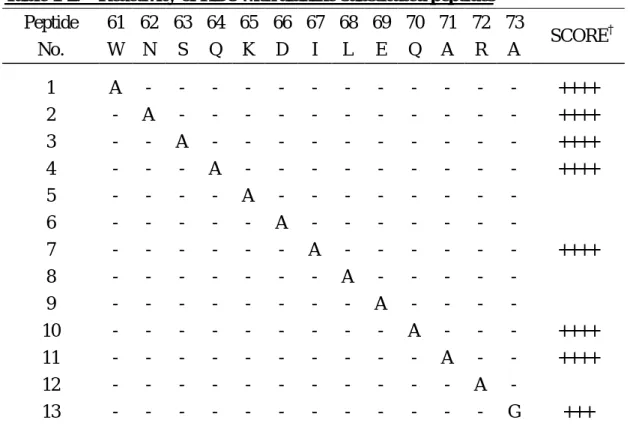
To clarify how HD8 recognizes this highly polymorphic region of the HLA-DR
chain, we identified the critical residues in the 61-WNSQKDILEQARA-73 epitope by
substituting each consecutive residue with alanine. As shown in Table 1-2,
substitution of residues 61-64, 67, 70, 71, or 73 had no effect on HD8 reactivity,
whereas substitution of residues 65, 66, 68, 69 or 72 abrogated HD8 reactivity. All of
Figure 1-1 (a). Delineation of the HD8 epitope on HLA-DR
HD8 recognizes the chain of HLA-DR protein. Affinity-purified HLA-DR protein
from SKW6.4 cells was subjected twice to SDS-PAGE in non-reducing conditions.
One gel was silver stained (left) while the other was subjected to immunoblot blot
analysis with HD8 (right). The two HLA-DR protein bands detected by silver stain
were determined by N-terminal protein sequencing to be the (upper) and (lower)
Figure 1-1 (b). Delineation of the HD8 epitope on HLA-DR
Mapping of the HLA-DR epitope recognized by HD8. 94 13-mer peptides spanning
the entire extracellular domain (199 residues) of the HLA-DR chain allele
DRB1*15011 were spot-synthesized onto a cellulose membrane. The peptides
overlapped with their direct neighbors by 11 residues. The membrane was subjected to
immunoblot analysis with HD8 and the relative chemiluminescence intensities are
shown. The sequences of the #31-#33 peptides that are strongly recognized by HD8
Figure 1-1 (c). Delineation of the HD8 epitope on HLA-DR
Peptide #31 (61-73) competes with HD8 for binding to the Raji lymphoma cell line.
2×10⁵ Raji cells were incubated with Alexa488-labeled HD8 (black bars) or L243
(white bars) antibody and various concentrations (1000, 100, 10 and 0 µg/mL) of
peptide #31. The mean fluorescence intensity (MFI) of FL-1 was analyzed by flow
Table 1-2. Reactivity of HD8 with alanine-substituted peptides Peptide No. 61 W 62 N 63 S 64 Q 65 K 66 D 67 I 68 L 69 E 70 Q 71 A 72 R 73 A SCORE † 1 2 3 4 5 6 7 8 9 10 11 12 13 A - - - - - - - - - - - - - A - - - - - - - - - - - - - A - - - - - - - - - - - - - A - - - - - - - - - - - - - A - - - - - - - - - - - - - A - - - - - - - - - - - - - A - - - - - - - - - - - - - A - - - - - - - - - - - - - A - - - - - - - - - - - - - A - - - - - - - - - - - - - A - - - - - - - - - - - - - A - - - - - - - - - - - - - G ++++ ++++ ++++ ++++ − − ++++ − − ++++ ++++ − +++
Each consecutive residue in the #31 (61-73) peptide was substituted with alanine and
the resulting 13 peptides were spot-synthesized on cellulose membranes and subjected
to immunoblot analysis with the HD8 antibody. '-' indicates the same amino acid as
the epitope peptide #31, WNSQKDILEQARA. †The relative intensity indices are
defined as follows: '++++', very strong; '+++', strong; '++', intermediate; '+', weak; '−',
In vitro binding of HD8 to the spectrum of different MHC class II alleles
We then tested the reactivity of HD8 with various MHC class II molecules by
synthesizing 25, 9, and 7 13-mer peptides that bear the 61-73-homologue sequences of
HLA-DR, HLA-DP, and HLA-DQ chain alleles, respectively. These peptides
together represent the homologue sequences of nearly all alleles apart from a few DR
and DP alleles. These peptides were subjected to immunoblot analyses using the HD8
antibody. The sequences of the peptides, the HD8 reactivity scores, and the number of
different alleles represented by each peptide sequence are shown in Table 1-3. This
analysis revealed that HD8 reacted with 21 of 25 HLA-DR peptides, all of which were
frequently used sequences. This means that HD8 can recognize 511 of 515 different
HLA-DR alleles (99.2%). HD8 also recognized all HLA-DP peptides and 5/7
Table 1-3. Reactivity of HD8 with peptides bearing the 61-73-homologue sequences of HLA-DR, HLA-DP and HLA-DQ chain alleles
MHC Type Peptide No. 61 W 62 N 63 S 64 Q 65 K 66 D 67 I 68 L 69 E 70 Q 71 A 72 R 73 A SCORE † No. of Alleles‡ DR 1 2 3 4 5 6 7 8 9 10 11 12 13 14 15 16 17 18 19 20 21 22 23 24 25 - - - - - - - - - - - - - - - - - - - - - - - - - - - - - - - - - - - - - - - - - - - - - - - - - - - - - - - - - - - - - - - - - - - - - - - - - - - - - - - - - - - - - - - - - - - - - - - - R - - - - - - - - - - - - - - - - - - - - - - - - - - - - - - - - - - - - - - - - - - - - - - N - - - - - - F L L - L - - L L F - - L F F L - - - F F - - L F - - - - - - - - - - - - - - - - - - - - - - - - - - - - - - - - - - - - - - - - - - - - - - - G - - D - - D R D - D - D D D D R - - - - - N D D D - D R K R E R R - R K E R K E R - - K K - R T R R R R - - - - - - - - - - - - - - - - - - - - - - - Q A - G - - - - - - - - G - - - - - G - - - - G - - - +++ +++ ++++ ++++ ++++ ++++ ++++ ++++ +++ +++ ++++ ++++ +++ ++++ ++++ ++++ ++++ +++ ++ ++ + − − − − 109 76 73 54 52 42 28 17 13 9 9 6 6 5 3 3 2 1 1 1 1 1 1 1 1 DP 26 27 28 29 30 - - - - - - - - - - - - - - - - - - - - - - - - - - - - - - - - L L L - - - - - - - - - - E E E E E K E K R E - - - - - - - - - - +++ ++++ ++++ ++++ ++++ 42 37 29 5 4
Table 1-3 (Continued). Reactivity of HD8 with peptides bearing the 61-73-homologue sequences of HLA-DR, HLA-DP and HLA-DQ chain alleles
DP 31 - - - F - - E E - - +++ 1 32 33 34 - - - - - - - - - - - - - - - - - H F N - - - - - - - E E E K K K - - - - - - ++++ +++ + 1 1 1 DQ 35 36 37 38 39 40 41 - - - - - - - - - - - - - - - - - - - - - - - - - - - - - - - - - - - E E E - - - E V V V - - - D - - - - - - - - - - - - - - R G G R E R R T T - K D T T - - - - - - - - - - - - - - + − − +++ ++ +++ + 33 17 7 5 4 3 1 Cellulose membranes bearing 25, nine, and seven 13-mer peptides that represent most
of the 61-73-homologue sequences of human leukocyte antigen (HLA)-DR, HLA-DP
and HLA-DQ alleles, respectively, were generated and subjected to immunoblot
analysis with HD8. Shadowed rows indicate the critical residues for HD8 binding
determined by alanine substitution on Table 1-2. '-' indicates the same amino acid as
the epitope peptide #31, WNSQKDILEQARA. †The relative intensity indices are
defined as follows: '++++', very strong; '+++', strong; '++', intermediate; '+', weak; '−',
no signal. ‡The number of alleles represented by each peptide, as determined by
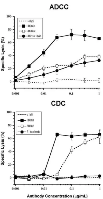
Ability of HD8 to induce in vitro cytotoxicity
To determine whether the HD8 antibody is tumoricidal, we first examined its ability
to induce ADCC and CDC against Raji in vitro (Figure 1-2). To maximize the
capacity of HD8 to induce ADCC and CDC by human effectors, IgG1-type HD8 was
constructed from the original IgG2-type of HD8 by subclass conversion. The
IgG1-type HD8 produced strong ADCC and CDC, both of which are stronger than
IgG2-type that produced weak ADCC and medium CDC. In contrast, rituximab
(IgG1) induced even weaker ADCC than IgG1-type HD8 and could not generate CDC
against Raji. The negative control antibody failed to induce either ADCC or CDC.
Anti-tumor activity of HD8 in lymphoma-xenografted murine models
To address the in vivo anti-tumor efficacy of HD8, scid mice were inoculated with
either the lymphoma cell line Raji or the lymphoblastoid cell line MC/CAR and injected
five days later with 5 or 50 µg/kg of the original IgG2-type HD8, rituximab, or control
human IgG (Figure 1-3). Scid mice that were xenografted with Raji and MC/CAR and
treated with control IgG died within 42 and 70 days of transplantation, respectively.
Rituximab significantly prolonged the life spans of Raji-bearing and MC/CAR-bearing
mice at doses of 5 and/or 50 µg/kg (p<0.05). HD8 treatment at both 5 and 50 µg/kg
100% of the Raji-bearing and MC/CAR-bearing animals were long-term survivors,
respectively.
To determine whether the host effector function contributes to HD8 efficacy, we
transplanted NOD/scid mice with Raji and treated them with 50 or 500 µg/kg HD8 on 5
days later. NOD/scid mice are known to possess less NK and complement activities
than scid mice (34). HD8 treatment significantly prolonged the life spans of the mice
compared to control antibody-treated mice (p<0.01 and p<0.001 for 50 and 500 µg/kg
HD8, respectively). However, a higher dose of HD8 was needed to achieve a similar
outcome in NOD/scid models relative to that observed in the scid models (Figure 1-3,
Figure 1-2. Abilities of IgG1- and IgG2-type HD8s to induce ADCC and CDC
51
Cr-labeled Raji cells were incubated with increasing concentrations of IgG1-type HD8
(■), original (IgG2-type) HD8 (□), the chimeric mouse-human IgG1 rituximab (●), or
the control antibody (no symbol, dotted line) together with human effector PBMCs
(ADCC assay, upper graph) or 5% complement sera (CDC assay, lower graph). The
specific lysis ratios were then calculated. The bars represent the SEMs of triplicates.
Figure 1-3. Anti-tumor activity of HD8 in lymphoma-xenograft mouse models
Scid (left and right graphs) and NOD/scid (middle graphs) mice were pretreated with
anti-asialo GM-1 antisera on day -1, inoculated with Raji (left and middle graphs) or
MC/CAR (right graphs) lymphoma cells intravenously on day 0, and treated with 5, 50
or 500 µg/kg of HD8 (top graphs), rituximab (bottom graphs), and control IgG on day 5.
The asterisks indicate significant differences relative to the survival of control
IgG-administered mice, as determined by log-rank tests (*p<0.05, **p<0.01,
Discussion
It has been known already for decades that many B-lineage tumor cells express
HLA-DR (35, 36). Consequently, this molecule is considered to be an attractive target
molecule for the treatment of B-cell malignancies. However, while two anti-HLA-DR
antibodies (Lym-1 and Hu1D10) have been brought to clinical trial, neither has yet been
approved for clinical application. One significant problem suffered by these
anti-HLA-DR antibodies is the limited spectrum of HLA-DR molecules that they can
recognize because of the extreme polymorphism of HLA-DR. The polymorphism of
HLA-DR is largely due to chain variability, as several hundred chain alleles exist.
Consequently, anti-HLA-DR chain antibodies like Lym-1 and Hu1D10 react with just
a proportion of HLA-DR alleles.
Here, we report the generation of HD8, a novel, fully-human antibody against
HLA-DR that is largely unaffected by HLA-DR polymorphism. Surprisingly, HD8
could react with highly-polymorphic HLA-DR chain. Moreover, while its epitope is
situated very close to the epitopes of Lym-1 and Hu1D10, all of which are located in a
highly polymorphic region of HLA-DR chain (26, 28, 37), HD8 could react with
99.2% of the 515 HLA-DRB alleles tested. That HD8 could nevertheless react with a
much wider range of HLA-DR alleles can be explained by the fact that the essential
among the various HLA-DRB alleles. Notably, since these residues are also highly
conserved in HLA-DPB alleles and moderately conserved in HLA-DQB alleles, HD8
could also recognize 100% and 66% of the HLA-DPB and HLA-DQB alleles,
respectively. That HD8 recognizes not only HLA-DR but also the other two MHC
class II molecules might enhance the cytotoxicity of this antibody, as it would increase
the absolute amount of antibodies bound to the target cell. In regard to this, the
expression levels of some target antigens have been shown to shape anti-tumor effector
functions (38). These favorable characteristics of HD8 may broaden its clinical
potential.
It is currently difficult to predict the pharmacological efficacy of a given antibody
on the basis of its binding characteristics, including its epitope location. However, the
variables that shape the efficacy of an antibody are gradually being elucidated. For
example, Polyak and Deans have reported that heterogeneity in the fine specificity of
anti-CD20 antibodies is due to marked differences in their ability to induce homotypic
cellular aggregation and translocation of CD20 to lipid rafts (39). Similarly, Cragg and
colleagues reported that anti-CD20 antibodies could be classified into two groups on the
basis of their ability to segregate CD20 into lipid rafts; notably, this segregating ability
anti-HLA-DR antibodies can be similarly categorized remains to be determined.
Our mapping analyses revealed that the essential epitope residues of HD8 are
residues 65, 66, 68, 69 and 72. Analysis of the tertiary structure of HLA-DR revealed
that these residues are located on the outer portion of the helix which forms the
antigen-presenting groove together with the HLA-DR chain (32, 33). This suggests
that HD8 could be immunosuppressive when used clinically, as it could interfere with
the antigen-presenting function of HLA-DR. However, we found HD8 is not
particularly immunosuppressive when tested in an allogenic mixed leukocyte reaction
assay (data not shown), which result may come from its epitope existing on “outer”
portion of the groove. How the pharmacological efficacy and immunosuppressive
capacity of HD8 relate to its epitope location should be studied further.
How HLA-DR antibodies induce tumor cell cytotoxicity is an important issue to be
clarified. Many papers have reported that such antibodies may induce HLA-DR
signaling including programmed cell death (26, 41-47). We also observed that HD8
induced programmed cell death in the same way of other anti-HLA-DR antibodies
(unpublished data, 2006). In addition, we found that IgG1-type HD8 induced strong
ADCC and CDC using human effectors in in vitro assays. We also showed in vivo
three models, HD8 was considerably more effective in prolonging murine life span than
rituximab, which difference of two antibodies may attribute to the expression levels of
the target proteins and the affinities of antibodies to the antigens (data not shown). We
also noticed that more HD8 was needed to prolong the lives of NOD/scid
Raji-xenografted mice compared to the amount needed to exert an equivalent effect in
scid Raji-xenografted mice. Since the NOD/scid mouse is known to have less NK and
complement activities than the scid mouse (34), this observation supports the notion that
the tumoricidal activity in vivo depends on ADCC and/or CDC, which notion should be
elucidated further. Notably, the clinical study of the humanized anti-HLA-DR IgG1
antibody Hu1D10 has suggested the existence of an additional pharmacological
mechanism, as some Hu1D10-responsive patients were found to develop anti-tumor
antibodies (48). Thus, it is possible that HLA-DR antibodies could somehow provoke
an immune response against the tumor. To elucidate how HD8 exerts its tumoricidal
functions, additional experiments are required.
There remains a critical issue which subclass of human antibody should be chosen
for anti-HLA-DR antibody. Kostney and colleagues reported Hu1D10 antibody with
IgG1-type (26), whereas Nazy and colleagues reported IgG4-type antibody in
antibody expressed stronger tumoricidal efficacy than IgG4-type HD8 in the
Raji-xenograft scid mouse model. Both IgG1- and IgG2-types HD8 possessed both
strong ADCC and CDC in vitro by using mouse effectors. In Figure 1-2 using human
effectors, IgG1-type expressed the strongest ADCC and CDC. Therefore, we chose
IgG1-type HD8 as a clinical candidate.
Besides, side effects of anti-HLA-DR antibodies should be elucidated, as HLA-DR
is expressed on a wide variety of hematopoietic cells, such as myeloid progenitor cells
and antigen presenting cells.
In conclusion, IgG1-type HD8 is promising as a potential immunotherapeutic agent
against HLA-DR-positive lymphocytic cancers, as its structure is completely human, it
exerts strong efficacies on in vitro cytotoxicity assays and in vivo xenograft models, as
References
1 Glennie MJ, Johnson PW. Clinical trials of antibody therapy. Immunol Today
2000; 21:403-10.
2 Grillo-Lopez AJ. AntiCD20 mAbs: modifying therapeutic strategies and outcomes
in the treatment of lymphoma patients. Expert Rev Anticancer Ther 2002; 2:323-9.
3 Weiner GJ, Link BK. Monoclonal antibody therapy of B cell lymphoma. Expert
Opin Biol Ther 2004; 4:375-85.
4 Forero A, Lobuglio AF. History of antibody therapy for non-Hodgkin's lymphoma.
Semin Oncol 2003; 30:1-5.
5 Dillman RO. Treatment of low-grade B-cell lymphoma with the monoclonal
antibody rituximab. Semin Oncol 2003; 30:434-47.
6 Lin TS, Lucas MS, Byrd JC. Rituximab in B-cell chronic lymphocytic leukemia.
Semin Oncol 2003; 30:483-92.
7 Shan D, Ledbetter JA, Press OW. Apoptosis of malignant human B cells by
ligation of CD20 with monoclonal antibodies. Blood 1998; 91:1644-52.
8 Taji H, Kagami Y, Okada Y et al. Growth inhibition of CD20-positive B lymphoma
cell lines by IDEC-C2B8 anti-CD20 monoclonal antibody. Jpn J Cancer Res 1998;
9 Jazirehi AR, Bonavida B. Cellular and molecular signal transduction pathways
modulated by rituximab (rituxan, anti-CD20 mAb) in non-Hodgkin's lymphoma:
implications in chemosensitization and therapeutic intervention. Oncogene 2005;
24:2121-43.
10 Emmanouilides C. Radioimmunotherapy for non-Hodgkin's lymphoma. Semin
Oncol 2003; 30:531-44.
11 Friedberg JW, Fisher RI. Iodine-131 tositumomab (Bexxar):
radioimmunoconjugate therapy for indolent and transformed B-cell non-Hodgkin's
lymphoma. Expert Rev Anticancer Ther 2004; 4:18-26.
12 Zelenetz AD. A clinical and scientific overview of tositumomab and iodine I 131
tositumomab. Semin Oncol 2003; 30:22-30.
13 Leonard JP. Targeting CD20 in Follicular NHL: Novel Anti-CD20 Therapies,
Antibody Engineering, and the Use of Radioimmunoconjugates. Hematology (Am
Soc Hematol Educ Program) 2005; 335-9.
14 Teeling JL, French RR, Cragg MS et al. Characterization of new human CD20
monoclonal antibodies with potent cytolytic activity against non-Hodgkin
lymphomas. Blood 2004; 104:1793-800.
anti-CD20 antibody PRO70769 in Macaca fascicularis. J Immunother 2005;
28:212-9.
16 Stein R, Qu Z, Chen S et al. Characterization of a new humanized anti-CD20
monoclonal antibody, IMMU-106, and Its use in combination with the humanized
anti-CD22 antibody, epratuzumab, for the therapy of non-Hodgkin's lymphoma. Clin
Cancer Res 2004; 10:2868-78.
17 Siegel AB, Goldenberg DM, Cesano A, Coleman M, Leonard JP. CD22-directed
monoclonal antibody therapy for lymphoma. Semin Oncol 2003; 30:457-64.
18 Stone MJ, Sausville EA, Fay JW et al. A phase I study of bolus versus continuous
infusion of the anti-CD19 immunotoxin, IgG-HD37-dgA, in patients with B-cell
lymphoma. Blood 1996; 88:1188-97.
19 Moreton P, Hillmen P. Alemtuzumab therapy in B-cell lymphoproliferative
disorders. Semin Oncol 2003; 30:493-501.
20 Dechant M, Bruenke J, Valerius T. HLA class II antibodies in the treatment of
hematologic malignancies. Semin Oncol 2003; 30:465-75.
21 Sweetenham JW. Diffuse large B-cell lymphoma: risk stratification and
management of relapsed disease. Hematology (Am Soc Hematol Educ Program).
22 Epstein AL, Marder RJ, Winter JN et al. Two new monoclonal antibodies, Lym-1
and Lym-2, reactive with human B-lymphocytes and derived tumors, with
immunodiagnostic and immunotherapeutic potential. Cancer Res 1987; 47:830-40.
23 Hu E, Epstein AL, Naeve GS et al. A phase 1a clinical trial of LYM-1 monoclonal
antibody serotherapy in patients with refractory B cell malignancies. Hematol Oncol
1989; 7:155-66.
24 Brown KS, Levitt DJ, Shannon M, Link BK. Phase II trial of Remitogen
(humanized 1D10) monoclonal antibody targeting class II in patients with relapsed
low-grade or follicular lymphoma. Clin Lymphoma 2001; 2:188-90.
25 Gingrich, RD, Dahle, CE, Hoskins, KF, Senneff, MJ. Identification and
characterization of a new surface membrane antigen found predominantly on
malignant B lymphocytes. Blood 1990; 75:2375-87.
26 Kostelny SA, Link BK, Tso JY et al. Humanization and characterization of the
anti-HLA-DR antibody 1D10. Int J Cancer 2001; 93:556-65.
27 Stockmeyer B, Schiller M, Repp R et al. Enhanced killing of B lymphoma cells by
granulocyte colony-stimulating factor-primed effector cells and Hu1D10--a
humanized human leucocyte antigen DR antibody. Br J Haematol 2002; 118:959-67.
polymorphic HLA-DR molecules. Mol Immunol 1999; 36:789-97.
29 Robinson J, Waller MJ, Parham P et al. IMGT/HLA and IMGT/MHC: sequence
databases for the study of the major histocompatibility complex. Nucleic Acids Res
2003; 31:311-4.
30 Ishida I, Tomizuka K, Yoshida H et al. Production of human monoclonal and
polyclonal antibodies in TransChromo animals. Cloning Stem Cells 2002; 4:91-102.
31 Kramer A, Keitel T, Winkler K, Stocklein W, Hohne W, Schneider-Mergener J.
Molecular basis for the binding promiscuity of an anti-p24 (HIV-1) monoclonal
antibody. Cell 1997; 91:799-809.
32 Jardetzky TS, Brown JH, Gorga JC et al. Three-dimensional structure of a human
class II histocompatibility molecule complexed with superantigen. Nature 1994;
368:711-8.
33 Dessen A, Lawrence CM, Cupo S, Zaller DM, Wiley DC. X-ray crystal structure of
HLA-DR4 (DRA*0101, DRB1*0401) complexed with a peptide from human
collagen II. Immunity 1997; 7:473-81.
34 Shultz LD, Schweitzer PA, Christianson SW et al. Multiple defects in innate and
adaptive immunologic function in NOD/LtSz-scid mice. J Immunol 1995;
35 Guy K, Krajewski AS, Dewar AE. Expression of MHC class II antigens in human
B-cell leukaemia and non-Hodgkin's lymphoma. Br J Cancer 1986; 53:161-73.
36 Ratech H. HLA-DR expression in B-cell non-Hodgkin's malignant lymphomas: a
multiparameter flow cytometry study. Hum Pathol 1990; 21:1275-82.
37 Rose LM, Gunasekera AH, DeNardo SJ, DeNardo GL, Meares CF.
Lymphoma-selective antibody Lym-1 recognizes a discontinuous epitope on the light
chain of HLA-DR10. Cancer Immunol Immunother 1996; 43:26-30.
38 Kono K, Takahashi A, Ichihara F, Sugai H, Fujii H, Matsumoto Y. Impaired
antibody-dependent cellular cytotoxicity mediated by herceptin in patients with
gastric cancer. Cancer Res 2002; 62:5813-7.
39 Polyak MJ, Deans JP. Alanine-170 and proline-172 are critical determinants for
extracellular CD20 epitopes; heterogeneity in the fine specificity of CD20
monoclonal antibodies is defined by additional requirements imposed by both amino
acid sequence and quaternary structure. Blood 2002; 99:3256-62.
40 Cragg MS, Morgan SM, Chan HT et al. Complement-mediated lysis by anti-CD20
mAb correlates with segregation into lipid rafts. Blood 2003; 101:1045-52.
41 Truman JP, Ericson ML, Choqueux-Seebold CJ, Charron DJ, Mooney NA.
1994; 6:887-96.
42 Ramirez R, Carracedo J, Mooney N, Charron D. HLA class-II-mediated homotypic
aggregation: involvement of a protein tyrosine kinase and protein kinase C. Hum
Immunol 1992; 34:115-25.
43 Tabata H, Matsuoka T, Endo F, Nishimura Y, Matsushita S. Ligation of HLA-DR
molecules on B cells induces enhanced expression of IgM heavy chain genes in
association with Syk activation. J Biol Chem 2000; 275:34998-5005.
44 Lokshin AE, Kalinski P, Sassi RR et al. Differential regulation of maturation and
apoptosis of human monocyte-derived dendritic cells mediated by MHC class II. Int
Immunol 2002; 14:1027-37.
45 Bains SK, Mone A, Yun Tso J et al. Mitochondria control of cell death induced by
anti-HLA-DR antibodies. Leukemia 2003; 17:1357-65.
46 Nagy ZA, Hubner B, Lohning C et al. Fully human, HLA-DR-specific monoclonal
antibodies efficiently induce programmed death of malignant lymphoid cells. Nat
Med 2002; 8:801-7.
47 Fransson J, Tornberg UC, Borrebaeck CA, Carlsson R, Frendeus B. Rapid
induction of apoptosis in B-cell lymphoma by functionally isolated human antibodies.
48 Link BK, Wang H, Byrd JC et al. Phase I Study of Hu1D10 Monoclonal Antibody
CHAPTER II
Complement activation plays a key role in antibody-induced
infusion toxicity in monkeys and rats
Summary
Infusion reactions are a major side effect of the administration of therapeutic
antibodies and are the result of complex immune reaction. We report here that
substitutions of Fc amino acids in the anti-HLA-DR antibody HD8 reduce its ability to
induce infusion reactions in rats and monkeys. We first showed that intravenous
administration of IgG1- and IgG2-subclass HD8 antibodies induces severe infusion
reactions in monkeys. These antibodies express strong complement-dependent
cytotoxicity (CDC), and in vivo depletion of complement in rats by pretreatment with
cobra venom factor abrogated the lethal infusion reactions generated by HD8-IgG1.
Thus, the infusion reactions appear to be largely driven by the complement system. To
reduce the CDC function of HD8-IgG1, its Fc region was modified by two amino acid
substitutions at Pro331Ser and Lys322Ala. The modified antibody was incapable of
expressing CDC in vitro and did not induce severe infusion reactions in rats and
monkeys, even at extremely high doses. The modified antibody retained its
antibody-dependent cellular cytotoxicity function as well as its anti-tumor activity in a
tumor-bearing mouse model. In summary, complement appears to drive infusion
reactions and modifications that eliminate the CDC activity of an antibody also reduce
Introduction
Infusion reactions are infusion-related side effects that occasionally occur especially
upon intravenous treatment with antibody-based drugs (e.g. rituximab and trastuzumab)
(1-3). Infusion reactions manifest themselves as flu-like or anaphylaxis-like symptoms
such as chills, asthenia, nausea, headache, rash, and vomiting. Several immune
mechanisms have been suggested to be responsible for the induction of infusion
reactions. First, the intensity of infusion reactions has been positively correlated with
the rapid release of large amounts of cytokines, especially inflammatory cytokines. For
example, the infusion of the anti-CD3 antibody OKT3 induces massive cytokine release
(4, 5). Second, several therapeutic antibodies have been suggested to activate the
complement system, resulting in the production of large amounts of C3a, C4a and C5a
which can act as anaphylatoxins and induce the release of chemical mediators such as
histamine and leukotriene (2, 6). Third, rituximab has also been suggested to induce
the acute lysis of a large number of tumor cells via antibody-dependent cellular
cytotoxicity (ADCC) and/or complement-dependent cytotoxicity (CDC); this in turn
induces an inflammatory reaction, i.e. infusion reaction, known as tumor lysis syndrome
(7-9). The infusion reactions can be ameliorated to some extent by administering
HLA-DR has been suggested to be a candidate target for antibody-based therapies
against hematological malignancies. It has been shown that while the IgG1-type
anti-HLA-DR antibody Hu1D10 induces infusion reactions (11-13), the IgG4-type
anti-HLA-DR antibody 1D09C3 does not (14). Since IgG4 antibodies do not evoke
either ADCC or CDC, these observations suggest that the infusion reactions occurred by
anti-HLA-DR antibodies like Hu1D10 are induced by effector functions such as ADCC
and/or CDC. In this chapter, we show that our anti-HLA-DR antibody HD8 (15) also
induces an infusion reaction, and that this reaction correlates positively with the ability
of this antibody to induce CDC. We also show that modifications of critical areas of
the Fc region of HD8 that decrease the CDC potential of this antibody also reduce the
risk of infusion reactions while retaining the ability of this antibody to induce ADCC