RESEARCH ARTICLE
Phosphatidic Acid Produced by Phospholipase
D Promotes RNA Replication of a Plant RNA
Virus
Kiwamu Hyodo1¤, Takako Taniguchi2, Yuki Manabe3, Masanori Kaido1, Kazuyuki Mise1,
Tatsuya Sugawara3, Hisaaki Taniguchi2, Tetsuro Okuno1*
1 Laboratory of Plant Pathology, Graduate School of Agriculture, Kyoto University, Kyoto, Japan, 2 Institute for Enzyme Research, University of Tokushima, Tokushima, Japan, 3 Laboratory of Marine Bioproducts Technology, Graduate School of Agriculture, Kyoto University, Kyoto, Japan
¤ Current address: Institute of Plant Science and Resources, Okayama University, Okayama, Japan *okuno@kais.kyoto-u.ac.jp
Abstract
Eukaryotic positive-strand RNA [(+)RNA] viruses are intracellular obligate parasites repli-cate using the membrane-bound replicase complexes that contain multiple viral and host components. To replicate, (+)RNA viruses exploit host resources and modify host metabo-lism and membrane organization. Phospholipase D (PLD) is a phosphatidylcholine- and phosphatidylethanolamine-hydrolyzing enzyme that catalyzes the production of phospha-tidic acid (PA), a lipid second messenger that modulates diverse intracellular signaling in various organisms. PA is normally present in small amounts (less than 1% of total phospho-lipids), but rapidly and transiently accumulates in lipid bilayers in response to different envi-ronmental cues such as biotic and abiotic stresses in plants. However, the precise functions of PLD and PA remain unknown. Here, we report the roles of PLD and PA in genomic RNA replication of a plant (+)RNA virus, Red clover necrotic mosaic virus (RCNMV). We found that RCNMV RNA replication complexes formed in Nicotiana benthamiana contained PLDα and PLDβ. Gene-silencing and pharmacological inhibition approaches showed that PLDs and PLDs-derived PA are required for viral RNA replication. Consistent with this, exoge-nous application of PA enhanced viral RNA replication in plant cells and plant-derived cell-free extracts. We also found that a viral auxiliary replication protein bound to PA in vitro, and that the amount of PA increased in RCNMV-infected plant leaves. Together, our findings suggest that RCNMV hijacks host PA-producing enzymes to replicate.
Author Summary
All characterized eukaryotic positive-strand RNA [(+)RNA] viruses replicate their ge-nomes using the viral replication complexes (VRCs), which contain multiple viral and host components, on intracellular membranes. Phospholipids are major constituents of cellular membranes; however, the function(s) of phospholipids in genome replication of (+)RNA viruses remains largely unknown. Here, we show that Red clover necrotic mosaic
OPEN ACCESS
Citation: Hyodo K, Taniguchi T, Manabe Y, Kaido M, Mise K, Sugawara T, et al. (2015) Phosphatidic Acid Produced by Phospholipase D Promotes RNA Replication of a Plant RNA Virus. PLoS Pathog 11(5): e1004909. doi:10.1371/journal.ppat.1004909 Editor: Aiming Wang, Agriculture and Agri-Food Canada, CANADA
Received: November 27, 2014 Accepted: April 23, 2015 Published: May 28, 2015
Copyright: © 2015 Hyodo et al. This is an open access article distributed under the terms of the Creative Commons Attribution License, which permits unrestricted use, distribution, and reproduction in any medium, provided the original author and source are credited.
Data Availability Statement: All relevant data are within the paper and its Supporting Information files. Funding: This study was supported by a Grant-in Aid for Science Research (A) (22248002) and by a Grant-in Aid for Science Research on Innovative Areas (25115509) from the Japan Society for the Promotion of Science. The funders had no role in study design, data collection and analysis, decision to publish, or preparation of the manuscript.
Competing Interests: The authors have declared that no competing interests exist.
virus (RCNMV), a plant (+)RNA virus, induces a high accumulation of phosphatidic acid (PA) in infected plant leaves. PA-producing enzymes, phospholipase Dα (PLDα) and PLDβ, are associated with RCNMV VRCs. PA interacts with the viral replication protein and enhances the viral replication by upregulating the activity/assembly of the VRCs in vitro. In summary, RCNMV alters cellular lipid metabolism via PLD to establish a suitable environment for viral replication.
Introduction
Positive-strand RNA [(+)RNA] viruses are the most abundant plant viruses, and include many viruses economically important in agriculture. (+)RNA plant viruses have a limited coding ca-pacity. To replicate and achieve successful infection in their hosts, they need to use host pro-teins, membranes, lipids, and metabolites. All characterized eukaryotic (+)RNA viruses replicate their genomes using viral replication complexes (VRCs), which contain multiple viral and host components on intracellular membranes [1–6]. A growing number of studies have suggested that plant viruses have evolved ways to hijack plant host factors and reprogram host cell metabolism for their successful infection [6,7]. Conversely, plants have evolved the ability to recognize viruses through specific interaction with viral proteins or viral double-stranded RNA intermediates for restricting virus infection [8,9]. Viruses must circumvent or suppress such surveillance systems and host defense mechanisms. Thus, viruses must be evolved to achieve a good balance between hijacking/reprogramming host factors for efficient viral repli-cation and avoiding the danger of stimulating antiviral defense responses.
Red clover necrotic mosaic virus (RCNMV) is a (+)RNA plant virus and a member of the genus Dianthovirus in the family Tombusviridae. The genome of RCNMV consists of RNA1 and RNA2. RNA1 encodes a p27 auxiliary replication protein, p88polRNA-dependent RNA polymerase (RdRp), and a coat protein [10]. RNA2 encodes a movement protein that is re-quired for viral cell-to-cell movement [10,11]. p27, p88pol, and host proteins form a 480-kDa replicase complex, which is a key player in the viral RNA replication [12]. p27 and p88pol colo-calize at the endoplasmic reticulum (ER) [13,14], where RCNMV replication takes place [15]. Our previous studies showed that RCNMV uses host heat shock proteins (HSPs), HSP70 and HSP90 [16], and ADP-ribosylation factor 1 (Arf1) [15] for the formation of the 480-kDa repli-case complex and p27-induced ER membrane alterations. Arf1 is a small GTPase that regulates COPI vesicle formation. Sar1, another small GTPase that regulates COPII vesicle-mediated trafficking, and Arf1 are recruited from their original subcellular locations to RCNMV replica-tion sites via p27, and p27 interferes with host membrane trafficking pathway in plant cells [15,
17]. Mammalian and yeast Arf1 recruits and/or stimulates its effector proteins, including a coatomer, phosphatidylinositol 4 kinase IIIβ (PI4KIIIβ), and phospholipase D (PLD) [18]. Arf1 can activate mammalian PLD1 and PLD2 directly. PLD hydrolyses structural phospholip-ids such as phosphatidylcholine (PC) and phosphatidylethanolamine (PE) to produce phos-phatidic acid (PA) and remaining headgroups. PA production resulting from Arf1-mediated PLD activation has been proposed to be associated with vesicle formation [19].
The 12 different PLD isoforms encoded in the Arabidopsis thaliana genome are classified into six groups (α, β, γ, δ, ε, and z) based on sequence similarity and in vitro activity [20]. PLDz1 and z2 have N-terminal phox homology (PX) and pleckstrin homology (PH) domains and share high sequence similarities to two PX/PH-PLDs in mammals. The remaining PLDs contain the Ca2+-dependent phospholipid-binding C2 domain and are unique to plants.
PA is normally present in small amounts (less than 1% of total phospholipids), but rapidly and transiently accumulates in lipid bilayers in response to different abiotic stresses such as de-hydration, salt, and osmotic stress [20–22]. PA also accumulates in response to several mi-crobe-associated molecular patterns (MAMPs) in plant cells and positively regulates salicylic acid (SA)-mediated defense signaling [23–27]. Moreover, effector proteins of bacterial and fun-gal pathogens, such as Cladosporium fulvum Avr4 and Pseudomonas syringae AvrRpm1 and AvrRpt2, trigger PA accumulation in their host cells, and multiple PLD isoforms contribute to AvrRpm1-triggered resistance in Arabidopsis thaliana [28–30]. PLDδ plays a positive role in
MAMPs-triggered cell wall based immunity and nonhost resistance against Blumeria graminis f. sp. hordei [31]. Moreover, overexpression of rice diacylglycerol (DAG) kinase, which cata-lyzes the conversion of DAG to PA, enhances resistance against tobacco mosaic virus and Phy-tophthora parasitica infections in tobacco plants [32]. In accordance with this, direct
application of PA to leaves has been shown to induce the expression of pathogenesis-related (PR) genes and cell death [28,33]. These findings indicate that PA is a positive regulator in plant defense against pathogens. In contrast, PLDβ1 acts like a negative regulator of the genera-tion of reactive oxygen species (ROS), the expression of PR genes, and plant defenses against biotrophic pathogens in rice and Arabidopsis [34–36].
In this study, using two-step affinity purification and liquid chromatography–tandem mass spectrometry (LC/MS/MS) analysis, we identified Nicotiana benthamiana PLDα and PLDβ as interaction partners of RCNMV replication protein, p88pol. Gene-silencing and pharmacologi-cal inhibition approaches show that PLDs-derived PA plays a positive role in viral RNA repli-cation. Consistent with this role, direct application of PA to plant cells or plant-derived cell-free extracts enhanced RCNMV RNA replication and negative-strand RNA synthesis, respec-tively. We found that p27 auxiliary replication protein interacted with PA in vitro and that the accumulation of PA increased in RCNMV-infected plant leaves. Together, our findings suggest that RCNMV hijacks host PA-producing enzymes to achieve successful RNA replication.
Results
To identify putative host proteins associated with the RCNMV replicase complex, we expressed six-His/FLAG-tagged p27 and p88polreplication proteins (p27-HF and p88pol-HF, respective-ly) together with an RNA2 replication template via agroinfiltration in N. benthamiana. Two-days after infiltration (dai), RCNMV replication proteins were purified via sequential affinity purification using nickel-agarose beads and FLAG-affinity resins as described in the Materials and Methods section. Note that p27-HF and p88pol-HF replication proteins can support RNA2 replication in N. benthamiana plants, although their activities were low compared with those of non-tagged p27 and p88pol(S1 Fig).
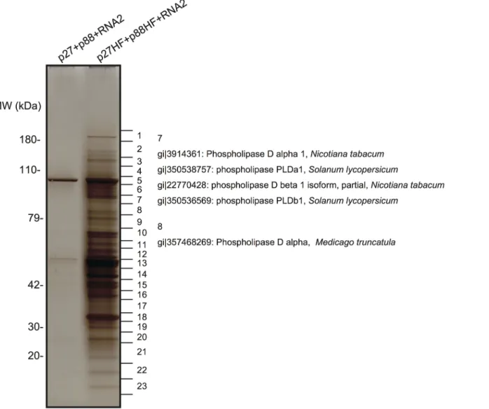
The purified fraction was subjected to sodium dodecyl sulfate-polyacrylamide gel electro-phoresis to separate the proteins that copurified with RCNMV replication proteins. A silver-stained gel showed the presence of many protein bands that were absent in the control fraction prepared from Agrobacterium-infiltrated N. benthamiana leaves expressing non-tagged p27 and p88poltogether with RNA2 (Fig 1). LC/MS/MS analysis of the isolated proteins excised from gels led to the identification of many host proteins (Fig 1andS1 Table). These proteins included PLDα and PLDβ, and several Arf1 effector proteins, such as coatomer subunits and clathrin heavy chain, in addition to previously identified host factors, HSP70 and HSP90 [16], and Arf1 [15]. It is known that the activities of yeast and mammalian PLDs are stimulated by Arf1 [37] and Arf1 is an essential host factor in RCNMV RNA replication [15]. Therefore, we investigated whether PLDα and PLDβ are also required for RCNMV RNA replication.
We isolated cDNAs encoding PLDα and PLDβ from N. benthamiana leaves. Deduced amino acid sequences and peptide sequences identified from LC/MS/MS analysis are presented inS2 Fig. To confirm the association of N. benthamiana (Nb)PLDα and NbPLDβ with
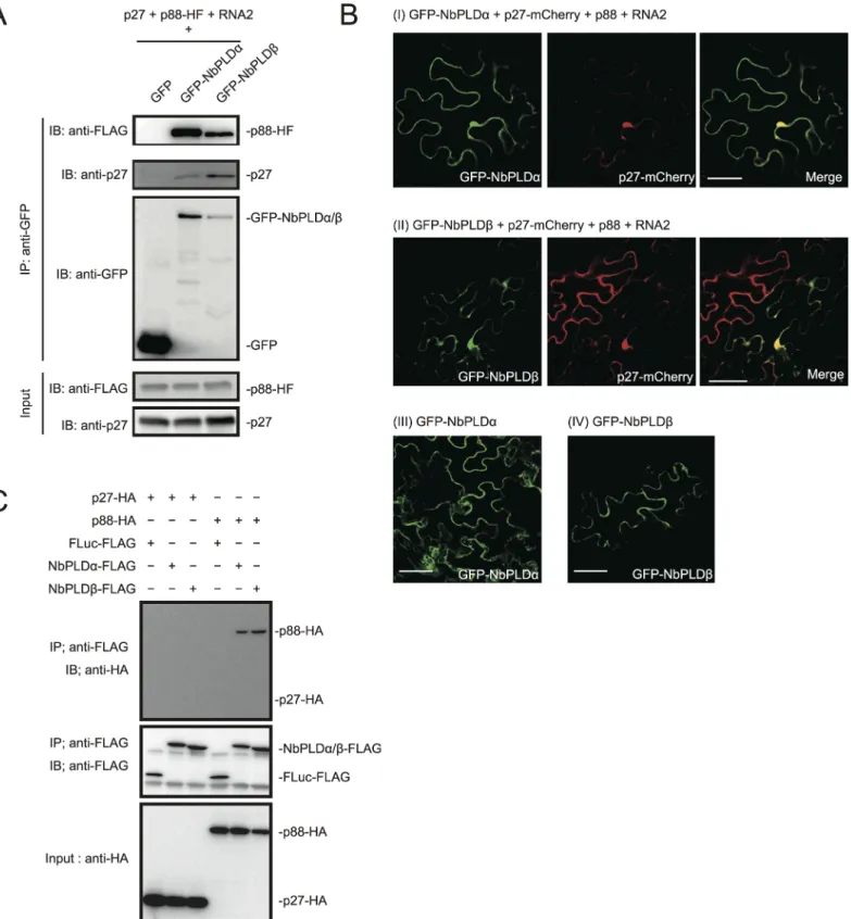
RCNMV replication proteins, we performed a co-immunoprecipitation (co-IP) assay in N. benthamiana plants using green fluorescent protein (GFP)-fused NbPLDα or NbPLDβ as bait proteins. We co-expressed p88pol-HF together with p27 and RNA2, because p88polcan be de-tected by immunoblot in N. benthamiana only when viral RNA replication takes place [12]. Both p27 and p88pol-HF were co-immunoprecipitated with GFP-NbPLDα or GFP-NbPLDβ, but not with GFP (Fig 2A), confirming the association of these PLDs with RCNMV replication proteins.
To investigate whether PLDs localize at VRCs, GFP-NbPLDα or GFP-NbPLDβ was co-ex-pressed with mCherry-fused p27 (as a marker of VRC), p88pol, and RNA2 via agroinfiltration
Fig 1. Identification of proteins copurified with RCNMV replication proteins. The solubilized total fractions prepared from Agrobacterium-infiltrated leaves expressing p27 plus p88 plus RNA2 or p27-His-FLAG (p27-HF) plus p88-His-FLAG (p88-HF) plus RNA2 were subjected to affinity purification with Ni-NTA agarose beads, followed by anti-FLAG affinity resins. The affinity-purified fractions were subjected to SDS-PAGE and stained using MS-compatible silver staining. Protein bands were excised, subjected to in-gel digestion, and analyzed by tandem mass spectrometry. Some of the proteins that were identified in LC/MS/MS analysis are listed inS1 Table.
Fig 2. RCNMV replication proteins interact with NbPLDα and NbPLDβ. (A) RCNMV replication proteins interacted with NbPLDα and NbPLDβ in a co-IP assay in N. benthamiana. Green fluorescent protein (GFP)-fused NbPLDα (GFP-NbPLDα) or GFP-NbPLDβ was coexpressed with p27, six-His/FLAG-tagged p88pol(p88pol-HF), RNA2, and p19 of RNA silencing suppressor of Tomato bushy stunt virus in N. benthamiana leaves. Total protein was extracted
and subjected to immunoprecipitation of GFP-fused proteins by anti-GFP antibody. Proteins were analyzed by immunoblot using antibodies as indicated. (B) NbPLDα and NbPLDβ were colocalized with p27 during viral replication. GFP-NbPLDα or GFP-NbPLDβ was coexpressed with C-terminally mCherry-fused p27 (p27-mCherry), p88pol, RNA2, and p19 (panel I and II) or coexpressed with empty vector and p19 (panel III and IV) in N. benthamiana leaves by
in N. benthamiana. At 4 dai, the fluorescence of GFP-NbPLDα and GFP-NbPLDβ was partially merged with the fluorescence of p27-mCherry in large aggregate structures (Fig 2Bpanels I and II), which are induced by p27 and are thought to be the site of RCNMV RNA replication [13,15,16]. The expression of GFP-NbPLDα or GFP-NbPLDβ alone did not induce such aggre-gated structures (Fig 2Bpanels III and IV), suggesting that both NbPLDα and NbPLDβ are
re-cruited to viral replication sites during RCNMV replication.
To investigate whether p27 or p88polinteract with NbPLDα and NbPLDβ, we performed a co-IP assay in cell lysates prepared from evacuolated tobacco BY-2 protoplasts (BYL) [38]. C-terminally HA-tagged viral replication protein (p27-HA or p88pol-HA) was coexpressed with C-terminally FLAG-tagged PLD (NbPLDα-FLAG or NbPLDβ-FLAG) from capped transcripts in BYL. In BYL, p88pol-HA was easily detected by immunoblot using anti-HA antibody in the absence of other viral components after 2 hour of in vitro translation (Fig 2C). After in vitro translation, the extracts were subjected to immunoprecipitation using FLAG-affinity resin. Im-munoblot analysis showed that p88pol-HA, but not p27-HA, was copurified with both
NbPLDα-FLAG and NbPLDβ-FLAG (Fig 2C). p88pol-HA was not copurified with C-terminal-ly FLAG-tagged firefC-terminal-ly luciferase (FLuc-FLAG) that was used as a negative control, excluding the possibility that p88pol-HA binds to FLAG-affinity resin nonspecifically. In reciprocal co-IP experiments using HA antibody, we also found copurification of NbPLDα-FLAG with p88pol -HA, but not with FLuc-HA or p27-HA (S3 Fig). However, as NbPLDβ-FLAG was co-purified
not only with p88pol-HA but also with FLuc-HA or p27-HA (S3 Fig), we could not confirm the specific interaction of p88polwith NbPLDβ in BYL.
Both PLDα and PLDβ are required for RCNMV infection
To test whether NbPLDα and NbPLDβ are required for infection of host plants with RCNMV, endogenous transcript levels of NbPLDα or NbPLDβ were downregulated using Tobacco rattle virus (TRV)-based virus-induced gene silencing (VIGS) in N. benthamiana plants. A TRV vec-tor harboring a partial fragment of NbPLDα (TRV:NbPLDα) or NbPLDβ (TRV:NbPLDβ) was expressed via Agrobacterium-mediated expression. An empty TRV vector (TRV:00) was ex-pressed as a control. Newly developed leaves were inoculated with RCNMV RNA1 and RNA2 at 18 dai. Two days after RCNMV inoculation, three inoculated leaves from three different plants were harvested and mixed, and total RNA was extracted. No morphological defects such as chlorotic and stunted phenotypes were observed at this stage (S4 Fig). Semiquantitative re-verse-transcription-PCR (RT-PCR) or quantitative RT-PCR (RT-qPCR) analyses confirmed the specific reduction of NbPLDα or NbPLDβ mRNAs in plants infiltrated with TRV:NbPLDα or TRV:NbPLDβ, respectively (Figs3andS5). Northern blot analyses using ribonucleotide probes specifically recognizing RCNMV RNA1 or RNA2 showed that the accumulation of RCNMV RNAs was dramatically reduced in both NbPLDα- and NbPLDβ-knockdown plants compared with control plants (Fig 3).
It is known that, in PLDβ1 knockdown transgenic rice plants, the generation of ROS and the expression of defense-related genes are induced even in the absence of pathogen infection [35]. Therefore, it is possible that the poor viral infection in NbPLDβ-knokedown plants was
due to activated defense responses. To address this possibility, we tested the effects of NbPLD α-or NbPLDβ-knockdown by TRV-mediated VIGS on the expression of defense-related genes by
Agrobacteriuminfiltration. Fluorescence was visualized by confocal microscopy at 4 days after infiltration. The merging of the green and red fluorescence is shown as a yellow color. Bars, 50μm. (C) p88polinteracted with NbPLDα and NbPLDβ in a co-IP assay in BYL. Appropriate combinations of capped transcripts were added to BYL. After in vitro translation at 25°C for 2 hours, the extract was solubilized and subjected to immunoprecipitation of FLAG-tagged proteins by anti-FLAG antibody. Proteins were analyzed by immunoblot using antibodies as indicated.
RT-qPCR analysis. The defense-related genes analyzed here were SA-signaling marker genes (PR-1, PR-2, and PR-5) [39,40], jasmonic acid (JA)-signaling marker genes (LOX1, PR-4, and PDF1.2.) [39,41], ROS-detoxification enzymes (APX, GST, and SOD) [39], MAMP-triggered immunity marker genes (CYP71D20 and ACRE132) [42], and mitogen-activated protein ki-nases (WIPK and SIPK) [42]. The expression levels of these defense-related genes were not sig-nificantly increased in NbPLDα- or NbPLDβ-knockdown plants compared with those in TRV control plants (S5 Fig), excluding the possibility that the reduced viral infection in NbPLDα- or
NbPLDβ-knockdown plants are due to activated defense responses in these plants. Some genes including PR-1, PR-2, and CYP71D20 genes were even repressed in NbPLDα- or NbPLDβ-knockdown plants (S5 Fig), consistent with the positive roles of PLDs and PLD-derived PA on plant defense signaling [24–30]. Altogether, these results suggest that both NbPLDα and
NbPLDβ play a positive role in RCNMV infection.
PA promotes viral RNA replication
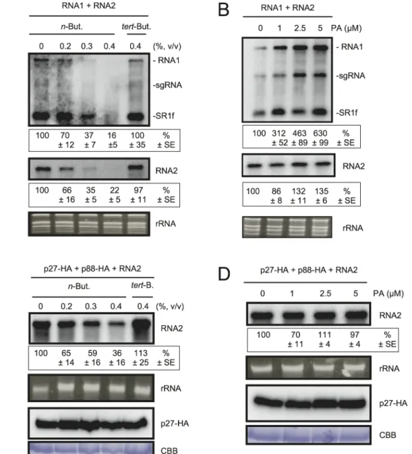
To test the possible contribution of PLD-derived PA to viral RNA replication, we exploited the transphosphatidylation activity of PLDs, which uses primary alcohols as substrates to form an artificial phosphatidyl alcohol. The preferential formation of this compound impairs PA pro-duction [43]. Thus, we tested the effect of n-butanol that inhibits PA production by PLDs on RCNMV RNA replication. N. benthamiana protoplasts were inoculated with RCNMV RNA1 and RNA2 and incubated with n-butanol or tert-butanol, an alcohol with no inhibitory effect on PA production, and viral RNA accumulation was determined by northern blot analysis. In-creasing n-butanol concentration caused a progressive reduction of viral RNA accumulation (Fig 4A). By contrast, viral RNA accumulation was only moderately reduced in protoplasts treated with tert-butanol compared with the water control. Note that n-butanol did not affect the accumulation of rRNA (Fig 4A). The inhibitory effect of n-butanol on RCNMV replication was also observed in tobacco BY-2 protoplasts (S6 Fig).
We also tested the effect of n-butanol and tert-butanol on defense-related gene expressions in N. benthamiana protoplasts. n-butanol did not increase the expression of defense-related genes (S7 Fig). This result corresponds well with the finding that n-butanol has no effects on the basal transcription of the PR-1 gene in Arabidopsis seedlings [27]. In contrast, tert-butanol treatment caused the induction of CYP71D20, ACRE132, and WIPK genes (S7 Fig). This may explain weak negative effect of tert-butanol on RCNMV replication (Figs4AandS6). Altogeth-er, these results suggest that PLD-derived PA plays a positive role in viral RNA replication.
To verify further the importance of PA in viral RNA replication, commercially available soy-derived PA was supplied to RCNMV-inoculated N. benthamiana protoplasts and viral
Fig 3. Knockdown ofNbPLDα and NbPLDβ mRNAs levels via gene silencing inhibits the accumulation of RCNMV RNAs inN. benthamiana. The tobacco rattle virus (TRV) vector harboring a partial fragment of N. benthamiana NbPLDα and NbPLDβ mRNAs (TRV:NbPLDα and TRV:NbPLDβ, respectively) was expressed in N. benthamiana via Agrobacterium infiltration. The empty TRV vector (TRV:00) was used as a control. The newly developed leaves were inoculated with RCNMV RNA1 and RNA2 at 18 days after agroinfiltration. Total RNA was extracted from the mixture of three independent inoculated leaves 2 days after virus inoculation. Accumulation of RCNMV RNA was analyzed by northern blotting. Ethidium bromide-stained ribosomal RNAs (rRNAs) are shown below the northern blots, as loading controls. The numbers below the images represent the relative accumulation levels (means± standard error) of viral RNAs (RNA1 and RNA2, respectively) using the Image Gauge program (Fuji Film), which were calculated based on three independent experiments. NbPLDα and NbPLDβ mRNA levels were assessed by RT-PCR using primers that allow the amplification of the region of coding regions that are not present in TRV vectors. RT-PCR results for the RbcS gene demonstrated that equal amounts of total RNA were used for the RT and showed equivalent efficiency of the RT reaction in the samples. sgRNA, subgenomic RNA; SR1f, a small RNA fragment that derived from non-coding region of RNA1 [47].
RNA accumulation was determined by northern blot analysis. Exogenously added PA en-hanced the accumulation of RNA1 in a dose-dependent manner (up to 6-fold increase by 5μM
Fig 4. The effects ofn-butanol and exogenously supplied PA on RCNMV RNA replication in N. benthamiana protoplasts. (A) An inhibitor of PLDs impairs RCNMV RNA replication in a single cell. N. benthamiana protoplasts were inoculated with in vitro transcribed RNA1 and RNA2. The inoculated protoplasts were incubated at 20°C for 18 hours in the presence of n-butanol. (B) An exogenously supplied phosphatidic acid (PA) enhances RCNMV RNA replication. N. benthamiana protoplasts were inoculated with in vitro transcribed RNA1 and RNA2. The inoculated protoplasts were incubated at 20°C for 18 hours in the presence of progressively increasing concentrations of PA. (C) An inhibitor of PLDs impairs replication of RNA2 in a single cell. N. benthamiana protoplasts were inoculated with in vitro transcribed RNA2 together with plasmids expressing p27-HA and p88-HA. The inoculated protoplasts were incubated at 20°C for 18 hours in the presence of n-butanol. (D) The effect of an exogenously supplied PA on the replication of RNA2. N. benthamiana protoplasts were inoculated with in vitro transcribed RNA2 together with plasmids expressing p27-HA and p88-HA. The inoculated protoplasts were incubated at 20°C for 18 hours in the presence of progressively increasing concentrations of PA. Total RNA was analyzed by northern blotting using ribonucleotide probes that recognize specifically RCNMV RNA1 and RNA2, respectively. Ethidium bromide-stained rRNAs were used as loading controls and are shown below the northern blots. The numbers below the images represent the relative accumulation levels (means± standard error) of viral RNAs (RNA1 and RNA2, respectively) using the Image Gauge program, which were calculated based on three independent experiments. In (C) and (D), total protein was analyzed by Immunoblot using anti-HA antibody. Coomassie Brilliant Blue (CBB) staining served as a loading control. sgRNA, subgenomic RNA; SR1f, a small RNA fragment that derived from non-coding region of RNA1 [47].
doi:10.1371/journal.ppat.1004909.g004
PA), whereas the effect of PA on the accumulation of RNA2 was negligible (Fig 4B). Neither exogenously supplied PC nor PE significantly affected the accumulation of viral RNA in N. benthamiana protoplasts (S8 Fig). These results suggest that PA plays an important role in RCNMV RNA replication, and that the requirement for PA may differ between RNA1 and RNA2.
Replication of RNA2 depends entirely on RNA1 simply because RNA2 uses replication pro-teins supplied from RNA1. The negative impact of n-butanol on the accumulation of RNA2 (Fig 4A) may be the indirect action of this primary alcohol through inhibition of RNA1 replica-tion. Therefore, it is possible that RNA2 replication does not require any PLD-derived PA. To investigate this possibility, we inoculated N. benthamiana protoplasts with RNA2 and plasmids expressing p27 and p88pol, as suppliers of the replication proteins. The accumulation of RNA2 was decreased by n-butanol in a dose-dependent manner (Fig 4C). Note that in this experi-ment, the accumulation of p27 replication protein was not significantly changed (Fig 4C). These results indicate that PLD-derived PA was also required for the replication of RNA2 as in the case of RNA1. However, the replication of RNA2 was not enhanced by exogenously sup-plied PA (Fig 4D), suggesting that the threshold of PA requirement for RNA2 replication is lower than that for RNA1. The differential requirement for PA in the replication of RNA1 and RNA2 is discussed later.
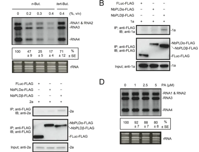
Next, we investigated the effect of n-butanol on RNA replication of Brome mosaic virus (BMV), another plant (+)RNA virus, which is unrelated to RCNMV. Increasing n-butanol con-centration caused progressive reduction in the accumulation of BMV RNA (Fig 5A). Co-IP ex-periments showed interactions between BMV replication proteins and NbPLDβ-FLAG (Fig 5B and 5C). These results suggest that PLD-derived PA is also important for BMV RNA replica-tion. However, exogenously added PA did not affect the accumulation of BMV RNA (Fig 5D) as similarly seen for RCNMV RNA2.
PA stimulates VRC activity in vitro
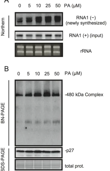
PA acts as a second messenger in signal transduction during multiple biotic and abiotic stress responses and plays multiple roles including that for transcriptional reprogramming [20–22]. To investigate whether PA has a direct role in viral RNA replication, we took advantage of BYL, a nucleus-depleted (therefore, the effects of PA on transcriptional reprogramming are negligible) in vitro translation/replication system that has been used successfully to recapitulate the negative-strand RNA synthesis of RCNMV [12,44–49]. Addition of PA into BYL stimulat-ed the accumulation of newly synthesizstimulat-ed negative-strand RNA (Fig 6A), and moderately en-hanced the accumulation of the 480-kDa replicase complex (Fig 6B), suggesting that PA stimulates the activity and/or assembly of the viral replicase complex in a direct manner. The stimulation effects of PA on the accumulation of the 480-kDa replicase complex was less obvi-ous than that on the accumulation of newly-synthesized viral RNA at the highest concentration of PA used in this experiment (50μM). This result may also support a direct role for PA in the enhancement of RdRP activity rather than the formation of the replicase complex.
p27 replication protein binds to PA in vitro
Because PA directly stimulated the viral negative-strand RNA synthesis in BYL, we hypothe-sized that p27, the multifunctional RCNMV replication protein, has an affinity for PA. To in-vestigate this possibility, we conducted a lipid overlay assay using bacterially expressed, purified C-terminally FLAG-tagged p27 (p27-FLAG) [49]. Purified p27-FLAG protein was in-cubated with phospholipid-spotted nitrocellulose membranes, and the interaction between p27-FLAG and phospholipids was detected using anti-FLAG antibody. p27 gave strong PA
binding signals on the blot (Fig 7A and 7B). p27 also exhibited weak binding to phosphatidyli-nositol-4-phosphate (PI4P), phosphatidylinositol (3,5)-bisphosphate, phosphatidylinositol (4,5)-bisphosphate, and phosphatidylinositol (3,4,5)-trisphosphate, but negligible binding to other lipids, including PC and PE (Fig 7A and 7B). These results were consistent with the find-ings that neither exogenously supplied PC nor PE promoted RCNMV replication in N. benthamiana protoplasts (S8 Fig). N- or C-terminal halves of p27 fragments did not give PA binding signals (Fig 7C and 7D), suggesting that overall protein conformation may be impor-tant for p27–PA binding.
Fig 5. BMV RNA replication requires PLD-derived PA inN. benthamiana protoplasts. (A) An inhibitor of PLDs impairs BMV RNA replication in a single cell. N. benthamiana protoplasts were inoculated with in vitro transcribed BMV RNA1, RNA2, and RNA3. The inoculated protoplasts were incubated at 20°C for 18 hours in the presence of n-butanol. (B)-(C) Brome mosaic virus replication proteins 1a (B) and 2apol(C) interact with NbPLDβ. Appropriate
combinations of capped transcripts were added to BYL. After in vitro translation at 25°C for 2 h, the extract was solubilized and mixed with a 10μl bed volume of anti-FLAG M2 antibody agarose and incubated further at 4°C for 1 hours. After washing, proteins copurified with FLAG-tagged proteins were analyzed by immunoblotting with appropriate antibodies. (D) The effect of an exogenously supplied PA on the replication of BMV. N. benthamiana protoplasts were inoculated with BMV RNA1, RNA2, and RNA3 transcribed in vitro. The inoculated protoplasts were incubated at 20°C for 18 hours in the presence of progressively increasing concentrations of PA. Total RNA was analyzed by northern blotting using ribonucleotide probes that specifically recognize the 3 ’-UTR of BMV RNAs. Ethidium bromide-stained rRNA was used as a loading control and is shown below the northern blots. The numbers below the images represent the relative accumulation levels (means± standard error) of viral RNAs (RNA1 + RNA2 + RNA3 + RNA4) using the Image Gauge program, which were calculated based on three independent experiments.
doi:10.1371/journal.ppat.1004909.g005
RCNMV promotes PA accumulation in planta
Next we investigated whether RCNMV infection affects the amount of PA in plant leaves. N. benthamiana leaves were inoculated with RCNMV via agroinfiltration. At 2 dai, lipids were ex-tracted and the amount of PA was analyzed by thin layer chromatography (TLC). Compared with control plant leaves infiltrated with Agrobacterium harboring an empty vector, the signal intensity of a lipid spot that showed a migration similar to that of soy-PA was increased in RCNMV-infected plant leaves (about 3-fold higher) (Fig 8A and 8B). To identify the lipid spe-cies of the spot, the same samples were again subjected to TLC, and the spot was scratched out from Coomassie Brilliant Blue R-250 stained TLC plates and subjected to LC/MS analysis. The lipid was identified as PA (Fig 8C–8E). We concluded that RCNMV infection upregulated PA accumulation in plants. It is known that expressions of PLD genes were induced by pathogen infection [50]. Therefore, the enhanced accumulation of PA in RCNMV-infected plants could
Fig 6. PA enhances accumulation of negative-strand RNA1 and the 480-kDa replicase complex in BYL. BYL was incubated for 4 h at 17°C with in vitro-transcribed RNA1 (300 ng) in the presence of various concentrations of PA as indicated. Accumulation of negative-strand RNA1 was detected by northern blotting (A). Total protein solubilized from BYL was subjected to blue native (BN)- and SDS-PAGE analyses, followed by immunoblotting with anti-p27 antisera (B). EtBr-stained rRNA and Coomassie Brilliant Blue (CBB)-stained cellular proteins are shown as loading controls.
be due to elevated accumulation of PLD through induction of PLD gene expression. To exam-ine this possibility, we investigated whether mRNA levels of NbPLDα and NbPLDβ were upre-gulated in RCNMV-infected plants by RT-qPCR analysis. The accumulation levels of NbPLDα and NbPLDβ transcripts in RCNMV-infected plants were about 1.2- and 1.9-fold higher, re-spectively, than those in control plants, although the increase in NbPLDα transcripts was insig-nificant by the Student’s t-test (S9 Fig).
Fig 7. p27 interacts with PAin vitro. p27-FLAG or its variant proteins expressed in E. coli BL21 (DE3) were purified by Ni-NTA agarose beads. Lipid overlay analysis of various lipids with full-length p27-FLAG (A)-(B), N-terminal half of p27-FLAG (p27N-FLAG) (C), or C-N-terminal half of p27-FLAG (p27C-FLAG) (D). Purified protein (500 ng) was used, followed by immunoblotting with anti-FLAG antibody. LysoPA, lysophosphatidic acid; LysoPC, lysophosphochorine; PI, phosphatidylinositol; PI(3)P, phosphatidylinositol 3-phosphate; PI(4)P, phosphatidylinositol 4-phosphate; PI(5)P, phosphatidylinositol 5-phosphate; PE, phosphatidylethanolamine; PC, phosphatidylcholine; S1P, sphingosine-1-phosphate; PI(3,4)P2, phosphatidylinositol (3,4)-bisphosphate; PI(3,5)P2, phosphatidylinositol (3,5)-bisphosphate; PI(4,5)P2, phosphatidylinositol (4,5)-bisphosphate; PI (3,4,5)P3, phosphatidylinositol (3,4,5)-trisphosphate; PA, phosphatidic acid; PS, phosphatidylserine; DAG, diacylglycerol; PG, phosphatidylglycerol; SM, sphingomyelin.
doi:10.1371/journal.ppat.1004909.g007
NbPLD
α- or NbPLDβ-knockdown plants exhibit reduced PA
accumulation
We compared the accumulation of endogenous PA in NbPLDα or NbPLDβ knockdown plants with that in TRV control plants by TLC analysis. As expected from their predicted function, NbPLDα- or NbPLDβ-knockdown plants showed reduced accumulation of PA compared with TRV control plants (S10 Fig), suggesting that these PLDs contribute to PA production in N. benthamiana. These results further supported the idea that RCNMV-induced PLDs-derived PA plays an important role in RCNMV replication.
Discussion
A growing number of studies have suggested that PLD and PLD-derived PA play vital roles in environmental responses in plants [19–22,50]. The properties of PA (i.e., normally present in small amounts, and rapidly and transiently accumulates in response to various environmental cues) seem to be suitable for its function in biotic and abiotic responses in which plants need to rapidly accommodate their surrounding environments. Indeed, PA has been shown to accu-mulate in response to several MAMPs and pathogen effector proteins, or SA that is a key hor-mone involved in plant resistance against biotrophic pathogens [23–29]. PA induces PR gene expression and cell death [28,33,34], and has been proposed to act as an important component in resistance to biotrophic pathogens such as tobacco mosaic virus and Phytophthora parasitica. Plants have diverse numbers of PLD isoforms and they appear to have distinct but somewhat overlapping functions in cellular responses [50]. In Arabidopsis, it is proposed that multiple PLD isoforms cooperatively contribute to AvrRpm1-triggered resistance [30]. In rice, PLDβ1
acts like a negative regulator of defense signaling because PLDβ1-knockdown rice plants exhib-it constexhib-itutive ROS production, expression of PR genes, and enhanced resistance against patho-gens [35]. In this study, we showed that PLD and PA are essential for and play a key role in RCNMV replication. Poor infection of RCNMV to NbPLDα- or NbPLDβ-knockdown plants was not due to constitutively activated defense responses, indicating that both NbPLDα and NbPLDβ act as essential host factors in RCNMV replication. The findings reveal novel aspects of PLD and PA in their roles during biotic stress responses in plants.
The replication of Tomato bushy stunt virus is enhanced by deletion of the PAH1 gene en-coding a PA phosphatase, which converts PA into DAG in yeast [51]. Moreover, ectopic ex-pression of Arabidopsis PA phosphatase, Pah2 in N. benthamiana results in the inhibition of tombusviruses and RCNMV infection [51]. These findings suggest that PA is positively in-volved in the life cycles of these viruses. However, whether viruses manipulate PA production for viral replication has been unknown. In the current study, we showed that RCNMV replica-tion proteins interacted with PA-producing enzymes, NbPLDα and NbPLDβ. RCNMV infec-tion induced a high accumulainfec-tion of PA in plant tissues, suggesting that RCNMV alters cellular lipid metabolism to establish a suitable environment for viral replication. It is known
Fig 8. RCNMV stimulates the accumulation of PA in a plant tissue. (A) The extracted lipids from Agrobacterium-infiltrated leaves expressing empty vector or RCNMV RNA1 plus RNA2 were subjected to thin layer chromatography (TLC) and phospholipids were visualized by CuSO4staining. The same
samples were again subjected to TLC, and the spot was scratched out from Coomassie Brilliant Blue R-250 (CBB) stained TLC plates and subjected to LC/ MS analysis. (B) Relative PA accumulation was quantified using ImageJ software. Bars represent means and standard error of values obtained from three independent biological samples. (C) Total ion and major extracted ion chromatograms of the TLC separated bands. The broken lines indicate the extracts from empty vector-expressed leaves, and the solid lines indicate the ones from the leaves expressing RCNMV RNA1 plus RNA2. (D)-(E), MS2spectra produced by CID of m/z 669.4 (D) and m/z 691.4 (E). The signal at m/z 153.0, 255.2, and 277.2 indicate the dehydrated glycerol phosphate, carboxylate anion of C16:0and C18:3, respectively. Ions of m/z 391.2 and 413.2 arose from the neutral loss of C18:3,and m/z 409.2 and 431.2 arose from the C18:3neutral
loss as ketene from the each precursor ion. The other ions, m/z 671.5, 693.4, 695.5, and 697.5 also produced the similar MS2spectra as described above, and were confirmed as phosphatidic acid.
doi:10.1371/journal.ppat.1004909.g008
that transcription of PLD genes is induced by pathogen infection [50]. RCNMV infection in-creased the accumulation of NbPLDα and NbPLDβ transcripts (S9 Fig). Currently, whether the enhancement of PA accumulation observed in RCNMV-infected plants is due to the upregula-tion of PLD gene expression remains unknown. PLDs may be activated through direct or indi-rect interaction with RCNMV replication proteins. Although we failed to show the interaction between p88poland NbPLDα or NbPLDβ in vivo because p88polaccumulates below detection limits in the absence of viral RNA replication in N. benthamiana [12], p88polinteracted with PLDs, at least with NbPLDα in a co-IP assay in BYL. The interaction of p88polwith PLDs
seems to make sense for viral replication strategy because it brings them to the sites of replica-tion. This strategy could increase PA only at VRCs and not at other cellular membranes where PA might affect cellular metabolism or activate the SA-mediated defense responses that is det-rimental for successful viral infection. This is critical for the viral life cycle because PA interacts with p27 auxiliary replication protein (Fig 7) and enhances viral replication through upregulat-ing the activity and/or assembly of the 480-kDa replicase complex (Fig 6). To our knowledge, this is the first demonstration of a functional role for PA in (+)RNA virus replication. It is likely that PA is involved in RNA replication of many (+)RNA viruses. Indeed, the replication pro-teins, 1a and 2apolof BMV, a virus unrelated to RCNMV, also interacted with NbPLDβ, and BMV RNA replication was sensitive to n-butanol, which is an inhibitor of PLDs-derived PA production (Fig 5). However, exogenously added PA did not affect the accumulation of BMV RNA (Fig 5), implying that the threshold of PA requirement for BMV RNA replication seems to be lower than that for RCNMV. The differential PA requirement may be explained by very weak affinity of BMV replication proteins with NbPLDα, which is more active in PA produc-tion than NbPLDβ (S10 Fig). Moreover, Dengue virus induces the accumulation of several lip-ids in infected mosquito cells, including PA [52]. It has been reported that Coxsackievirus B3 and mouse hepatitis coronavirus replication is insensitive to n-butanol [53,54]. However, be-cause PA can also be formed through the combined action of phospholipase C and DAG kinase [55], it is unknown whether replication of these viruses depends on PA or not.
How does PA affect viral RNA replication? PA binding to proteins modulates the catalytic activity of target proteins, tethers proteins to the membranes, and promotes the formation and/or stability of protein complexes [55]. We found that exogenous PA enhanced the accu-mulation of newly synthesized viral RNA and the formation of 480-kDa replicase complexes in BYL in vitro translation/replication systems, and that p27 had affinity for PA in vitro (Figs6
and7). Therefore, PA could promote viral replicase activity and/or assembly directly. The 480-kDa replicase complexes contain p27 oligomer. Therefore, it is possible that PA-binding to p27 in the replicase complexes assists the conformational change of p27 that is suitable for RNA synthesis. Alternatively, PA could serve as an assembly platform for host PA-binding proteins. PA binds to various proteins, including transcription factors, kinases, phosphatases, enzymes involved in central metabolism, and proteins involved in vesicular trafficking and cy-toskeletal rearrangements [20,21]. Several known cellular PA-binding proteins were also iden-tified in our LC/MS/MS analysis (S1 Table). These include NADPH oxidase [56],
glyceraldehyde-3-phosphate dehydrogenase (GAPDH) [57,58], and SNF1-related kinase [58]. RCNMV-induced accumulation of high PA levels may facilitate the recruitment of these PA-binding proteins to viral replication sites. One of the candidate proteins is GAPDH-A. Al-though GAPDH-A is not required for RCNMV replication, it recruits RCNMV movement protein to viral replication sites and plays an important role in virus cell-to-cell movement [59]. Therefore, PA may also function in bridging viral replication and cell-to-cell movement. RCNMV induces large ER aggregates in infected cells, which are thought to be viral RNA repli-cation factories [13–16]. PA may play a direct role in the formation of RNA replication facto-ries. In support of this hypothesis, deletion of pah1 in yeast causes the expanded ER
membranes and leads to enhanced TBSV replication on these membranes, although TBSV rep-licates normally at peroxisomes [51]. In addition, it is also possible that PA might affect un-known cellular factors that are involved in viral RNA replication. Further studies are needed to elucidate the molecular functions of PA in viral RNA replication.
Our results suggest that RNA1 and RNA2 have differential PA requirements in RNA repli-cation. How does this difference contribute to RCNMV infection? RNA1 requires a larger amount of PA for maximum efficiency of its replication than that required by RNA2. This may reflect differences in the translation and replication mechanisms between RNA1 and RNA2. Translation of MP from RNA2 couples with RNA replication: only progeny RNA2 generated de novo through the RNA replication pathway could function as mRNA [60]. Poor enhance-ment of RNA2 replication by PA may be beneficial for switching from replication to transla-tion. By contrast, because RNA1 has a cap-independent translation enhancer that is an effective recruiter of translation factors [61], newly synthesized RNA1 serves as a template for further translation of the replication proteins that would activate PA production in infected cells. Moreover, RNA1 also serves as a template for transcription of subgenomic RNA, which encodes CP. Therefore, PA-mediated enhancement of RNA1 replication may be suitable for the production of CP subgenomic RNA during the late stage of viral infection. High PA re-quirement for maximum RNA1 replication may explain the use of both NbPLDα and NbPLDβ as essential host factors.
A growing number of studies have suggested that multiple lipid species affect virus replica-tion and that (+)RNA viruses employ a multifaceted strategy to rewire host machinery involved in lipid transport and synthesis [62]. HCV infection stimulates the production of cellular PC, PE, and PI4P and HCV co-opts PI4KIIIα for replication [63,64]. In addition, HCV infection stimulates the accumulation of cellular sphingomyelin, which binds to and activates HCV NS5B polymerase [65,66]. Enterovirus utilizes PI4KIIIβ for RNA replication and viral RdRP 3Dpolbinds to PI4P [67]. Enteroviruses also upregulate cellular uptake of fatty acids, which are channeled toward highly upregulated PC synthesis in infected cells [68]. The elevated PC has been proposed to serve as a building block for the formations of the viral replication factory [68,69]. Recently, it has been shown that PE and PC stimulate TBSV RdRP activity in vitro [70]. RCNMV p27 showed affinity, not only for PA but also for other phospholipids such as PI4P in vitro. However, our LC/MS/MS analysis failed to detect any PI4P-producing enzymes, such as PI4K. Combined lipidomics, proteomics, and transcriptome analysis will be helpful for a comprehensive understanding of lipid species involved in viral RNA replication.
Materials and Methods
Gene cloning and plasmid construction
Plasmids given the prefix‘‘pBIC” were used for Agrobacterium infiltration, ‘‘pUC”, ‘‘pRC” and ‘‘pR” were used for in vitro transcription, ‘‘pCold” was used for protein expression in Escheri-chia coli. pUCR1 [71] and pRC2_G [72] are full-length cDNA clones of RNA1 and RNA2 of the RCNMV Australian strain, respectively. pB1TP3, pB2TP5, and pB3TP8 are full-length cDNA clones of RNA1, RNA2, and RNA3 of the BMV M1 strain, respectively [73] (generous gift from Paul Ahlquist). The constructs described previously used in this study include pBICp27 [71], pBICp88 [71], pBICR2 [71], pBICR1R2 [71], pBICp19 [71], pCOLDIp27-FLAG [49], pCOLDIp27N-FLAG [49], and pCOLDIp27C-FLAG [49]. pUC118 was purchased from Takara Bio Inc. (Shiga, Japan). Escherichia coli DH5α was used for the construction of all plas-mids. All plasmids constructed in this study were verified by sequencing.
RNA extraction from Nicotiana benthamiana leaves was performed using an RNeasy Plant Mini Kit (Qiagen, Hilden, Germany). Reverse-transcription-PCR (RT-PCR) was catalyzed by
Superscript III reverse transcriptase (Invitrogen) using oligo (dT) [16]. Primers to amplify cod-ing sequences of NbPLDα or NbPLDβ were designed based on the N. benthamiana RNA seq data (Transcriptome version 5:http://sydney.edu.au/science/molecular_bioscience/sites/ benthamiana/) [74].
pBICp27-His-FLAG. A PCR fragment from pBICp27 was amplified using primers 5'-CGGGGATCCATGGGTTTTATAAATCTTTCG-3' and 5'-CGGGGATCCCTACTTGTCA TCGTCGTCCTTGTAATCATGATGATGATGATGATGAAAATCCTCAAGGGATTTG-3'. The amplified fragment was digested with BamHI and inserted into the corresponding region of pBICP35.
pBICp88-His-FLAG. A PCR fragment from pBICp88 was amplified using primers 5'-CCGGGTACCATGGGTTTTATAAATCTTTCG-3' and 5'- CGGGGTACCTTACTTGTCAT CGTCGTCCTTGTAATCATGATGATGATGATGATGTCGGGCTTTGATTAGATCTTT G-3'. The amplified fragment was digested with KpnI and inserted into the corresponding re-gion of pBICP35.
pBYLp27-HA. A PCR fragment from pUBp27-HA [16] was amplified using primers 5'-CTGGCGCGCCATGGGTTTTATAAATCTTTCGCTTTTTGATGTGG-3' and 5'-CTGGC GCGCCTTAAGCGTAATCTGGAACATCGTATGGGTA-3'. The amplified fragment was di-gested with AscI and inserted into the corresponding region of pBYL2 [12].
pBYLp88-HA. A PCR fragment from pUBp88-HA [16] was amplified using primers 5'-CTGGCGCGCCATGGGTTTTATAAATCTTTCGCTTTTTGATGTGG-3' and 5'-CTGGC GCGCCTTAAGCGTAATCTGGAACATCGTATGGGTA-3'. The amplified fragment was di-gested with AscI and inserted into the corresponding region of pBYL2.
pBYLNbPLDα-FLAG. A PCR fragment from cDNA derived from N. benthamiana was amplified using primers 5'-CTGGCGCGCCATGGCTCAGATTCTGCTTCATGGAACTC TCC-3' and 5'-CTGGCGCGCCCTACTTGTCATCGTCGTCCTTGTAATCTGTAGTGAGG ATAGGAGGAAGG-3'. The amplified fragment was digested with AscI and inserted into the corresponding region of pBYL2.
pBYLSP6NbPLDβ-FLAG. A PCR fragment from cDNA derived from N. benthamiana was amplified using primers 5'-CTGGCGCGCCATGGCTCATTTGTCTTATTCTGATTC TATTAG-3' and 5'-CTGGCGCGCCTCACTTGTCATCGTCGTCCTTGTAATCGATATC TGGAAAGGTTTCACAGCCAGGGAG-3'. The amplified fragment was digested with AscI and inserted into the corresponding region of pBYL2 to generate pBYLNbPLDβ-FLAG. A PCR fragment from pBYLNbPLDβ-FLAG was amplified using primers 5'-GAGGATCCCCGATTT AGGTGACACTATAGAGACCCAAGCTGGCGCGCCATGGCTCATTTGT-3' and 5'-AGT CCCGGGCACACCCTTATACTCGTGATCTCCTCC-3'. The amplified fragment was di-gested with BamHI and XmaI, and inserted into the corresponding region of
pBYLNbPLDβ-FLAG.
pBYLAtPAH1-HA. A PCR fragment from cDNA derived from A. thaliana was amplified using primers 5'-CTGGCGCGCCATGAGTTTGGTTGGAAGAGTTGGGAGTTTGATTT-3' and 5'-GCTGGCGCGCCTCAAGCGTAATCTGGAACATCGTATGGGTATTCAACCTC TTCTATTGGCAGTTTCC-3'. The amplified fragment was digested with AscI and inserted into the corresponding region of pBYL2.
pBYLFLuc-HA. A PCR fragment from pLUCpA60 [75] was amplified using primers 5'-C TGGCGCGCCATGGAAGACGCCAAAAACATAAAGAAAGGCCC-3' and 5'-CTGGCGC GCCTTAAGCGTAATCTGGAACATCGTATGGGTACACGGCGATCTTTCCGCCCTTC TTGGCC-3'. The amplified fragment was digested with AscI and inserted into the correspond-ing region of pBYL2.
pBYLFLuc-FLAG. A PCR fragment from pLUCpA60 [75] was amplified using primers 5'-CTGGCGCGCCATGGAAGACGCCAAAAACATAAAGAAAGGCCC-3' and 5'-CTGGC
GCGCCTTACTTGTCATCGTCGTCCTTGTAATCCACGGCGATCTTTCCGCCCTTC TTGGCC-3'. The amplified fragment was digested with AscI and inserted into the correspond-ing region of pBYL2.
pBICp27-mCherry. A PCR fragment from pUBp27-mCherry [15] was amplified using primers AGGATCCGGATGGGTTTTATAAATCTTTCGCTTTTTGATGTGG-3' and 5'-GGGGTACCCTACTTGTACAGCTCGTCCATGCCGCCGGTGG-3'. The amplified fragment was digested with BamHI and KpnI, and inserted into the corresponding region of pBICP35 [76].
pBICsGFP-NbPLDα. A PCR fragment from pBYLNbPLDα-FLAG was amplified using primers CTGGCGCGCCATGGCTCAGATTCTGCTTCATGGAACTCTCC-3' and 5'-CTGGCGCGCCCTATGTAGTGAGGATAGGAGGAAGGTAGTCAG-3'. The amplified frag-ment was digested with AscI and inserted into the corresponding region of pBICsGFP [13].
pBICsGFP-NbPLDβ. A PCR fragment from pBYLNbPLDα-FLAG was amplified using primers CTGGCGCGCCATGGCTCATTTGTCTTATTCTGATTCTATTAG-3' and 5'-CTGGCGCGCCTCAGATATCTGGAAAGGTTTCACAGCCAGGGAG-3'. The amplified fragment was digested with AscI and inserted into the corresponding region of pBICsGFP.
pTVNbPLDα. A PLDα cDNA fragment was amplified from cDNA derived from N. benthamiana (NbPLDα) RNA using primers 5'-ATCCCCCGGGAGTTCCTTGTAGCTCTG TGACATCCCC-3' and 5'-GCAGCCCGGGTCAGCCAACATAAACCAGAGATCGATGG ACGG-3'. The generated PCR product was then cloned in the antisense orientation into the XmaI site of pTV00 [77].
pTVNbPLDβ. A PLDβ cDNA fragment was amplified from cDNA derived from N. benthamiana (NbPLDβ) RNA using primers 5'-CCCCCGGGTCGACTTTTCTGACTGAGTT CCTGAGGTGTAGCAGG-3' and 5'-AGCCCGGGTTGATACCAATGGAGATTGCTCT AAAAATTGCC-3'. The generated PCR product was then cloned in the antisense orientation into the XmaI site of pTV00.
Plant growth conditions
N. benthamiana plants were grown on commercial soil (Tsuchi-Taro, Sumirin-Nosan-Kogyo Co. Ltd.) at 25 ± 2°C and 16 h illumination per day.
RNA preparation
RCNMV RNA1 and RNA2 were transcribed from SmaI-linearized pUCR1 and pRC2_G, re-spectively, using T7 RNA polymerase (TaKaRa Bio, Inc). BMV RNAs were transcribed from EcoRI-linearized pB plasmids using T7 RNA polymerase and capped with a ScriptCapm7G capping system (Epicentre Biotechnology). Capped mRNAs were transcribed from NotI-line-arized pBYL plasmids using T7 or SP6 RNA polymerase (TaKaRa Bio, Inc) and capped with a ScriptCapm7G capping system (Epicentre Biotechnology). All transcripts were purified with a Sephadex G-50 fine column (Amersham Pharmacia Biotech). RNA concentration was deter-mined spectrophotometrically, and its integrity was verified by agarose gel electrophoresis.
Tandem affinity purification
Four-week-old N. benthamiana plants were agroinfiltrated as described previously [15]. At 2 days postinfiltration (dpi), total proteins were extracted from 6 g of leaves in 10 ml of buffer A (50 mM HEPES, 150 mM NaCl, 0.1% 2-mercaptoethanol, 0.5% Triton X-100, 5% glycerol, pH 7.5) containing 30 mM imidazole and a cocktail of protease inhibitors (Roche). Following the removal of cell debris by filtering the mixture through cheesecloth, the extract was centrifuged at 21,000g at 4°C for 10 min and the supernatant was mixed with Ni-NTA beads (400μl)
(Qiagen, Hilden, Germany) and incubated for 1 h at 4°C with gentle mixing. The beads were washed three times with 1 ml of buffer A containing 100 mM imidazole. The bound proteins were eluted with 1 ml of buffer A containing 500 mM imidazole. The eluted proteins were mixed with 50μl of ANTI-FLAG M2-Agarose Affinity Gel (Sigma-Aldrich) and incubated for overnight at 4°C with gentle mixing. The beads were washed 3 times with 1 ml of buffer A. The bound proteins were eluted with 300μl of buffer A containing 150 μg/ml 3 × FLAG peptides (Sigma-Aldrich). The eluted proteins were concentrated by acetone precipitation and dissolved in 1 × NuPAGE sample buffer (Invitrogen). The purified proteins were separated by sodium dodecyl sulfate (SDS)-PAGE (NuPAGE 3%–12% bis-Tris gel: Invitrogen) and visualized by sil-ver staining (Wako, Osaka, Japan). Proteins in excised gel pieces were subjected to digestion with trypsin, LC–MS/MS analysis, and MASCOT searching as described previously [12].
Silencing of PLD
α and PLDß in N. benthamiana
Appropriate combinations of silencing vectors were expressed via Agrobacterium infiltration in 3- to 4-week-old N. benthamiana plants as described previously [16]. At 18 dpi, the leaves lo-cated above the infiltrated leaves were inoculated with in vitro transcribed RNA1 and RNA2 (500 ng each). At 2 days after inoculation, three inoculated leaves from three different plants infected with the same inoculum were pooled, and total RNA was extracted using RNA extrac-tion reagent (Invitrogen), treated with RQ1 RNase-free DNase (Promega, Madison, WI), puri-fied by phenol–chloroform and chloroform extractions, and precipitated with ethanol. Viral RNAs were detected by northern blotting, as described previously [16]. The mRNA levels of NbPLDα and NbPLDß were examined by RT-PCR using primer pairs 50-TATCAAGGTAG AGGAGATAGGTGC-30and 50-TACATCATCTCCATCGTTCTCCTC-30, and 50-GAAGG CTTCAAAGCGCCATG-30and 50-CTTAGGCAAGGGACATCAGC-30, respectively. As a control to show the equal amounts of cDNA templates in each reaction mixture, the ribulose 1,5-biphosphate carboxylase small subunit gene (RbcS), a gene that is constitutively expressed, was amplified by RT-PCR as described previously [16].
Protoplasts experiments
N. benthamiana protoplasts were prepared according to Kaido et al. (2014) [59]. N. benthami-ana protoplasts were inoculated with RCNMV RNA1 (1.5μg) and RNA2 (0.5 μg) and incubat-ed with various concentrations of n-butanol (Sigma-Aldrich), tert-butanol (Sigma-Aldrich), phosphatidic acid (PA) (Soy-derived; Avanti Polar Lipid), phosphatidyl choline (PC) (Soy-de-rived; Avanti Polar Lipid) or phosphatidyl ethanolamine (PE) (Soy-de(Soy-de-rived; Avanti Polar Lipid) at 20°C for 18 h. Phospholipids were dissolved in dimethylsulfoxide. Total RNA was ex-tracted and subjected to northern blotting, as described previously [15]. Each experiment was repeated at least three times using different batches of protoplasts.
BYL experiments
The preparation of BYL was as described previously [38,46]. The BYL translation/replication assay was performed essentially as described previously [46]. Briefly, 300 ng of RNA1 was added to 30μL of BYL translation/replication mixture in the presence of various concentra-tions of PA. The mixture was incubated at 17°C for 240 min. Aliquots of the reaction mixture were subjected to northern and immunoblotting analyses, as described previously [45,46,48].
Coimmunopurification assay
Four-week-old N. benthamiana plants were agroinfiltrated as described previously [15]. At 4 days postinfiltration (dpi), total proteins were extracted from 0.33 g of leaves in 1 ml of buffer A containing a cocktail of protease inhibitors (Roche). Following the removal of cell debris by centrifugation at 21,000g at 4°C for 10 min, the supernatant was mixed with GFP-Trap agarose beads (10μl) (ChromoTek) and incubated for 1 h at 4°C with gentle mixing. The beads were washed 3 times with 1 ml of buffer A. The bound proteins were eluted by addition of 1 × SDS gel loading buffer, followed by incubation for 3 min at 95°C. Protein samples were subjected to SDS-PAGE, followed by immunoblotting with appropriate antibodies.
FLAG- or HA-tagged proteins were expressed in BYL by adding an in vitro transcript. After incubation at 25°C for 120 min, a 10-μl bed volume of anti-HA Affinity Matrix (Roche) or ANTI-FLAG M2-Agarose Affinity Gel (Sigma-Aldrich) was added to the BYL and further in-cubated for 60 min with gentle mixing. The resin was washed three times with 200μl of TR buffer [38] supplemented with 150 mM NaCl and 0.5% Triton X-100. The bound proteins were eluted by addition of 1 × SDS gel loading buffer, followed by incubation for 3 min at 95°C. Protein samples were subjected to SDS-PAGE, followed by immunoblotting with appro-priate antibodies.
Subcellular localization assays
Appropriate combinations of fluorescent protein-fused proteins were expressed in N. benthamiana leaves by Agrobacterium infiltration. Fluorescence of GFP and mCherry was vi-sualized with confocal microscopy at 4 dai as described previously [59].
Protein purification and lipid overlay assay
Protein expression in E. coli BL21 (DE3) and subsequent purification were done as described previously [15]. The concentration of purified protein was measured using a Coomassie Pro-tein Assay Kit (Thermo Fisher Scientific, Waltham, MA). The purified proPro-tein was subjected to SDS-PAGE and visualized with Coomassie brilliant blue R-250 to check its purity.
Lipid overlay assays were conducted as recommended by the manufacture’s protocol. Brief-ly, the membrane (PIP Strips or Membrane Lipid Arrays; Echelon Bioscience Inc) was incubat-ed in 3% fatty acid free BSA (Sigma-Aldrich) in a mixture of phosphate-bufferincubat-ed saline and 0.1% Tween 20 (PBST) for 1 h at room temperature (RT) and then incubated in the same solu-tion containing 500 ng of purified recombinant protein for 1 h at RT. After washing three times with PBST, the membrane was incubated with a mouse anti-FLAG antibody (1:10000; Sigma-Aldrich) for 1 h at RT, followed by three washes with PBST. An anti-mouse IgG conju-gated with horseradish peroxidase (1:10000; KPL) was used as a secondary antibody. Binding of proteins to phospholipids was visualized by incubation with a chemiluminescent substrate.
Lipid extraction and TLC
Four-week-old N. benthamiana plants were inoculated with RCNMV via agroinfiltration. At 2 dai, 0.33 g of infiltrated leaves were ground in liquid nitrogen and extracted in 900μl of water. Total lipids were extracted by adding 3 ml CHCl3/CH3OH (2:1, v/v) to each sample. The
sam-ples were vortexed and centrifuged at 1690g, at 4°C for 10 min. The organic phase was recov-ered and dried under nitrogen gas stream. Lipids were dissolved in 100μl CHCl3/CH3OH (2:1,
v/v). Subsequently, 5μl of the samples were analyzed on TLC plates (Merck, Germany). The chromatography was performed using CHCl3/CH3OH/formic acid/acetic acid (12:6:0.6:0.4, v/
v). Plates were air-dried, soaked in 10% CuSO4, and charred at 180°C for 10 min to visualize lipids.
To identify lipid species, the air-dried TLC plates were stained for an hour in a 0.03% Coo-massie Brilliant Blue R-250 solution containing 20% of methanol and 0.5% of acetic acid. De-staining of the plates was performed with 20% methanol containing 0.5% acetic acid for 5 min. After drying the plates for a few minutes, the blue bands of interest were scratched out and transferred to glass tubes. The scratched silica gels were mixed with chloroform/methanol (3/7, v/v), followed by the centrifugation at 1,690g, at 4°C for 10 min. The supernatants were sub-jected to the LC-MS analysis using LCMS-IT-TOF mass spectrometer (Shimadzu, Kyoto, Japan). A TSK gel ODS-100Z column (2.0 × 150 mm, 5μm, Tosoh, Tokyo, Japan) was eluted isocratically with acetonitrile/methanol/2-propanol/water (6/131/110/3, by volume) containing 19.6 mM of ammonium formate and 0.2% of formic acid at a flow rate of 0.2 mL/min. The MS was performed using an electrospray ionization interface operated in negative ion mode, under the following conditions: CDL temperature, 200°C; block heater temperature, 200°C; nebuliz-ing gas (N2) flow, 1.5 L/min. The MS data were acquired in the range of m/z 600 to 1,000 using
10 msec ion accumulation time. The MS2data were acquired in the range of m/z 125 to 500, using 50 msec ion accumulation time and automatic precursor ion selection in the range of m/ z 650 to 750. CID parameters were follows: energy, 50%; collision gas (argon) 50%.
Quantitative RT-PCR analysis
Total RNA extracted from N. benthamina leaves or protoplasts were subjected to reverse tran-scription using PrimeScript RT reagent Kit (Takara) using oligo-dT and random primers ac-cording to manufacturer’s protocol. Real-time PCR was carried out using SYBR Premix Ex Taq (RR420A, Takara). Primers used for real-time PCR analysis were listed inS2 Table. Quantita-tive analysis of each mRNA was performed using a Thermal cycler Dice Real Time System TP800 (Takara).
Accession numbers
NbPLDα and NbPLDβ were registered through DDBJ and accession numbers LC033851 and LC033852, respectively, were given on March 11 2015.
Supporting Information
S1 Table. LC/MS/MS analysis of proteins copurified with the tagged RCNMV replication proteins.Each piece of silver-stained gel slices inFig 1was subjected to LC/MS/MS analysis. (EPS)
S2 Table. List of primers used in RT-qPCR analysis. (EPS)
S1 Fig. Six-His- and FLAG-tagged p27 (p27-HF) and p88pol(p88pol-HF) support RNA2 replication inN. benthamiana. Total RNA was extracted from Agrobacterium-infiltrated leaves expressing p27 plus p88 plus RNA2 or p27-HF plus p88-HF plus RNA2 at 2 days after infiltration, and the accumulations of positive- or negative-stranded RNA2 were analyzed by northern blotting. Ethidium bromide-stained rRNA was used as a loading control and is shown below the northern blots.
S2 Fig. The deduced amino acid sequences of NbPLDα (A) and NbPLDβ (B). Peptide se-quences identified by LC/MS/MS analysis are highlighted in dark pink.
(TIFF)
S3 Fig. p88polinteracts with NbPLDα in a co-IP assay in BYL. Appropriate combinations of capped transcripts were added to BYL. After in vitro translation at 25°C for 2 hours, the extract was solubilized and subjected to immunoprecipitation of HA-tagged proteins by HA anti-body. Proteins were analyzed by immunoblot using antibodies as indicated.
(TIFF)
S4 Fig. NbPLDα- or NbPLDβ-knockdown plants do not exhibit any symptoms. The tobacco rattle virus (TRV) vector harboring a partial fragment of N. benthamiana PLDα (TRV:
NbPLDα) or PLDβ (TRV:NbPLDβ) was expressed in N. benthamiana by Agrobacterium infil-tration. The empty TRV vector (TRV:00) was used as a control. Pictures were taken at 20 days after infiltration (dai). Note that the infiltrated plants show no morphological defects at this stage.
(TIFF)
S5 Fig. Expression of defense-related and PLD genes in TRV:00, TRV:NbPLDα, or TRV: NbPLDβ infected N. benthaminana. The transcript accumulations of defense-related genes were analyzed by quantitative RT-PCR. TRV:NbPLDα or TRV:NbPLDβ was expressed in N. benthamiana by Agrobacterium infiltration. The empty TRV vector (TRV:00) was used as a control. Total RNA was extracted at 18 dai from the newly developed leaves. SA-signaling marker genes (PR-1, PR-2, and PR-5) (A), JA-signaling marker genes (LOX1, PR-4, and PDF1.2.) (B), ROS-detoxification enzymes (APX, GST, and SOD) (C), MAMP-triggered immu-nity (MTI) marker genes (CYP71D20 and ACRE132) (D), mitogen-activated protein kinases (MAPKs) (WIPK and SIPK) (E), and PLDs (PLDα and PLDβ) (F). UBQ3 was used as an inter-nal control. Bars represent means and standard error of values obtained from two independent biological samples. Three technical replicates for each biological sample were examined. Repli-cation of the experiment showed similar results.
(TIFF)
S6 Fig. An inhibitor of PLDs impairs RCNMV RNA replication in tobacco BY-2 proto-plasts.Protoplasts were inoculated with in vitro transcribed RNA1 and RNA2. The inoculated protoplasts were incubated at 20°C for 18 hours in the presence of n-butanol. Accumulation of RCNMV RNAs was analyzed by northern blotting. Ethidium bromide-stained ribosomal RNAs (rRNAs) are shown below the northern blots, as loading controls. sgRNA, subgenomic RNA; SR1f, a small RNA fragment that derived from non-coding region of RNA1 [47]. (TIFF)
S7 Fig. Effects ofn-butanol and tert-butanol on defense-related gene expressions in N. benthamiana protoplasts. The isolated protoplasts were incubated at 20°C for 18 hours in the presence of n-butanol (0.4% v/v), or tert-butanol (0.4% v/v). The transcript accumulations of defense-related genes were analyzed by quantitative RT-PCR. SA-signaling marker genes (PR-1, PR-2, and PR-5) (A), JA-signaling marker genes (LOX(PR-1, PR-4, and PDF1.2.) (B), ROS-detoxi-fication enzymes (APX, GST, and SOD) (C), MAMP-triggered immunity marker (MTI) genes (CYP71D20 and ACRE132) (D), and mitogen-activated protein kinases (MAPKs) (WIPK and SIPK) (E). UBQ3 was used as an internal control. Bars represent means and standard error of values obtained from three independent biological samples. Three technical replicates for each biological sample were examined. Replication of the experiment showed similar results. (TIFF)
S8 Fig. An exogenously supplied phosphatidyl choline (PC) or phosphatidyl ethanolamine (PE) has no effects on RCNMV RNA replication.N. benthamiana protoplasts were inoculat-ed with in vitro transcribinoculat-ed RNA1 and RNA2. The inoculatinoculat-ed protoplasts were incubatinoculat-ed at 20°C for 18 hours in the presence of phospholipids (0, 1, 2.5, or 5μM). Accumulation of RCNMV RNA was analyzed by northern blotting. Ethidium bromide-stained RNAs (rRNA) is shown below the northern blots, as loading controls. The numbers below the images represent the relative accumulation levels (means ± standard error) of viral RNAs (RNA1 and RNA2, re-spectively) using the Image Gauge program, which were calculated based on three independent experiments. sgRNA, subgenomic RNA; SR1f, a small RNA fragment that derived from non-coding region of RNA1 [47].
(TIFF)
S9 Fig. RCNMV upregulates mRNA levels ofNbPLDα and NbPLDβ. The transcript accu-mulations of PLD genes were analyzed by quantitative RT-PCR. The accuaccu-mulations of NbPLDα and NbPLDβ transcripts increased about 1.2- and 1.9-fold, respectively in RCNMV-infected plants compared with those in empty vector-expressing control plants. Asterisk indi-cates a significant (P<0.05; Student’s t-test) difference compared with the accumulation level of the transcript in the leaves from empty vector expressing N. benthamiana. UBQ3 was used as an internal control. Bars represent means and standard error of values obtained from three independent biological samples. Three technical replicates for each biological sample were ex-amined. Replication of the experiment showed similar results.
(TIFF)
S10 Fig. NbPLDα- and NbPLDβ-knockdown N. benthamiana plants exhibit reduced accu-mulation of PA.TRV:NbPLDα or TRV:NbPLDβ was expressed in N. benthamiana by Agro-bacterium infiltration. The empty TRV vector (TRV:00) was used as a control. Total lipids were extracted at 20 dai from the newly developed leaves. The extracted lipids were subjected to thin layer chromatography and phospholipids were visualized by CuSO4staining. PA,
phosphatidic acid. (TIFF)
Acknowledgments
We thank S. A. Lommel for providing plasmids pRC1|G, and pRC2|G. We thank the reviewers of the paper for their valuable comments.
Author Contributions
Conceived and designed the experiments: KH TO. Performed the experiments: KH TT YM. Analyzed the data: KH TT YM MK KM TS HT TO. Contributed reagents/materials/analysis tools: KH TT TS MK KM HT TO. Wrote the paper: KH TO.
References
1. den Boon JA, Ahlquist P. Organelle-like membrane compartmentalization of positive-strand RNA virus replication factories. Annu Rev Microbiol. 2010; 64: 241–256. doi:10.1146/annurev.micro.112408. 134012PMID:20825348
2. Hyodo K, Kaido M, Okuno T. Host and viral RNA-binding proteins involved in membrane targeting, repli-cation and intercellular movement of plant RNA virus genomes. Front Plant Sci. 2014; 5: Article 321 3. Laliberté JF, Zhang H. Viral manipulation of plant host membranes. Annu Rev Virol. 2014; 1: 237–259. 4. Miller S, Krijnse-Locker J. Modification of intracellular membrane structures for virus replication. Nat
5. Mine A, Okuno T. Composition of plant virus RNA replicase complexes. Curr Opin Virol. 2012; 2: 669– 675. doi:10.1016/j.coviro.2012.09.014PMID:23083891
6. Nagy PD, Pogany J. The dependence of viral RNA replication on co-opted host factors. Nat Rev Micro-biol. 2012; 10: 137–149. doi:10.1038/nrmicro2692PMID:22183253
7. Hyodo K, Okuno T. Host factors used by positive-strand RNA plant viruses for genome replication. J. Gen. Plant Pathol. 2014; 80: 123–135.
8. Mandadi KK, Scholthof KB. Plant immune responses against viruses: how does a virus cause disease? Plant Cell. 2013; 25: 1489–1505 doi:10.1105/tpc.113.111658PMID:23709626
9. Pumplin N, Voinnet O. RNA silencing suppression by plant pathogens: defence, counter-defence and counter-counter-defence. Nat Rev Microbiol. 2013; 11: 745–760. doi:10.1038/nrmicro3120PMID: 24129510
10. Okuno T, Hiruki C. Molecular biology and epidemiology of Dianthoviruses. Adv Virus Res. 2013; 87: 36–71.
11. Kaido M, Tsuno Y, Mise K, Okuno T. Endoplasmic reticulum targeting of the Red clover necrotic mosaic virusmovement protein is associated with the replication of viral RNA1 but not that of RNA2. Virology. 2009; 395: 232–242. doi:10.1016/j.virol.2009.09.022PMID:19819513
12. Mine A, Takeda A, Taniguchi T, Taniguchi H, Kaido M, Mise K, et al. Identification and characterization of the 480-kilodalton template-specific RNA-dependent RNA polymerase complex of Red clover ne-crotic mosaic virus. J Virol. 2010; 84: 6070–6081. doi:10.1128/JVI.00054-10PMID:20375154 13. Kusumanegara K, Mine A, Hyodo K, Kaido M, Mise K, Okuno T. Identification of domains in p27
auxilia-ry replicase protein essential for its association with the endoplasmic reticulum membranes in Red clo-ver necrotic mosaic virus. Virology. 2012; 433: 131–141. doi:10.1016/j.virol.2012.07.017PMID: 22898643
14. Turner KA, Sit TL, Callaway AS, Allen NS, Lommel SA. Red clover necrotic mosaic virus replication proteins accumulate at the endoplasmic reticulum. Virology. 2004; 320: 276–290. PMID:15016550 15. Hyodo K, Mine A, Taniguchi T, Kaido M, Mise K, Taniguchi H, et al. ADP ribosylation factor 1 plays an
essential role in the replication of a plant RNA virus. J Virol. 2013; 87: 163–176. doi:10.1128/JVI. 02383-12PMID:23097452
16. Mine A, Hyodo K, Tajima Y, Kusumanegara K, Taniguchi T, Kaido M, et al. Differential roles of Hsp70 and Hsp90 in the assembly of the replicase complex of a positive-strand RNA plant virus. J Virol. 2012; 86: 12091–12104. doi:10.1128/JVI.01659-12PMID:22933272
17. Hyodo K, Kaido M, Okuno T. Traffic jam on the cellular secretory pathway generated by a replication protein from a plant RNA virus. Plant Signal Behav. 2014; 9: e28644. PMID:24714629
18. Donaldson JG, Jackson CL. ARF family G proteins and their regulators: roles in membrane transport, development and disease. Nat Rev Mol Cell Biol. 2011; 12: 362–375. doi:10.1038/nrm3117PMID: 21587297
19. Jang JH, Lee CS, Hwang D, Ryu SH. Understanding of the roles of phospholipase D and phosphatidic acid through their binding partners. Prog Lipid Res. 2012; 51: 71–81 doi:10.1016/j.plipres.2011.12. 003PMID:22212660
20. Li M, Hong Y, Wang X. Phospholipase D- and phosphatidic acid-mediated signaling in plants. Biochim Biophys Acta. 2009; 1791: 927–935. doi:10.1016/j.bbalip.2009.02.017PMID:19289179
21. Testerink C, Munnik T. Molecular, cellular, and physiological responses to phosphatidic acid formation in plants. J Exp Bot. 2011; 62: 2349–2361. doi:10.1093/jxb/err079PMID:21430291
22. Hong Y, Zhang W, Wang X. Phospholipase D and phosphatidic acid signalling in plant response to drought and salinity. Plant Cell Environ. 2010; 33: 627–635. doi:10.1111/j.1365-3040.2009.02087.x PMID:19968827
23. Bargmann BO, Laxalt AM, Riet BT, Schouten E, van Leeuwen W, Dekker HL, et al. LePLDbeta1 activa-tion and relocalizaactiva-tion in suspension-cultured tomato cells treated with xylanase. Plant J. 2006; 4: 358–368.
24. Kirik A, Mudgett MB. SOBER1 phospholipase activity suppresses phosphatidic acid accumulation and plant immunity in response to bacterial effector AvrBsT. Proc Natl Acad Sci U S A. 2009; 106: 20532– 20537. doi:10.1073/pnas.0903859106PMID:19918071
25. van der Luit AH, Piatti T, van Doorn A, Musgrave A, Felix G, Boller T, et al. Elicitation of suspension-cul-tured tomato cells triggers the formation of phosphatidic acid and diacylglycerol pyrophosphate. Plant Physiol. 2000; 123: 1507–1516. PMID:10938366
26. Yamaguchi T, Minami E, Ueki J, Shibuya N. Elicitor-induced activation of phospholipases plays an im-portant role for the induction of defense responses in suspension-cultured rice cells. Plant Cell Physiol. 2005; 46: 579–587. PMID:15695430