Natural Science Research Univ. Tokushima (Peer-Reviewed Paper) Vol. 26, No.2 (2012) p.7-10.
-7-
Further analysis on lidocaine-induced increase in intracellular Zn
2+concentration: Cytometric model study using FluoZin-3,
5-chloromethylfluorescein, and rat thymocytes
Yumiko Nishimura
1)• Yasuo Oyama
2)* 1) Dental Department, Hiroshima University Hospital, Hiroshima 734-8551, Japan.2) Graduate School of Integrated Arts and Sciences, The University of Tokushima, Tokushima 770-8502, Japan *Corresponding author: Yasuo Oyama. E-mail: oyamay@tokushima-u.ac.jp
Abstract
Lidocaine augmented FluoZin-3 fluorescence of rat thymocytes, suggesting the increase in intracellular Zn2+
concentration. However, the mechanism for lidocaine-induced increase in the intensity of FluoZin-3 fluorescence was not elucidated. In this study, in order to reveal the mechanism, the effects of lidocaine on FluoZin-3 and 5-chloromethylfluorescein (5-CMF) fluorescence were examined under various conditions. 5-CMF fluorescence was used as a parameter for cellular content of nonprotein thiols. The lidocaine-induced augmentation of FluoZin-3 fluorescence was observed under external Ca2+- and/or Zn2+-free conditions,
suggesting that lidocaine induced intracellular Zn2+ release. Lidocaine attenuated 5-CMF fluorescence,
suggesting the decrease in cellular content of nonprotein thiols. Since some peptides consisting of SH-group have been proposed to store Zn2+, the decrease in nonprotein thiol content by lidocaine reduced the cellular
ability to store Zn2+, leading to an increase in intracellular Zn2+ concentration. Key words: Lidocaine; Intracellular Zn2+ level; FluoZin-3
1. Introduction
Both lidocaine and tetracaine are well known as representative local anesthetics. In our previous studies, lidocaine augmented FluoZin-3 fluorescence of rat thymocytes (Nishimura and Oyama, 2009) whereas tetracaine attenuated it (Kimura et al., 2011). These results suggested that lidocaine increased intracellular Zn2+ concentration while tetracaine
decreased it. Tetracaine was supposed to attenuate an influx of external Zn2+, resulting in the decrease in intracellular Zn2+ concentration (Kimura et al., 2011).
However, the mechanism for the lidocaine-induced augmentation of FluoZin-3 fluorescence (presumably, the lidocaine-induced increase in intracellular Zn2+
concentration) was not elucidated.
In this study, the effect of lidocaine on FluoZin-3 fluorescence was examined under Ca2+- and/or
Zn2+-free conditions to reveal the contribution of intracellular Zn2+ release to the lidocaine-induced augmentation of FluoZin-3 fluorescence. Furthermore, the lidocaine-induced change in 5-chloromethylfluorescein (5-CMF) fluorescence, a parameter for cellular content of nonprotein thiols, was examined to clarify the source of released Zn2+ because there was negative correlation between the increase in intracellular Zn2+ concentration and the decrease in cellular thiol content during oxidative
stress (Matsui et al., 2010)
2. Materials and methods 2.1. Chemicals
NaCl, CaCl2, MgCl2, KCl, glucose, HEPES,
NaOH, and dimethyl sulfoxide (DMSO) were obtained from Wako Pure Chemicals (Osaka, Japan). FluoZin-3-AM, propidium iodide, and 5-CMF diacetate (5-CMF-DA) were obtained from Molecular Probes Inc. - Invitrogen (Eugene, Oregon, USA). Propidium iodide was dissolved with 50 % ethanol. FluoZin-3-AM and 5-CMF-DA were dissolved in DMSO. The final concentrations of solvents (0.1 % DMSO and 0.05% ethanol) in the cell suspension did not affect the cell viability and any fluorescence. Diethylenetriamine-N,N,N′,N′′,N′′-pentaacetic acid (DTPA), a chelator for external Zn2+, was obtained from Dojin Chemical Laboratory (Kumamoto, Japan). 2.2. Animals and cell preparation
This study was approved by the Committee for Animal Experiments at the University of Tokushima (No. 05279). The cell suspension was prepared in a similar manner to that reported previously (Chikahisa et al., 1996; Matsui et al., 2008; Kinazaki et al., 2011). In brief, thymus glands dissected from
Lidocaine-induced increase in intracellular Zn2+ concentration
-8- ether-anesthetized rats were sliced at a thickness of 1–2 mm with a blade under cold conditions (3–4°C). The slices were triturated by gently shaking in Tyrode's solution to dissociate the thymocytes. Thereafter, the Tyrode's solution containing the cells was passed through a mesh (diameter 10 µm) to prepare the cell suspension. The beaker containing the cell suspension was incubated in a water bath at 36–37°C for 1 h before the experiment. Although the Tyrode's solution did not contain ZnCl2, the cell
suspension generally contained 200–230 nM zinc derived from the cell preparation (Sakanashi et al., 2009).
2.3. Fluorescence measurements of cellular and membrane parameters
The methods for measuring the cellular and membrane parameters by using a flow cytometer equipped with an argon laser (CytoACE-150; JASCO, Tokyo, Japan) and fluorescent probes were similar to those described previously (Chikahisa et al., 1996; Matsui et al., 2008). The fluorescence was analyzed by JASCO software (Version 3.06; JASCO). To assess cell lethality, propidium iodide was added to the cell suspensions to a final concentration of 5 µM. Because propidium stains dead cells, the measurement of propidium fluorescence from the cells provided information on lethality. The fluorescence was measured 2 min after the application of propidium iodide by a flow cytometer. The excitation wavelength used for propidium was 488 nm and the emission was detected at 600 ± 20 nm. FluoZin-3-AM (Gee et al., 2002) was used as an indicator of intracellular Zn2+. The cells were
incubated with 500 nM FluoZin-3-AM for 60 min before any fluorescence measurements were taken to estimate the change in the intracellular Zn2+ level of the rat thymocytes with intact membranes. FluoZin-3 fluorescence was measured in the cells that were not stained with propidium iodide (Matsui et al., 2008). The excitation wavelength used for FluoZin-3 was 488 nm and the emission was detected at 530 ± 20 nm. 5-CMF-DA was used to monitor changes in the cellular content of nonprotein thiols (Chikahisa et al., 1996). The cells were incubated with 1 µM 5-CMF-DA for 30 min before any fluorescence measurements were taken. 5-CMF fluorescence was measured in the cells that were not stained with propidium iodide. The excitation wavelength used for 5-CMF was 488 nm, and the emission was detected at 530 ± 15 nm.
2.4. Statistics
Values were expressed as the mean ± standard deviation of 4 experiments. Statistical analysis was performed with Scheffe's multivariate analysis. A P value of <0.05 was considered significant.
3. Results
3.1. The change in FluoZin-3 fluorescence of rat thymocytes by lidocaine
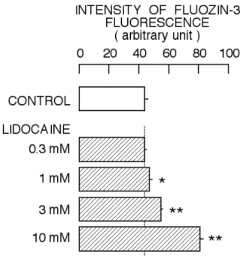
The incubation with 0.3 mM lidocaine for 1 h did not affect the FluoZin-3 fluorescence. Lidocaine at 1 mM started to increase the intensity of FluoZin-3 fluorescence. The increase by 1 mM lidocaine was slight, but statistically-significant. When the cells were incubated with 3 mM or 10 mM lidocaine for 1 hr, further augmentation of FluoZin-3 fluorescence was observed. The results were summarized in Fig. 1. Thus, the incubation with lidocaine at concentrations ranging from 1 mM to 10 mM concentration-dependently increased the intensity of FluoZin-3 fluorescence.
Figure 1. Concentration-dependent change in FluoZin-3 fluorescence by lidocaine. Effect was examined 60 min after the start of application of lidocaine. Columns and bars indicate the means and standard deviations of 4 experiments, respectively. The symbols (*, **) indicate significant differences (P < 0.05, P < 0.01) between the control group and the lidocaine-treated groups.
3.3. The change in FluoZin-3 fluorescence by lidocaine under external Ca2+-free and Zn2+-free conditions
In previous study (Nishimura and Oyama, 2010), lidocaine at 10 mM significantly increased intracellular Ca2+ concentration and this increase by
lidocaine was greatly dependent on extracellular Ca2+.
To remove the contribution of the lidocaine-induced increase in intracellular Ca2+ concentration to the
lidocaine-induced augmentation of FluoZin-3 fluorescence, the effect of lidocaine was examined under nominal Ca2+-free condition.
Removal of CaCl2 from external solution
significantly increased the intensity of FluoZin-3 fluorescence and lidocaine at 10 mM further augmented the FluoZin-3 fluorescence (Fig. 2). The
Yumiko Nishimura • Yasuo Oyama
-9- augmentative action of lidocaine on the FluoZin-3 fluorescence was similarly observed under nominal Ca2+-free condition. The incubation solution
contained small amount (200–230 nM) of Zn2+ that
was delivered from the cell preparation (Sakanashi et al., 2009). The experiment was conducted to examine the contribution of external Zn2+ to the lidocaine-induced augmentation of FluoZin-3 fluorescence. The application of 3 µM DTPA, a chelator for extracellular Zn2+, decreased the intensity
of FluoZin-3 fluorescence under nominal Ca2+-free condition (Fig. 2). Further application of 10 mM lidocaine significantly increased the intensity of FluoZin-3 fluorescence in the presence of DTPA. Thus, the augmentation of FluoZin-3 fluorescence by lidocaine was observed under Ca2+- and Zn2+-free
condition.
Figure 2. Effect of lidocaine on FluoZin-3 fluorescence under Ca2+-free and/or Zn2+-free conditions. Effect was
examined 60 min after the start of application of lidocaine. Columns and bars indicate the means and standard deviations of 4–8 experiments, respectively. The symbol (**) indicates significant difference (P < 0.01) between the control group and the lidocaine-treated group under respective condition.
3.3. The change in 5-CMF fluorescence of rat thymocytes by lidocaine
From the results of Fig. 2, lidocaine was supposed to increase intracellular Zn2+ concentration, independent from external Zn2+. Some agents that decreased cellular content of nonprotein thiols were supposed to increase intracellular Zn2+ concentration
(Hashimoto et al., 2009; Oyama et al., 2009; Kinazaki et al., 2011; Tamura et al., 2011). It was reminiscent of the possibility that lidocaine induced the release of Zn2+ from nonprotein zinc-binding thiols. To test the possibility, the change in 5-CMF fluorescence by
lidocaine was examined. As shown in Fig. 3, the incubation with 10 mM lidocaine significantly decreased the intensity of 5-CMF fluorescence. Thus, lidocaine augmented the FluoZin-3 fluorescence whereas it attenuated the 5-CMF fluorescence.
Figure 3. Effect of lidocaine on 5-CMF fluorescence. The cells were treated with lidocaine for 60 min and thereafter 5-CMF-DA was applied to the cell suspension. Effect was examined 30 min after the start of application of 5-CMF-DA. Therefore, the time for lidocaine treatment was 90 min. Columns and bars indicate the means and standard deviations of 4 experiments, respectively. The symbol (**) indicates significant difference (P < 0.01) between the control group and the lidocaine-treated group. 4. Discussion
Milimolar concentrations (1–10 mM) of lidocaine significantly increased the intensity of FluoZin-3 in a concentration-dependent manner (Fig. 1). The result indicated the lidocaine-induced increase in intracellular Zn2+ concentration (Nishimura and
Oyama, 2010; Kimura et al., 2011). Removal of external Ca2+ increased the intensity of FluoZin-3
fluorescence under control condition (Fig. 2). The mechanism for the augmentation of FluoZin-3 fluorescence by external Ca2+ removal was not
elucidated in this study. Lidocaine further increased the intensity of FluoZin-3 fluorescence under nominal Ca2+-free condition (Fig. 2). Thus, external Ca2+
was not involved in the lidocaine-induced augmentation of FluoZin-3 fluorescence. It was noted that the cell suspension contained 200–230 nM Zn2+ that was delivered from cell preparation
although there was no ZnCl2 in the composition of
Tyrode's solution (Sakanashi et al., 2009). The application of DTPA, a chelator for external Zn2+, decreased the intensity of FluoZin-3 fluorescence under nominal Ca2+-free condition (Fig. 2). Thus,
the Zn2+ influx was supposed to maintain the level of
intracellular Zn2+. Lidocaine also increased the intensity of FluoZin-3 fluorescence under external Ca2+- and Zn2+-free condition (Fig. 2), indicating that external Zn2+ was not involved in the lidocaine-induced increase in the intensity of FluoZin-3 fluorescence. Therefore, it was suggested
Lidocaine-induced increase in intracellular Zn2+ concentration
-10- that lidocaine increased intracellular Zn2+ concentration that was independent from external Zn2+.
Oxidative stress by hydrogen peroxide increased the intensity of FluoZin-3 fluorescence of rat thymocytes (Matsui et al., 2010). The application of hydrogen peroxide greatly attenuated the 5-CMF fluorescence (Chikahisa et al., 1996). There was also reverse correlation between the attenuation of 5-CMF fluorescence and the augmentation of FluoZin-3 fluorescence in the cases of thimerosal, tri-n-butyltin, N-ethylmaleimide, and triclosan (Hashimoto et al., 2009; Oyama et al., 2009; Kinazaki et al., 2011; Tamura et al., 2011). In this study,
lidocaine decreased the intensity of 5-CMF fluorescence whereas it increased that of FluoZin-3 fluorescence. It is presumably suggested that lidocaine decreases the cellular content of nonprotein thiols, resulting in the release of Zn2+ that increases intracellular Zn2+ concentration.
Acknowledgment
This study was supported by a Grant-in-Aid for Scientific Research (C23510078) from the Japan Society for the Promotion of Science.
References
Chikahisa, L., Oyama, Y., Okazaki, E., Noda, K., 1996. Fluorescent estimation of H2O2-induced
changes in cell viability and cellular nonprotein thiol level of dissociated rat thymocytes. Jpn. J. Pharmacol. 71, 299–305.
Gee, K.R., Zhou, Z.L., Qian, W.J., Kennedy, R., 2002. Detection and imaging of zinc secretion from pancreatic beta-cells using a new fluorescent zinc indicator. J. Am. Chem. Soc. 124, 776–778. Hashimoto, E., Oyama, T.B., Oyama, K., Nishimura,
Y., Oyama, T.M., Ueha-Ishibashi, T., Okano, Y., Oyama, Y., 2009. Increase in intracellular Zn2+
concentration by thimerosal in rat thymocytes: Intracellular Zn2+ release induced by oxidative stress. Toxicol. In Vitro. 23, 1092–1099.
Kimura, K., Nishimura, Y., Oyama, K., Kawanai, T., Hashimoto, E., Oyama, Y., 2011. Tetracaine decreases intracellular Zn2+ concentration by
inhibiting Zn2+ influx in rat thymocytes. Nat. Sci. Res. Univ. Tokushima 25, 7–13.
Kinazaki, A., Chen, H., Koizumi, K., Kawanai, T., Oyama, T.M., Satoh, M., Ishida, S., Okano, Y., Oyama, Y., 2011. Putative role of intracellular Zn2+ release during oxidative stress: A trigger to
restore cellular thiol content that is decreased by oxidative stress. J. Physiol. Sci. 61, 403–409. Matsui, H., Sakanashi, Y., Oyama, T.M., Oyama, Y.,
Yokota, S., Ishida, S., Okano, Y., Oyama, T.B., Nishimura, Y., 2008. Imidazole antifungals, but not triazole antifungals, increase membrane Zn2+
permeability in rat thymocytes: Possible contribution to their cytotoxicity. Toxicology. 248, 142–150.
Matsui, H., Oyama, T.M., Okano, Y., Hashimoto, E., Kawanai, T., Oyama, Y., 2010. Low micromolar zinc exerts cytotoxic action under H2O2-induced
oxidative stress: Excessive increase in intracellular Zn2+ concentration. Toxicology. 276, 27–32. Nishimura, Y., Oyama, Y., 2009. Cytotoxic actions of
lidocaine at sublethal concentrations: A model in vitro experiment using rat thymocytes. Nat. Sci. Res. Univ. Tokushima. 24, 1–5.
Oyama, T.B., Oyama, K., Kawanai, T., Oyama, T.M., Hashimoto, E., Satoh, M., Oyama, Y., 2009. Tri-n-butyltin increases intracellular Zn2+
concentration by decreasing cellular thiol content in rat thymocytes. Toxicology. 262, 245–249. Sakanashi, Y., Oyama, T.M., Matsuo, Y., Oyama,
T.B., Nishimura, Y., Ishida, S., Imai, S., Okano, Y., Oyama, Y., 2009. Zn2+, derived from cell preparation, partly attenuates Ca2+-dependent cell
death induced by A23187, calcium ionophore, in rat thymocytes. Toxicol. In Vitro. 23, 338–345. Tamura, I., Kanbara, Y., Saito, M., Horimono, K.,
Satoh, M., Yamamoto, H., Oyama, Y., 2011. Triclosan, an antibacterial agent, increases intracellular Zn2+ concentration in rat thymocytes:
Its relation to oxidative stress. Chemosphere. 86, 70–75.
Article History:
Received MS – 18 April 2012
Received revised MS – 25 April 2012 Accepted MS – 27 April 2012

