I
n 2018, there were an estimated 403,000 new cases of kidney cancer worldwide [1]. Renal cell carci-noma (RCC) accounts for 3% of all malignancies, and over the last two decades the annual incidence of RCC has increased by 2% [2]. The majority of RCCs occur sporadically, but 2-4% of all RCC cases are known to have a heritable basis, for example, von Hippel-Lindau disease (VHL) [3]. VHL is the most common heredi-tary renal cancer syndrome, occurring in approx. 1 of every 36,000 births [4,5]. It is an autosomal dominant inherited syndrome caused by germline mutations in the VHL tumor suppressor gene. VHL is characterizedby tumors in multiple organs: e.g., retinal and central nerve system (CNS) hemangioblastomas, pheochro-mocytomas, pancreatic neuroendocrine tumors, and multiple clear-cell RCC [6,7].
RCC is the most common cancer that develops in individuals with VHL; approx. 40% of individuals with VHL have multifocal, bilateral RCC, and the lesions will be solid, cystic, or both [8]. The reported average life expectancy in patients with VHL is roughly 50 years because of a high prevalence and recurrence rate of RCC [3]. RCC is the major cause of death in patients with VHL. One report demonstrated that nearly 30% of patients with VHL died due to RCC [9]. The
manage-CopyrightⒸ 2020 by Okayama University Medical School.
http ://escholarship.lib.okayama-u.ac.jp/amo/
Case Report
Combined Laparoscopic and CT Monitoring of the Ice-Ball Margin
during Cryoablation for Renal Cell Carcinoma Associated with
von Hippel-Lindau Disease: First Case
Takanori Sekito
a, Motoo Araki
a*, Takao Hiraki
b, Mayu Uka
b,
Toshiyuki Komaki
b, Yusuke Matsui
b, Toshihiro Iguchi
b, Satoshi Katayama
a,
Kasumi Yoshinaga
a, Shogo Watari
a, Yuki Maruyama
a, Yosuke Mitsui
a,
Risa Kubota
a, Takuya Sadahira
a, Shingo Nishimura
a, Koichiro Wada
a,
Atsushi Takamoto
a, Kohei Edamura
a, Tomoko Sako
a, Yasuyuki Kobayashi
a,
Toyohiko Watanabe
a, Susumu Kanazawa
b, and Yasutomo Nasu
aDepartments of aUrology, and bRadiology Okayama University Graduate School of Medicine, Dentistry and Pharmaceutical Sciences, Okayama 700-8558, Japan
We report a 47-year-old Japanese female with 10 previous treatments for multiple bilateral renal cell carcinoma (RCC) associated with von Hippel-Lindau disease. The 14-mm right lower pole renal tumor was in contact with the right ureter. Laparoscopic cryoablation was performed to protect the ureter wrapped with gauze. Computed tomography (CT) monitoring was used to confirm the precise ≥6 mm ice-ball margin. There was no local progression at 6-months post-surgery. The serum creatinine has been stable. This is apparently the first report of combined laparoscopic and CT monitoring of an ice-ball formation and its margin during cryoabla-tion for RCC.
Key words: laparoscopic cryoablation, multiple renal masses, nephron-sparing surgery, renal cell carcinoma, von
Hippel-Lindau disease
Received April 13, 2020 ; accepted June 23, 2020.
*Corresponding author. Phone : +81-86-235-7287; Fax : +81-86-231-3986
ment of RCC in patients with VHL is thus very import-ant.
Historically, the surgical treatment options for RCC are radical nephrectomy and partial nephrectomy (PN). More recently, ablative techniques including radiofre-quency ablation (RFA) and cryoablation (CA) became valid alternatives to nephron-sparing surgery via a min-imally invasive approach. CA was approved by Japan’s national health insurance system in 2011 at the limited number of institutions. The modality used for CA is either percutaneous or laparoscopic.
Percutaneous CA has been preferred in Japan because of its lower cost, shorter procedure time, and lesser invasiveness than laparoscopic CA (‘lap CA’). Lap CA requires the use of many disposable devices such as lap-aroscopic ports. Percutaneous hydrodissection is some-times used to separate tumors and important organs in order to prevent injury [10]. In some cases however, a safety margin between the tumor and an organ cannot be achieved even with hydrodissection [11]. Lap CA is useful in these cases since important organs can be pro-tected laparoscopically. To our knowledge, the present case report is the first to describe combined laparo-scopic and computed tomography (CT) monitoring of ice-ball formation and its precise margin during cryoablation for renal cell carcinoma.
Case Report
A 47-year-old Japanese female was referred to our institution for the treatment of bilateral multiple RCC associated with VHL. She had undergone 10 prior treatments for bilateral multiple RCC. Briefly, her treatment had begun with left PNs performed 18 and 21 years earlier at another hospital. The 2nd PN was for local recurrence. She was then referred to our interven-tional radiologist. She had undergone two percutane-ous RFAs for two right RCCs 11 years earlier, and four percutaneous CA for 6 different left tumors and one right tumor 1,2,4 and 7 years prior to her referral. Two of the lesions had been treated by transcatheter arterial embolization (TAE) using the mixture of ethanol and iodized oil (Lipiodol, Guerbet, Villepinte, France): one on the right 2 years ago and the other on the left 1 year ago. The left lesion was very small, and it was dif-ficult to perform cryoablation of the right lesion due to the tumor’s location. Thus, ablation therapy was not applied to those lesions, which were then observed
carefully.
Twenty-one years after the patient’s first diagnosis of RCC, CT revealed the enlarged lesion in the lower pole of the right kidney, which was considered the recur-rence of the RCC after the TAE (Fig.1A). The tumor had grown from 10 mm to 14 mm in 2 years. The tumor was attached to the right ureter. A safety margin between the tumor and the ureter could not have been achieved even with hydrodissection during the percuta-neous technique. Therefore, lap CA was deemed safer and the patient was referred to us by our interventional radiologist.
Her medical history was significant for cerebellar hemangioblastoma, and uterine myoma. She was tak-ing no medications. Her father and younger brother also had VHL. Laboratory test results were normal. Her serum creatinine (Cr) level was 0.77 mg/dL. The treatment options were thoroughly discussed with the patient, including percutaneous CA, lap CA, robot-as-sisted PN (RAPN), and radical nephrectomy. The patient selected lap CA to maximize the possibility of preserving renal function.
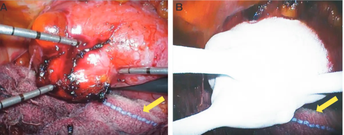
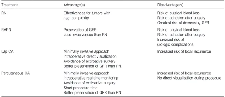
The procedure was performed in a room equipped with a sliding gantry CT scanner (Aquilion CX64, Canon Medical Systems, Otawara, Japan), with the patient under general anesthesia in the supine position. First, the right ureteral stent was placed to help identify the ureter. The patient was then changed to right decu-bitus position. Five ports were used. The ascending colon was mobilized, and the right kidney and the tumor were exposed. The right ureter was attached to the tumor due to the effect of the previous TAE. Special care was taken to dissect the ureter to prevent ischemia of the ureter. The renal artery and vein were intention-ally not dissected; this was to avoid postoperative adhesion because the patient might someday require a PN or nephrectomy for local recurrence considering the nature of VHL. CA was performed by interventional radiologists using an argon-based cryoablation system (Cryo-Hit, Galil Medical, Youknum, Israel) and three 17G cryoprobes (IceSeed, Galil Medical) under laparo-scopic vision. Three cryoprobes were inserted from the 12-mm and 5-mm laparoscopic ports. The right ureter was protected by being wrapped three times with gauze (Fig.2A). CT scanning was performed before the abla-tion to check the probe posiabla-tions, followed by adjust-ment of the probe positions. The ablation protocol included 10- to 15-min freezing cycles separated by
2 min of passive thawing (Fig.2B). Special care was also taken in order not to injure the ureter during the freez-ing and thawfreez-ing. Durfreez-ing the freezfreez-ing, the CT scan was repeated to confirm an ice-ball margin of ≥6 mm (Fig.1B).
The operative time was 4 h and 15 min; the time for CA was 2 h and 3 min. The estimated blood loss was
minimal. Good hemostasis was obtained without the need for parenchymal suturing, a fibrin sealant patch, or fibrin glue.
The follow-up CT scan at 6 months revealed no local progression (Fig.1C). The patient’s serum Cr level has remained at the baseline at 0.73 mg/dL, and the patient
does not shown proteinuria in urinalyses. *
A
B
C
A B
Fig. 2 A, Intraoperative image of the laparoscopic cryoablation. Three cryoprobes were inserted from the laparoscopic ports; B, The freezing phase. Arrow: Gauze. The right ureter was protected by being wrapped three times with gauze.
Fig. 1 A, Computed tomography (CT) with intravenous contrast medium showed the enlarged lesion (black dotted line) in the lower pole of the right kidney beside the lipiodol deposition (asterisk), which was considered local progression of the renal cell carcinoma after the patientʼs transcatheter arte-rial embolization. The diameter of the right tumor was 14 mm. The tumor was attached to the right ureter (arrow); B, Plain CT at the end of the sec-ond freeze cycle demonstrated three cryoprobes (arrows) positioned within the renal mass. The ice-ball was detected as a well-demarcated region of hypodensity in the surrounding tissue (arrowhead); C, CT with intravenous medium 6 months after laparoscopic cryoablation shows no local progression of the tumor. Black dotted line: The laparoscopic cryoablation treatment area.
Discussion
We have reported a successful lap CA procedure in a patient with VHL who had undergone 10 treatments for her bilateral RCC. The tumor could not have been treated with percutaneous CA due to its proximity to the ureter. CT monitoring was also used to confirm the precise ≥6 mm ice-ball margin. To our knowledge, this is the first report of combined laparoscopic and CT monitoring of the formation of an ice-ball margin during cryoablation for RCC.
In patients with VHL, several interventions includ-ing surgical and ablative treatments are available to prevent disease progression and for the maintenance of renal function. Matin et al. reported that 88% of patients with VHL and RCC required some interven-tions, and 80% of the interventions were nephron- sparing approaches at a median follow-up of 41 months. The metastasis-free survival was 93% and the overall survival (OS) was 88% [12].
The treatment of localized RCC has shifted from radical nephrectomy to PN and ablative therapies over the years. Ablative interventions include percutaneous or lap RFA/CA. Table 1 provides an overview of the management strategies used for patients with localized RCC, including the advantages and disadvantages of
each approach [13]. Ablative techniques can offer a minimally invasive approach, and their use helps avoid extirpative surgery and provides shorter hospital stays and faster recovery [14]. The current available guide-lines note that ablative techniques as treatment options are confined to patients who are at risk of renal function loss (e.g., single kidney or genetic predisposition for multiple tumors) [2].
CA has become an important therapeutic option as a nephron-sparing modality. The effectiveness of CA for treating T1a renal tumors is well established [15]. CA therapy is a potential treatment alternative for patients with T1a renal tumors that are not suitable for extirpa-tive surgery [16]. Lap CA was introduced in 1998 [17]. From a short-term perspective, lap CA is associated with lower perioperative complications compared to open surgery [18,19]. Larcher et al. reported that lap CA leads to safe long-term cancer control in patients who have a single T1a small renal tumor [20]. Nielsen et al. reported that the rates of 5- and 10-year disease- free survival after lap CA were 90% and 80%, and the 5- and 10-year OS rates were 83% and 64%, respec-tively [16].
We selected lap CA for our patient because the tumor was too close to the right ureter. Percutaneous hydrodissection is frequently used to separate a tumor
Table 1 Treatments options used for localized renal cell carcinoma
Treatment Advantage(s) Disadvantage(s)
RN Effectiveness for tumors with
high complexity Risk of surgical blood lossRisk of adhesion after surgery Greatest risk of decreasing GFR
RAPN Preservation of GFR
Less invasiveness than RN Risk of surgical blood lossRisk of adhesion after surgery Increased risk of
urologic complications Lap CA Minimally invasive approach
Intraoperative direct visualization Avoidance of extirpative surgery Better preservation of GFR than PN
Increased risk of local recurrence
Percutaneous CA Minimally invasive approach Intraoperative real-time monitoring Avoidance of extirpative surgery Short procedure time
Better preservation of GFR than PN
Increased risk of local recurrence No direct visualization during procedure
Source. —Reference 13.
Note. — RN, radical nephrectomy; RAPN, robot assisted partial nephrectomy; Lap CA, laparoscopic cryoablation; PN, partial nephrec-tomy; CA, cryoablation; GFR, glomerular filtration rate.
and an important organ to prevent injury [10]. However, we speculated that hydrodissection could not have worked well in case because the right ureter was adhered firmly to the tumor due to the effect of the pre-vious TAE treatment. A laparoscopic lysis of the adhe-sion was required. We also added a step by wrapping the right ureter 3 times with gauze and lifting the entire right kidney to protect the ureter from injury during the freezing and thawing.
Lap CA is usually monitored by ultrasound whereas percutaneous CA is usually monitored by ultrasound or CT. CT is more accurate for monitoring ice-ball forma-tion and its precise margin [21]. It can be difficult to monitor a lap CA procedure by CT because most oper-ation rooms are not equipped with CT. We used CT to assist the lap CA procedure in the present case, and we observed that unlike direct vision through laparoscopy and ultrasound monitoring, CT was useful to confirm the ice-ball formation and its precise margin inside the kidney and thereby to secure complete tumor ablation.
RAPN was another option in the present case. We have performed over 200 RAPNs. RAPN was not selected for our present patient, for the following two reasons. (1) A systematic review and meta-analysis comparing CA and PN in clinical stage T1 renal masses demonstrated a decreased estimated glomerular filtra-tion rate and increased Cr level [22]. The authors of that meta-analysis indicated that CA could preserve renal function significantly better compared to PN, and they speculated that CA could preserve better renal function by being combined with laparoscopic or CT/ultrasound vision. We felt that CA was more appropriate than a PN for our patient in light of her young age (47 years) and the high possibility of RCC recurrence due to the nature of VHL. (2) RAPN was feasible. Our patient’s R.E.N.A.L. nephrometry score (a scoring system used to assess the difficulty of nephron-sparing surgery [23]) was 5a, which indicates a low risk. However, RAPN requires renal hilum dissection and usually uses a fibrin sealant patch or fibrin glue for hemostasis. These procedures cause adhesion around the renal hilum, making the next surgery very difficult. Considering our patient’s age and the high recurrence rate of VHL, it is entirely possible that she could have a further recurrence and require multiple treatments over the rest of her life. We felt that RAPN should be saved for when there are no other options (including CA).
The lap CA was successful without complications. It
was especially useful to protect the ureter. The CT monitoring was also useful to confirm the precise ≥6 mm ice-ball margin. At 6 months post-surgery, renal pres-ervation and good cancer control were confirmed after the minimally invasive procedure.
References
1. Ferlay J, Colombet M, Soerjomataram I, Mathers C, Parkin DM, M Piñeros, Znaor A and Bray F: Estimating the global cancer inci-dence and mortality in 2018: GLOBOCAN sources and methods. Int J Cancer (2019) 144: 1941-1953.
2. Ljungberg B, Albiges L, Abu-Ghanem Y, Bensalah K, Dabestani S, Fernández-Pello S, Giles RH, Hofmann F, Hora M, Kuczyk MA, Kuusk T, Lam TB, Marconi L, Merseburger AS, Powles T, Staehler M, Tahbaz R, Volpe A and Bex A: European Association of Urology Guidelines on Renal Cell Carcinoma: The 2019 Update. Eur Urol (2019) 75: 799-810.
3. Bausch B, Jilg C, Gläsker S, Vortmeyer A, Lützen N, Anton A, Eng C and Neumann HP: Renal cancer in von Hippel-Lindau dis-ease and related syndromes. Nat Rev Nephrol (2013) 9: 529-538. 4. Maher ER, Iselius L, Yates JR, Littler M, Benjamin C, Harris R,
Sampson J, Williams A, Ferguson-Smith MA and Morton N: Von Hippel-Lindau disease: a genetic study. J Med Genet (1991) 28: 443-447.
5. Neumann HP and Wiestler OD: Clustering of features of von Hippel-Lindau syndrome: evidence for a complex genetic locus. Lancet (1991) 337: 1052-1054.
6. Lonser RR, Glenn GM, Walther M, Chew EY, Libutti SK, Linehan WM and Oldfield EH: von Hippel-Lindau disease. Lancet (2003) 361: 2059-2067.
7. Neumann HP, Lips CJ, Hsia YE and Zbar B: Von Hippel-Lindau syndrome. Brain Pathol (1995) 5: 181-193.
8. Gallou C, Chauveau D, Richard S, Joly D, Giraud S, Olschwang S, Martin N, Saquet C, Chrétien Y, Méjean A, Correas JM, Benoît G, Colombeau P, Grünfeld JP, Junien C and Béroud C: Genotype-phenotype correlation in von Hippel-Lindau families with renal lesions. Hum Mutat (2004) 24: 435-436.
9. Binderup ML, Jensen AM, Budtz-Jørgensen E and Bisgaard ML: Survival and causes of death in patients with von Hippel-Lindau disease. J Med Genet (2017) 54: 11-18.
10. Cheng Z, Yu X, Han Z, Liu F, Yu J and Liang P: Ultrasound-guided hydrodissection for assisting percutaneous microwave ablation of renal cell carcinomas adjacent to intestinal tracts: a preliminary clinical study. Int J Hyperthermia (2018) 34: 315-320.
11. Bodily KD, Atwell TD, Mandrekar JN, Farrell MA, Callstrom MR, Schmit GD and Charboneau JW: Hydrodisplacement in the percu-taneous cryoablation of 50 renal tumors. Am J Roentgenol (2010) 194: 779-783.
12. Matin SF, Ahrar K, Wood CG, Daniels M and Jonasch E: Patterns of intervention for renal lesions in von Hippel-Lindau disease. BJU Int (2008) 102: 940-945.
13. Ryan DW, Hajime T, Steven CC and Erick MR: 2017 AUA Renal Mass and Localized Renal Cancer Guidelines: Imaging Implica-tions. Radiographics (2018) 38: 2021-2033.
14. Pessoa RR, Autorino R, Laguna MP, Molina WR, Gustafson D, Nogueira L, Silva RD, Werahera PN and Kim FJ: Laparoscopic versus percutaneous cryoablation of small renal mass: systematic review and cumulative analysis of comparative studies. Clin
Genitourin Cancer (2017) 15: 513-519.
15. Campbell SC, Novick AC, Belldegrun A, Blute ML, Chow GK, Derweesh IH, Faraday MM, Kaouk JH, Leveillee RJ, Matin SF, Russo P and Uzzo RG: Guideline for management of the clinical T1 renal mass. J Urol (2009) 182: 1271-1279.
16. Nielsen TK, Lagerveld BW, Keeley F, Lughezzani G, Sriprasad S, Barber NJ, Hansen LU, Buffi NM, Guazzoni G, Zee JA, Ismail M, Farrag K, Emara AM, Lund L, Østraat Ø and Borre M: Oncological outcomes and complication rates after laparoscopic-assisted cryoablation: a European Registry for Renal Cryoablation (EuRECA) multi-institutional study. BJU Int (2017) 119: 390-395. 17. Gill IS, Novick AC, Soble JJ, Sung GT, Remer EM, Hale J and
OʼMalley CM: Laparoscopic renal cryoablation: initial clinical
series. Urology (1998) 52: 543-551.
18. Klatte T, Grubmüller B, Waldert M, Weibl P and Remzi M: Laparoscopic cryoablation versus partial nephrectomy for the treat-ment of small renal masses: systematic review and cumulative analysis of observational studies. Eur Urol (2011) 60: 435-443. 19. Klatte T, Shariat SF and Remzi M: Systematic review and
meta-analysis of perioperative and oncologic outcomes of
laparo-scopic cryoablation versus laparolaparo-scopic partial nephrectomy for the treatment of small renal tumors. J Urol (2014) 191: 1209-1217.
20. Larcher A, Fossati N, Mistretta F, Lughezzani G, Lista G, DellʼOglio P, Abrate A, Sun M, Karakiewicz P, Suardi N, Lazzeri
M, Montorsi F, Guazzoni G and Buffi N: Long-term oncologic out-comes of laparoscopic renal cryoablation as primary treatment for small renal masses. Urol Oncol (2015) 33: 22.
21. Allen BC and Remer EM: Percutaneous cryoablation of renal tumors: patient selection, technique, and postprocedural imaging. Radiographics (2010) 30: 887-900.
22. Deng W, Chen L, Wang Y, Liu X, Wang G, Liu W, Zhang C, Zhou X, Li Y and Fu B: Cryoablation versus Partial Nephrectomy for Clinical Stage T1 Renal Masses: A Systematic Review and Meta-Analysis. J Cancer (2019) 10: 1226-1236.
23. Chang X, Liu T, Zhang F, Cheng Q, Changwei J, Xiaozhi Z, Guangxiang L and Hongqian G: The comparison of R.E.N.A.L., PADUA and centrality index score in predicting perioperative out-comes and complications after laparoscopic radio frequency abla-tion of renal tumors. J Urol (2015) 194: 897-902.