リファンピシンおよびエタンブトールにより急性
汎発性発疹性膿疱症を発症した肺結核の 1 例
渡邉 裕文 宍戸雄一郎 鈴木 貴人 野口 理絵
三枝 美香 赤松 泰介 山本 輝人 森田 悟
朝田 和博 白井 敏博
緒 言 抗結核薬治療中に出現する皮疹をしばしば経験する。 結核治療を継続するためには,原因薬剤の同定や皮疹の コントロールは重要である。今回,尋常性乾癬を基礎に もつ肺結核患者の治療中に膿疱を伴う皮疹が出現し,重 症薬疹である急性汎発性発疹性膿疱症(acute generalized exanthematous pustulosis : AGEP)を発症した肺結核の症 例を経験したため報告する。 症 例 症 例:53 歳,女性。 主 訴:胸部異常影。 既往歴:33 歳,肺結核(詳細不明)。23 歳より尋常性 乾癬(保湿剤のみ外用)。52 歳時に下行結腸癌手術,人 工肛門造設(受診時テガフール・ウラシル内服中)。 家族歴:特記事項なし 生活歴:喫煙歴なし。定期的な検診歴はなし。 職業歴:20 歳から 24 歳まで販売業に従事。 アレルギー:なし。 内服薬:テガフール・ウラシル。 現病歴:X−20 年に肺結核と診断されたが,治療歴や 入院歴の詳細は不明であった。X 年 4 月に胸部異常陰影 を指摘され近医に受診した。胃液抗酸菌塗抹検査で2+, 結核菌 PCR 法陽性であり,肺結核の診断で同年 5 月 30 日(第 1 病日)に当院に入院となる。 入 院 時 身 体 所 見: 身 長 146.8 cm,体 重 32.5kg,血 圧 127/86 mmHg,脈拍 73 回 ⁄分・整,体温 36.5℃,SpO2 96% (室内気),眼瞼結膜貧血なし,頸部リンパ節腫脹なし, 心音純,呼吸音は右上肺で減弱あり,腹部は平坦・軟, 正中にストマあり,四肢体幹に褐色の色素痕あり。 入院時検査所見(Table):CRP 1.08 mg/dl と高値を認め, 静岡県立総合病院呼吸器内科 連絡先 : 渡邉裕文,静岡県立総合病院呼吸器内科,〒 420 _ 8527 静岡県静岡市葵区北安東 4 _ 27 _ 1 (E-mail : [email protected])(Received 7 Aug. 2017 / Accepted 4 Oct. 2017)
要旨:症例は 53 歳女性。既往に尋常性乾癬,大腸癌がある。肺結核(学会分類 bⅡ2)で当科入院と なった。イソニアジド(INH),リファンピシン(RFP),エタンブトール(EB),ピラジナミド(PZA) で治療を開始したが,治療開始 9 日後に発熱,全身の紅斑と膿疱が出現し,血液検査で CRP の上昇を 認めたため抗結核薬を中止した。皮膚生検では好中球浸潤と角層下海綿状膿疱の所見であった。その 後薬剤を順次投与し,発熱と全身の紅斑,膿疱の原因は RFP と EB の 2 剤と判明した。上記皮疹が薬 剤中止後 1 日で消退傾向を示したことから薬疹である急性汎発性発疹性膿疱症(AGEP)と診断した。 その後 INH から再開し,PZA,レボフロキサシン(LVFX),ストレプトマイシン(SM)を順次追加し たが皮疹の出現はみられなかった。 4 剤での継続が可能と判断し,塗抹の陰性を確認したため第 108 病日に自宅退院となった。抗結核薬による AGEP は INH,RFP では既報告があるが,EB での報告や抗 結核薬 2 剤での報告はなく,貴重な症例と考えた。
キーワーズ:肺結核,急性汎発性発疹性膿疱症(acute generalized exanthematous pustulosis : AGEP),尋
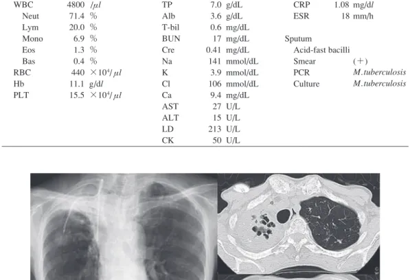
Fig. 1 Chest X ray and CT scan on admission. Consolidation with cavity can be seen, especially
in right upper lobe. A CV port is planted on her body.
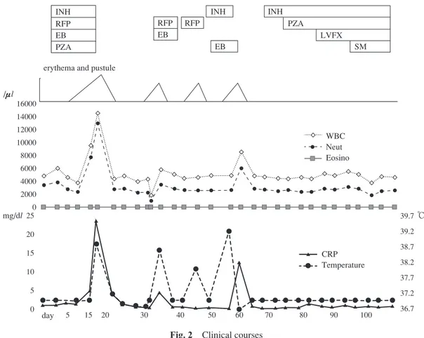
Table Laboratory fi ndings Hematology WBC Neut Lym Mono Eos Bas RBC Hb PLT 4800 71.4 20.0 6.9 1.3 0.4 440 11.1 15.5 /μl % % % % % ×104/μl g/dl ×104/μl Biochemistry TP Alb T-bil BUN Cre Na K Cl Ca AST ALT LD CK 7.0 3.6 0.6 17 0.41 141 3.9 106 9.4 27 15 213 50 g/dL g/dL mg/dL mg/dL mg/dL mmol/dL mmol/dL mmol/dL mg/dL U/L U/L U/L U/L Serology CRP 1.08 ESR 18 Sputum Acid-fast bacilli Smear PCR Culture mg/dl mm/h (+) M. tuberculosis M. tuberculosis 既報告 ¹)により INH による尋常性乾癬の増悪と考え皮膚 生検後ステロイド剤外用および活性化ビタミン D3剤の 外用を開始した。しかし翌日には高熱となり炎症データ も上昇したため第 16 病日に抗結核薬はすべて中止し, エトレチナート30 mg内服を開始した。膿疱は培養陰性, また組織学的には角層下膿疱と表皮内に好中球膿疱と海 綿状態を示す Kogoj 海綿状膿疱を認めた(Fig. 4)。第 28 病日には皮膚所見が落ち着いたためエトレチナートは中 止した。皮疹と熱型安定後の第 32 病日より RFP,EB で 投与再開した。結核菌の薬剤耐性の獲得を危惧し 2 剤で 再開としたが,投与後 3 時間後に全身の紅斑が出現した ため 2 剤を中止した。 2 日後には消退傾向を示し,膿疱 は落屑を示した。皮疹消退後,第 40 病日に RFP 単剤で再 開したが,同様に数時間で全身の紅斑が出現し,RFP が 被疑薬と考えられた。第 44 病日よりINH 単剤で再開した が,皮疹の出現は認めず,第 57 病日より EB を追加した。 血沈 18 mm/h と亢進していた。喀痰抗酸菌塗抹で 2 +, 液体培養と小川培養でそれぞれ 1 週,4 週で結核菌が証 明された。薬剤感受性検査ではいずれの薬剤に対しても 耐性は示さなかった。 胸部画像(Fig. 1):胸部 X 線検査では右上肺野の空洞 を伴う浸潤影と両側肺に気道散布影あり(学会分類bⅡ2), また CV ポートも確認できる。胸部 CT では右上葉を中 心に空洞を伴うコンソリデーションを確認できる。 臨床経過(Fig. 2):第 3 病日よりイソニアジド(INH) 200 mg ⁄日,リファンピシン(RFP)300 mg ⁄日,エタンブ トール(EB)500 mg ⁄日,ピラジナミド(PZA)900 mg ⁄ 日の 4 剤で治療を開始したが,第 12 病日に発熱と全身 の紅斑と膿疱が出現し(Fig. 3),血液検査で CRP の上昇 を認めたため皮膚科にコンサルテーションした。皮疹は 全身に鱗屑を付す紅斑,紅色局面を呈し,紅斑上には毛 孔に一致しない膿疱が多発し一部膿海を形成していた。
Fig. 3 Clinical appearance of the back : a : diffuse erythema with scales. b : military pustules inconsistent
with pores in the skin. 16000 14000 12000 10000 8000 6000 4000 2000 0 25 20 15 10 5 0 39.7 39.2 38.7 38.2 37.7 37.2 36.7 ℃ WBC Neut Eosino CRP Temperature mg/dl /μl
INH INH INH
RFP RFP RFP EB EB EB PZA PZA LVFX SM erythema and pustule
day 5 15 20 30 40 50 60 70 80 90 100 ◆ ◆ ◆ ◆ ◆ ◆ ◆ ◆ ◆ ◆ ◆ ◆ ◆ ◆ ◆ ◆ ◆ ◆ ◆ ◆ ◆ ◆ ◆ ◆ ◆ ◆ ◆ ◆ ◆ ◆ ◆ ◆ ● ● ● ● ● ● ● ● ● ● ● ● ● ● ● ● ● ● ● ● ● ● ● ● ● ● ● ● ● ● ● ● ● ● ● ● ● ● ● ● ● ● ● ● ● ● ● ● ● ● ● ● ● ● ● ● ● ● ● ● ● ● ● ▲ ▲ ▲ ▲ ▲ ▲ ▲ ▲ ▲ ▲ ▲ ▲ ▲ ▲ ▲ ▲ ▲ ▲ ▲ ▲ ▲ ▲ ▲ ▲ ▲ ▲ ▲ ▲ ▲ ▲
Fig. 2 Clinical courses
INH : isoniazid, RFP : rifampicin, EB : ethambutol, PZA : pyrazinamide LVFX : levofl oxacin, SM : streptomycin
a b ところが EB 追加後 3 時間程度で全身の紅斑と発熱,膿 疱が出現したため,2 剤を中止した。尋常性乾癬はある ものの,薬剤投与後に短期間で発熱と膿疱を伴う紅斑が 出現し,中止後 1 日から消退傾向を認めたことから薬疹 のひとつである急性汎発性発疹性膿疱症(AGEP)と診 断した。INH,RFP,EB に対する薬剤誘発性リンパ球刺 激 試 験(drug-induced lymphocyte stimulation test : DLST)
は陰性,INH,RFP,EB,PZA を用い皮膚パッチテストを 施行したが結果は陰性であった。また,膿疱化を繰り返 したため汎発性膿疱性乾癬の可能性も考慮し,IL36RN 遺伝子変異を検索したが,陰性であった。入院前の尋常 性乾癬の経過としては,20 歳代に尋常性乾癬発症時に感 染性関節炎を併発し現在も手指の変形は残存するがそれ まで一度も膿疱化した既往はなく,入院時は四肢体幹に
Fig. 4 Histological fi ndings of skin (hematoxylin and eosin stain a :×40, b :×100) : There are numerous
neutrophils in the epidermis and subcorneal pustules. Kogoj’s spongiform pustule can be seen.
a b 軽度角化性紅斑はみられるもののワセリン外用のみで対 処できていた程度であった。また,解熱し膿疱が消退し た後既存の角化性紅斑がみられるところからも皮膚生検 を施行しているが,組織学的には典型的な尋常性乾癬の 所見であった。被疑薬である RFP,EB は使用せず,第 63 病日より INH を再開し,順次,第 70 病日に PZA,第 84 病日にレボフロキサシン(LVFX),第 91 病日にストレプ トマイシン(SM)を追加したが皮疹の出現はみられな かった。最終的に INH,PZA,LVFX,SM の 4 剤での継 続が可能となり,第 108 病日に自宅退院となった。 考 察 AGEP は重症薬疹の一つであり,薬剤などへの曝露後, 発熱,白血球増多とともに紅斑,膿疱を全身性に生じ, 短期間で軽快する病態として 1980 年 Beylot らが提唱し た名称である2)。原因の 90% が薬剤であり,なかでも抗 菌薬,特にペニシリン系やマクロライド系が多いとされ ている。Britschgi らは,薬剤特異的 T 細胞の活性化と表 皮への好中球浸潤が起こるアレルギー反応と報告してい る3)。Roujeau らは,AGEP 63 例中 11 例に乾癬の既往が あり,乾癬を背景にもつ者により生じやすい可能性を示 唆しており4),同論文では以下のような診断基準があげ られている。①汎発性の紅斑上に 5 mm 以下の非毛孔一 致性小膿疱の多発,②病理組織学的に表皮内,または角 層下膿疱で,真皮浮腫,血管炎,血管周囲の好酸球浸潤, 角化細胞の限局性壊死のいずれか,または複数を伴う, ③ 38℃を超える発熱,④末梢血の好中球増多(7000/μl 以上),⑤急な発症と 15 日以内の膿疱の自然消退,であ る。本症例はいずれの項目も満たしていた。乾癬素因の ある患者に全身に膿疱が多発した場合,汎発性膿疱性乾 癬と AGEP との鑑別は困難なことが多く,両者の鑑別は 臨床的な判断によるところが大きいとされるが,AGEP では急速な発症と速やかな消退を認めること,薬剤投与 歴があることがあげられている。なお,病理所見上は, 両者を鑑別することは困難とされている。膿疱性乾癬の 場合は,治療法はステロイド剤外用および活性化ビタミ ン D3剤外用やエトレチナート内服など,乾癬治療の強 化が必須となる。乾癬素因の患者に発生した AGEP の報 告が散見されるが5) ∼ 7),既報告では薬剤投与歴や皮疹の 速やかな改善といった経過から AGEP の診断に至ってい る。 汎発性膿疱性乾癬患者の多くが IL36RN 遺伝子変異を 有するとされ検索したが8),本症例は陰性であった。 既述の Roujeau らの診断基準のほか,診断に関しては, Euro SCAR study group が診断を的確に行うために皮膚症 状,組織所見,臨床経過をスコア化した診断基準を提唱 しているが9)(12 点満点,8 点∼12 点で確定となる),本 症例では 11 点と AGEP 確定と判断できた。AGEP の治療 は,原因薬剤の中止が原則であり,中止により 15 日以内 に自然に消退するが,症状が強ければステロイド内服治 療を行うこととされる。被疑薬の再投与は禁忌である。 本症例では,乾癬治療としてのエトレチナート投与も行 ったが,被疑薬中止後 1 日から膿疱は消退したため,薬 剤の中止が有効であったと判断した。これまで,抗結核 薬による AGEP の報告は INH,RFP,リファブチンで 1 例ずつ報告されており10)∼12),抗結核薬多剤内服中に発 症し原因薬剤同定不能であった症例の報告もある13)。 結核治療中に出現する皮疹のコントロールは重要であ る。 今 回,RFP と EB が 原 因 で あ っ た 重 症 薬 疹 で あ る AGEP の症例を経験した。汎発性膿疱性乾癬との鑑別が
重要であったが,同薬剤投与により,紅斑と膿疱,発熱 を繰り返し発症したため診断に至った。EB による AGEP の報告はなく,また抗結核薬 2 剤が原因と同定できた報 告もみられず,貴重な症例と考えられた。 謝 辞 本報告を作成するにあたり,診療時より多くのご助言 およびご指導をいただきました静岡県立総合病院皮膚 科 佐野悠子先生に深謝致します。 また,IL36RN 遺伝子変異に関して検索いただいた名 古屋大学医学部皮膚科 武市拓也先生に深謝致します。 著者の COI(confl icts of interest)開示:本論文発表内 容に関して特になし。
文 献
1 ) 大沼すみ, 池澤善郎, 西村百合香, 他:乾癬型薬疹―自 験15例の臨床的検討. 日本皮膚科学会雑誌. 1999 ; 109 : 1451 1460.
2 ) Beylot C, Bioulac P, Doutre MS, et al.: Pustuloses exan-thematiques aigues generalisees. Ann Dermatol Venereol. 1980 ; 107 : 37 48.
3 ) Britschgi M, Pichler WJ: Acute generalized exanthematous pustulosis, a clue to neutrophil-mediated infl ammatory processes orchestrated by T cells. Curr Opin Allergy Clin Immunol. 2002 ; 2 : 325 331.
4 ) Roujeau JC, Bioulac-Sage P, Bourseau C, et al.: Acute generalized exanthematous pustulosis: analysis of 63 cases.
Arch Dermatol. 1991 ; 127 : 1333 1338. 5 ) 加藤香澄, 加藤裕史, 坂井田高志, 他:膿疱性乾癬の患 者に発症した, アモキシシリンによる急性汎発性発疹 性膿疱症と考えられた症例. 皮膚病診療. 2016 ; 38 : 259 262. 6 ) 内平美穂, 濱本嘉昭, 武藤正彦:尋常性乾癬患者に生 じた塩酸ミノサイクリンによる急性汎発性発疹性膿疱 症(AGEP). 西日皮膚. 2005 ; 67 : 373 377. 7 ) 佐々木喜教, 笹井 収, 松永 純, 他:市販の解熱鎮痛 剤内服により生じた急性全身性発疹性膿疱症の 1 例. 臨床皮膚. 2002 ; 56 : 810 813.
8 ) Sugiura K, Takemoto A, Yamaguchi M, et al.: The Majority of Generalized Pustular Psoriasis without Psoriasis Vulgaris Is Caused by Defi ciency of Interleukin-36 Receptor Antag-onist. J Invest Dermatol. 2013 ; 133 : 2514 2521.
9 ) Sidoroff A, Halevy S, Bavinck JNB, et al.: Acute general-ized exanthematous pustulosis (AGEP) ― A clinical reaction pattern. J Cutan Pathol. 2001 ; 28 : 113 119.
10) Yamasaki R, Yamasaki M, Kawasaki Y, et al.: Generalized pustular dermatosis caused by isoniazid. Br J Dermatol. 1985 ; 112 : 504 506.
11) Azad A, Connely N : Case of rifampicin-induced acute generalized exanthematous pustulosis. Intern Med J. 2006 ; 36 : 619 622.
12) Chen CP, Hsu YH, Hong SJ, et al.: Acute generalized exanthematous pustulosis caused by rifabutin. Arch Dermatol. 2009 ; 145 : 1069 1070.
13) Cantisani C, Paradisi A, Richetta AG, et al.: Acute gen-eralized exanthematous pustulosis during antituberculosis therapy. Clin Ter. 2013 ; 164 : 137 138.
Abstract We present the case of a 53-year-old female with
pulmonary tuberculosis. She had psoriasis vulgaris and colon cancer. After admission, she was treated with four antituber-cular agents: isoniazid (INH), rifampicin (RFP), ethambutol (EB), and pyrazinamide (PZA). However, 9 days after the treatment, she developed fever up to 38.5℃, erythema and pustules on the whole body. Therefore, we discontinued agents. Histological examination of skin biopsy revealed Kogoj’s spongiform pustule and numerous neutrophils in the epider-mis. After skin eruption disappeared, drugs were adminis-tered sequentially, and it became clear that the cause of skin eruption were RFP and EB.
Because the skin eruption and fever tended to disappear after the discontinuation of these drugs in one day and pathological fi ndings showed Kogoj’s spongiform pustule, we diagnosed acute generalized exanthematous pustulosis (AGEP), one of the drug-induced eruption. After that, we restarted INH and then added PZA, levofl oxacin, and strepto-mycin sequentially, and the skin eruption didn’t appear. We concluded that the continuation of treatment with the latter four drugs is possible, then she was discharged from the hospital. Development of AGEP due to antitubercular agents,
such as INH and RFP, has been reported previously; how-ever, reports on AGEP due to EB have not been observed. And reports on AGEP due to two antitubercular agents in one patient have not been observed. Thus, this case is rare. We should consider AGEP when we use antitubercular agents.
Key words: Pulmonary tuberculosis, Acute generalized
exan-thematous pustulosis (AGEP), Psoriasis vulgaris, Drug-induced eruption, Pustular psoriasis
Division of Respirology, Shizuoka General Hospital
Correspondence to: Hirofumi Watanabe, Division of Respi-rology, Shizuoka General Hospital, 4_27_1, Kita-ando, Aoi-ku, Shizuoka-shi, Shizuoka 420_8527 Japan.
(E-mail: [email protected])
Nonmember cooperators: ¹Yuko SANO, ²Takuya TAKEICHI ¹Division of Dermatology, Shizuoka General Hospital, ²Department of Dermatology, Nagoya University School of Medicine
−−−−−−−−Case Report−−−−−−−−
A CASE OF ACUTE GENERALIZED EXANTHEMATOUS PUSTULOSIS
CAUSED BY RIFAMPICIN AND ETHAMBUTOL
IN A PULMONARY TUBERCULOSIS
Hirofumi WATANABE, Yuichiro SHISHIDO, Takahito SUZUKI, Rie NOGUCHI, Mika SAIGUSA, Taisuke AKAMATSU, Akito YAMAMOTO, Satoru MORITA,