九州大学学術情報リポジトリ
Kyushu University Institutional Repository
免疫学的な恒常性の維持に寄与する材料設計
杠, 和樹
http://hdl.handle.net/2324/4110397
出版情報:Kyushu University, 2020, 博士(工学), 課程博士 バージョン:
権利関係:
Material designs for maintenance of immunological homeostasis
Kazuki Yuzuriha
Doctor of Philosophy
Graduate School of Systems Life Sciences Kyushu University
2020
Abstract
Material designs for maintenance of immunological homeostasis
Kazuki Yuzuriha
Doctor of Philosophy
Graduate School of Systems Life Sciences Kyushu University
2020
Increase in the patient of diseases related to the disruption of immunological homeostasis is threatening issue worldwide. Many reasons have been proposed for the increment of the disruption of immunological homeostasis. One of the reasons is the dysbiosis of the intestinal microbiome which is caused by administration of antibiotics. To restore the disrupted immunological homeostasis, immunotherapy using an antigen is fundamental solution. However, usually this kind of therapy (desensitization) takes time for completion. In this paper, I approached the goal in two ways to maintain or restore the immunological homeostasis.
In Chapter 2, I developed vitamin-peptide conjugates as effective inducer of immunotolerance toward the antigen peptides. Here I selected ATRA and vD3 as inducer of tolerogenic dendritic cells. Selective modification of these vitamins on the N-terminus of the peptide was achieved by the scheme I developed here. The obtained conjugates
showed the functions of both vitamins and antigen peptides, i.e., anti-inflammatory effects resulting from ATRA and activation of antigen-specific T cells. The conjugates reported here can be expected to promote the induction of antigen-specific Tregs, which is essential to restore the disruption of immunological homeostasis.
In Chapter 3, I proposed an antibiotic-specific adsorbent to prevent dysbiosis caused by antibiotic treatment. Hydrophilic polyethyleneglycol-based microparticles were modified with peptide ligands in a high density which is a vancomycin-specific ligand. The microparticles showed high capacity and specificity to vancomycin and successfully captured vancomycin in vivo. The microparticles protected from the C.
difficile infection induced by vancomycin-induced dysbiosis.
Table of contents
CHAPTER 1 ... 1
General Introduction ... 1
1.1. Immunological homeostasis ... 1
1.1.1. Functions of dendritic cells in immunological homeostasis .... 2
1.1.2. Approaches to induce immune tolerance for allergic and autoimmune diseases ... 3
1.2 Contribution of microbiome to host homeostasis ... 6
1.2.1. Dysbiosis caused by antibiotics... 7
1.2.2. Utilizing -lactamase to protect microbiome ... 9
1.2.3. Activated carbon as antibiotics adsorbent ... 10
1.3. Overview of this thesis ... 11
1.4. References ... 12
CHAPTER 2 ... 18
Development of vitamin-peptide conjugate for induction of antigen- specific immunotolerance ... 18
2.1. Introduction ... 18
2.2. Materials and Methods ... 20
2.2.1. Synthesis of lysine residue-protected peptide ... 20
2.2.2. Synthesis of conjugate 1 and 2 ... 21
2.2.3. Cell line and culture ... 21
2.2.4. Cell viability ... 22
2.2.5. Alkaline phosphatase assay ... 22
2.2.6. Quantitative real-time-PCR assays (qRT-PCR) ... 23
2.2.7. Flow cytometry ... 23
2.2.8. Antigen presentation assay ... 23
2.3. Results and Discussion ... 25
2.3.1. Synthesis of vitamin-peptide conjugate ... 25
2.3.2. Cytotoxicity of conjugates ... 27
2.3.3. Alkaline phosphatase assay ... 28
2.3.4. Suppression of LPS-induced inflammatory response of DC by ATRA conjugate ... 29
2.3.5. Expression of ligand on surface of DC2.4 cells ... 31
2.3.6. Conjugate induction of antigen-specific immune responses ... 32
2.4. Conclusions ... 34
2.5. References ... 35
CHAPTER 3 ... 40
Protection of gut microbiome from antibiotics: development of a vancomycin-specific adsorbent with high adsorption capacity ... 40
3.1. Introduction ... 40
3.2. Materials and Methods ... 44
3.2.1. Preparation of MPs ... 44
3.2.2. Field emission scanning electron microscope observations ... 44
3.2.3. Adsorption of VCM to MPs ... 44
3.2.4. Evaluation of the minimal inhibitory concentration 50 value ... 45
3.2.5. Determination of the dose of MPs in vivo ... 46
3.2.6. C.difficile spore preparation ... 46
3.2.7. VCM and MP administration and challenge with C. difficile .... 47
3.2.8. 16s ribosomal DNA analysis ... 47
3.2.9. Evaluation of fecal CFU ... 48
3.2.10. ELISA of fecal lipocalin-2 ... 48
3.2.11. Statistical analysis ... 48
3.3. Results and Discussion ... 50
3.3.1. Preparation of ligand-modified MPs ... 50
3.3.2. Adsorption of VCM to MPs ... 52
3.3.3. Adsorption of VCM to MPs in vivo to protect the microbiome 55 3.3.4. Protection from C. difficile infection ... 59
3.4. Conclusions ... 61
3.5. References ... 62
CHAPTURE 4 ... 68
General Conclusions ... 68
Perspectives... 69
Accomplishments ... 70
Acknowledgments ... 73
CHAPTER 1
General Introduction
1.1. Immunological homeostasis
Immunological homeostasis is the state at which immune system maintains a strong defense against pathogens, while avoiding the pathogenic consequences of inappropriate responses to non-pathogenic antigens including auto-antigens [1]. Breakdown of immunological homeostasis, in association with defects in immunoregulatory mechanisms, lead to the development of an autoimmune disease and allergy. The appearance of breakdown of immunological homeostasis is caused by dietary factor, exposure to medications, and infections. Genes of antigen presenting molecules (HLA- DR, DQ) are known to determine the susceptibility to these diseases [2–5]. Antibody dugs and antigen specific immunotherapy have been developed to restore immunological homeostasis [6,7]. These therapies work for immune cells such as antigen presenting cells (APCs), B cells and T cells to achieve immunotolerance toward non-pathogenic antigens.
1.1.1. Functions of dendritic cells in immunological homeostasis
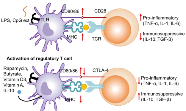
Dendritic cells (DCs) are representative APCs of the mammalian immune system. Their function is to present treated antigens on their cell surface, which are recognized by T cells. They act as messengers between the innate and the adaptive immune systems (Fig.1.3). Immature DCs are stimulated by ligands of toll-like receptors (TLRs) such as pathogen-associated molecular patterns (PAMPs) and inflammatory cytokine, tumor necrosis factor- (TNF-) thereby differentiated into mature DC [8–10]. Then the maturated DCs increase the expression of MHC II and costimulatory molecules, and secrete a wide variety of pro-inflammatory cytokine such as TNF- , IL-1 and IL-6.
Helper T cells which recognize MHC-antigens complexes presented on DCs and become activated. In contact, immature DCs compete with the above-mentioned activation of helper T cells. DCs are maintained in immature state by anti-inflammatory cytokines such as IL-10 and transforming growth factor- (TGF-) and immunomodulatory small molecules such as calcitriol (active form of vitamin D3) and retinoic acids [11]. The immature DCs avoid the activation of helper T cells by inducing anergic response or activating Treg [12]. Induction of Tregs via DCs is important for maitaining the immunological homeostasis.
Fig. 1.1. Antigen presentation by mature and immature dendritic cell.
1.1.2. Approaches to induce immune tolerance for allergic and autoimmune diseases
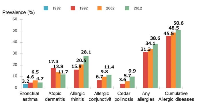
Patients of allergic and autoimmune diseases are increasing worldwide [13,14]. In Japan, the number of patients with autoimmune diseases is increasing year by year, and ulcerative colitis and Parkinson's disease are the most notably increasing (Fig.1.2). And it is known that 50% of Japanese suffer from some allergy (Fig.1.3). Immunological homeostasis is disrupted in the patients; causing an excessive immune response to harmless allergens and self-tissues. Induction of antigen-specific immunotolerance is a way to recover the immunological homeostasis to treat allergies and autoimmune diseases.
In the treatment of allergic diseases toward house mites and pollens, small amount allergens are administered continuously to induce allergen-specific Tregs or anergic response to allergen-specific helper T cells. However, this therapy takes long time, have risk of anaphylaxis .
To overcome the issues of conventional therapy, Kishimoto et al. reported that co-delivery of an immunomodulator and an allergen by encapsulating into nanoparticles (tNPs) improved the therapeutic effect (Fig.1.4) [17]. The nanoparticles engulfed by DCs maintain phenotype of DCs in immature state by the function of immunomodulators and promote differentiation of antigen-specific Tregs by antigen presented on DCs (Fig.1.5).
They succeeded in induce immunotolerance for allergy model mice [18]. Thus, material that achieving co-delivery of immunomodulators and antigens to DCs has high potential to treat diseases in which the balance of immunological homeostasis is disrupted.
Figure. 1.2. Prevalence of autoimmune disease [15]
Figure. 1.3. Prevalence of allergic disease [16]
Fig. 1.4. Different types of tolerogenic nanoparticles
Fig. 1.5. Comparison of A) whole-allergen immunotherapy and B) tNP immunotherapy.
1.2 Contribution of microbiome to host homeostasis
Living organisms coexist with many microorganisms. In humans, there are 10 to 100 trillion microorganisms for every 10 trillion human cells [19,20]. Most of these microorganisms live in the gut. Microbiomes effectively add vast amounts of genes to the human genome, potentially increasing up to 200-fold [21]. As a result, the composition of human microbiomes can be important in health and disease situations. Commensal bacteria interact with the immune system directly or through metabolites and suppresses activities that contribute to homeostatic mechanisms (Fig.1.6). However, the pathogenic bacteria interact specifically with the immune system, disrupt homeostasis, and promote a non-immunogenic hyper-inflammatory response that promotes various gastric diseases [22]. Intestinal lymph nodes contain about 60% of systemic immune cells, and intestinal immunity is also involved in neuron and brain immune function [23,24]. Like the spleen, intestinal lymph nodes function like an organ where immunity is educated [25]. Intestinal
bacteria induce immune tolerance by metabolites produced by metabolism or by direct stimulation. For example, Clostridium, Lactobacillus, Bifidobacterium and Bacteroides protect mice from experimental allergies and colitis by accumulating Tregs [26–28].
Fig. 1.6. Effect of microbiome-modulated metabolites on human health. Altered levels of microbiome-modulated metabolites have been associated with immune-mediated and immune-associated disease risk [29].
1.2.1. Dysbiosis caused by antibiotics
It is known that the intestinal flora are altered by various factors such as diet, exercise, and medication. Among them, administration of antibiotics significantly changes the composition of the intestinal flora and causes destruction (dysbiosis) of the
intestinal flora (Fig1.7) [30].
Dysbiosis caused by antibiotics reduces the number of commensal bacteria which is responsible for immune homeostasis, and increases the number of pathogenic bacteria [31]. As a result, the number of microorganisms that induce the differentiation of Tregs is reduced, so that immuological homeostasis is disrupted to cause allergies and autoimmune diseases. For example, continuous administration of vancomycin to pregnant mice resulted in excessive eosinophilia, elevated allergen-specific IgE levels, and increased airway hyper-reactivity [32]. Antibiotic treated patients also showed increased expression of IgE on basophils and IgE in serum and decreased numbers of Tregs [33].
The balance of the microbiome is one of the important factors for maintaining immunological homeostasis.
Fig. 1.7. Dysbiosis caused by medication and therapy [20].
1.2.2. Utilizing -lactamase to protect microbiome
In 2016, M. Kaleko et al. reported a method to protect microbiome from antibiotics by oral administration of β-lactamases [34,35]. β-Lactamase is an enzyme that degrades
penicillin-type antibiotics. In this method, β-lactamase is orally administered before and after intravenous administration of the antibiotic and delivered to the large intestine.
Thereby, a fraction of antibiotics reached the large intestine was degraded by β-lactamase to protect the intestinal flora. This method can protect the intestinal flora with a small amount of administration of the enzyme because of its high activity and specificity toward antibiotics. However, this promising approach is limited to penicillin-type antibiotics.
1.2.3. Activated carbon as antibiotics adsorbent
Activated carbon for medical use has been used to remove waste and drug overdose in patients with renal failure. In 2018, D. Gunzburg et al. reported a method to protect microbiome from antibiotics by using activated carbon [36,37]. In the method, activated carbon is orally administered before and after oral administration of antibiotics. The activated carbon reached to large intestine to trap antibiotics for protection of the gut flora.
Finally, the activated carbon is excreted as stool (Fig. 1.8). However, activated carbon adsorbs hydrophobic substances nonspecifically which may result in adsorption of essential biological substances such as vitamins and fatty acid.
Fig. 1.8. Activated carbon for adsorbing residual antibiotics in large intestine.
1.3. Overview of this thesis
As mentioned above, the immunological homeostasis is disrupted by several reasons including antibiotic treatment. The disruption of immunological homeostasis results in diseases such as allergy and autoimmune diseases. To rebalance this situation, antigen- specific immunotolerance against allergen and auto-antigen should be induced.
Chapter 2 introduces vitamin-peptide conjugates that achieve co-delivery of immunomodulatory vitamins and antigen peptides to APC. Vitamins were selective modified on the peptide N-terminus that will not affect T cell recognition and MHC binding. Among the designed conjugated, retinoic acid-peptide conjugate showed anti- inflammatory effects, presented antigens on dendritic cells to activate T cells. The conjugates have the potential to treat allergy and auto-immune diseases by efficiently inducing antigen-specific immune tolerance.
In Chapter 3, I designed an antibiotic specific adsorbent to protect microbiome.
Here I targeted vancomycin and utilizing a peptide ligand to capture vancomycin with relatively high affninity. The peptide ligands-loaded microparticles showed high selectivity and capacity for vancomycin, and exerted their effects in vivo to suppress dysbiosis and C. difficil infection associated with dysbiosis.
In Chapter 4, I summarized the conclusions of this paper. Then, I explained the prospects of my research conducted in this thesis.
1.4. References
[1] J. Ermann, C.G. Fathman, Autoimmune diseases: Genes, bugs and failed
regulation, Nat. Immunol. 2 (2001) 759–761. https://doi.org/10.1038/ni0901-759.
[2] J. A. Hemler, E. J. Phillips, M. D, S. A. Mallal, M. B. B. S, P. L. Kendall, The Evolving Story of HLA and the Immunogenetics of Peanut Allergy, Ann Allergy Asthma Immunol. 115 (2015) 471-476. http://doi: 10. 1016/j.anai.2015.10.008 [3] K.M. Spach, F.E. Nashold, B.N. Dittel, C.E. Hayes, IL-10 Signaling Is
Essential for 1,25-Dihydroxyvitamin D 3 -Mediated Inhibition of Experimental Autoimmune Encephalomyelitis , J. Immunol. 177 (2006) 6030–6037.
https://doi.org/10.4049/jimmunol.177.9.6030.
[4] F.E. Nashold, K.A. Hoag, J. Goverman, C.E. Hayes, Rag-1-dependent cells are necessary for 1,25-dihydroxyvitamin D3 prevention of experimental autoimmune encephalomyelitis, J. Neuroimmunol. 119 (2001) 16–29.
https://doi.org/10.1016/S0165-5728(01)00360-5.
[5] T.P. Wypych, B.J. Marsland, Antibiotics as Instigators of Microbial Dysbiosis:
Implications for Asthma and Allergy, Trends Immunol. 39 (2018) 697–711.
https://doi.org/10.1016/j.it.2018.02.008.
[6] L.S.K. Walker, EFIS Lecture: Understanding the CTLA-4 checkpoint in the maintenance of immune homeostasis, Immunol. Lett. 184 (2017) 43–50.
https://doi.org/10.1016/j.imlet.2017.02.007.
[7] D. Saadoun, M. Rosenzwajg, D. Landau, J.C. Piette, D. Klatzmann, P. Cacoub, Restoration of peripheral immune homeostasis after rituximab in mixed
cryoglobulinemia vasculitis, Blood. 111 (2008) 5334–5341.
https://doi.org/10.1182/blood-2007-11-122713.
[8] H. Tsujimoto, P.A. Efron, T. Matsumoto, R.F. Ungaro, A. Abouhamze, S. Ono,
H. Mochizuki, L.L. Moldawer, Maturation of murine bone marrow-derived dendritic cells with poly(I:C) produces altered TLR-9 expression and response to CpG DNA, Immunol. Lett. 107 (2006) 155–162.
https://doi.org/10.1016/j.imlet.2006.09.001.
[9] G. Hartmann, G.J. Weiner, A.M. Krieg, CpG DNA: A potent signal for growth, activation, and maturation of human dendritic cells, Proc. Natl. Acad. Sci. U. S.
A. 96 (1999) 9305–9310. https://doi.org/10.1073/pnas.96.16.9305.
[10] S. Schlickeiser, S. Stanojlovic, C. Appelt, K. Vogt, S. Vogel, S. Haase, T. Ritter, H.-D. Volk, U. Pleyer, B. Sawitzki, Control of TNF-Induced Dendritic Cell Maturation by Hybrid-Type N -Glycans , J. Immunol. 186 (2011) 5201–5211.
https://doi.org/10.4049/jimmunol.1003410.
[11] J.R. Gordon, Y. Ma, L. Churchman, S.A. Gordon, W. Dawicki, Regulatory dendritic cells for immunotherapy in immunologic diseases, Front. Immunol. 5 (2014) 1–19. https://doi.org/10.3389/fimmu.2014.00007.
[12] H. Hasegawa, T. Matsumoto, Mechanisms of tolerance induction by dendritic cells in vivo, Front. Immunol. 9 (2018).
https://doi.org/10.3389/fimmu.2018.00350.
[13] J. Ring, Davos Declaration: Allergy as a global problem, Allergy Eur. J. Allergy Clin. Immunol. 67 (2012) 141–143. https://doi.org/10.1111/j.1398-
9995.2011.02770.x.
[14] M. do C. Borralho, R.F. da Silva, A.S. Santana, R. de M. Caetano, Boas práticas da OMS para laboratórios de microbiologia farmacêutica de microbiologia farmacêutica, Série Rede Parf. 11 (2013) 1–37.
https://doi.org/10.1016/j.jaut.2009.09.008.Recent.
[15] Japan intractable diseases information center, Transition data from 1975 to 2004, https://www.nanbyou.or.jp/entry/1356#p01
[16] M. Nishima, H. Odashima, K. Ota, N. Ota, K. Okazaki, M. Kanaya, N. Hisada, T.
Kumamoto, T. Koga, N. Kobayashi, K. Satomi, Y. Shimada, M. Shimomura, M.
Suda, I. Sunagawa, S.H. Osaka, Y. Nagata, T. Nakamura, K. Nishikawa, K.
Hiraba, T. Fujino, T. Fujiwara, S. Honjo, T. Maeda, S. Matsumoto, T. Minami, Y. Miyazato, Prevalence Survey of Allergic Diseases in West Elementary School Children: Comparison in 1992, 2002, and 2012, Journal of Japanese Society of Pediatric Allergy, 27 (2013) 149-169, https://doi.org/10.3388/jspaci.27.149 [17] T.K. Kishimoto, R.A. Maldonado, Nanoparticles for the induction of antigen-
specific immunological tolerance, Front. Immunol. 9 (2018).
https://doi.org/10.3389/fimmu.2018.00230.
[18] R.A. Maldonado, R.A. LaMothe, J.D. Ferrari, A.H. Zhang, R.J. Rossi, P.N.
Kolte, A.P. Griset, C. O’Neil, D.H. Altreuter, E. Browning, L. Johnston, O.C.
Farokhzad, R. Langer, D.W. Scott, U.H. Von Andrian, T.K. Kishimoto, Polymeric synthetic nanoparticles for the induction of antigen-specific
immunological tolerance, Proc. Natl. Acad. Sci. U. S. A. 112 (2015) E156–E165.
https://doi.org/10.1073/pnas.1408686111.
[19] R.E. Ley, D.A. Peterson, J.I. Gordon, Ecological and evolutionary forces shaping microbial diversity in the human intestine, Cell. 124 (2006) 837–848.
https://doi.org/10.1016/j.cell.2006.02.017.
[20] C. Bruhn, Wohngemeinschaft Haut, Dtsch. Apotheker Zeitung. 157 (2017) e1002. https://doi.org/10.1371/journal.pbio.1002533.
[21] P.J. Turnbaugh, R.E. Ley, M. Hamady, C.M. Fraser-Liggett, R. Knight, J.I.
Gordon, The Human Microbiome Project, Nature. 449 (2007) 804–810.
https://doi.org/10.1038/nature06244.
[22] D. Toor, M.K. Wasson, P. Kumar, G. Karthikeyan, N.K. Kaushik, C. Goel, S.
Singh, A. Kumar, H. Prakash, Dysbiosis disrupts gut immune homeostasis and
promotes gastric diseases, Int. J. Mol. Sci. 20 (2019) 1–14.
https://doi.org/10.3390/ijms20102432.
[23] M. Yousaf, Inayatullah, A.R. Khan, N. Ahmad, S. Ali, The presentation pattern of otitis media with effusion, J. Med. Sci. 17 (2009) 53–55.
https://doi.org/10.1126/scitranslmed.3009759.The.
[24] E.Y. Hsiao, S.W. McBride, S. Hsien, G. Sharon, E.R. Hyde, T. McCue, J.A.
Codelli, J. Chow, S.E. Reisman, J.F. Petrosino, P.H. Patterson, S.K. Mazmanian, Microbiota modulate behavioral and physiological abnormalities associated with neurodevelopmental disorders, Cell. 155 (2013) 1451–1463.
https://doi.org/10.1016/j.cell.2013.11.024.
[25] Q. Zhao, C.O. Elson, Adaptive immune education by gut microbiota antigens, Immunology. 154 (2018) 28–37. https://doi.org/10.1111/imm.12896.
[26] T. Tanoue, K. Atarashi, K. Honda, Development and maintenance of intestinal regulatory T cells, Nat. Rev. Immunol. 16 (2016) 295–309.
https://doi.org/10.1038/nri.2016.36.
[27] K. Atarashi, T. Tanoue, K. Oshima, W. Suda, Y. Nagano, H. Nishikawa, S.
Fukuda, T. Saito, S. Narushima, K. Hase, S. Kim, J. V. Fritz, P. Wilmes, S. Ueha, K. Matsushima, H. Ohno, B. Olle, S. Sakaguchi, T. Taniguchi, H. Morita, M.
Hattori, K. Honda, Treg induction by a rationally selected mixture of Clostridia strains from the human microbiota, Nature. 500 (2013) 232–236.
https://doi.org/10.1038/nature12331.
[28] S.K. Lathrop, S.M. Bloom, S.M. Rao, K. Nutsch, C.W. Lio, N. Santacruz, D.A.
Peterson, T.S. Stappenbeck, C.S. Hsieh, Peripheral education of the immune system by colonic commensal microbiota, Nature. 478 (2011) 250–254.
https://doi.org/10.1038/nature10434.
[29] E. Blacher, M. Levy, E. Tatirovsky, E. Elinav, Microbiome-Modulated
Metabolites at the Interface of Host Immunity, J. Immunol. 198 (2017) 572–580.
https://doi.org/10.4049/jimmunol.1601247.
[30] L.E. Papanicolas, D.L. Gordon, S.L. Wesselingh, G.B. Rogers, Not Just
Antibiotics: Is Cancer Chemotherapy Driving Antimicrobial Resistance?, Trends Microbiol. 26 (2018) 393–400. https://doi.org/10.1016/j.tim.2017.10.009.
[31] Y. and T.H. Belkaid, Role of the Microbiota in Immunity and inflammation Yasmine, Cell. 157 (2015) 121–141.
https://doi.org/10.1016/j.cell.2014.03.011.Role.
[32] S.L. Russell, M.J. Gold, M. Hartmann, B.P. Willing, L. Thorson, M. Wlodarska, N. Gill, M.R. Blanchet, W.W. Mohn, K.M. McNagny, B.B. Finlay, Early life antibiotic-driven changes in microbiota enhance susceptibility to allergic asthma, EMBO Rep. 13 (2012) 440–447. https://doi.org/10.1038/embor.2012.32.
[33] S.L. Russell, M.J. Gold, B.P. Willing, L. Thorson, K.M. McNagny, B.B. Finlay, Perinatal antibiotic treatment affects murine microbiota, immune responses and allergic asthma, Gut Microbes. 4 (2013) 158–164.
https://doi.org/10.4161/gmic.23567.
[34] M. Kaleko, J.A. Bristol, S. Hubert, T. Parsley, G. Widmer, S. Tzipori, P.
Subramanian, N. Hasan, P. Koski, J. Kokai-Kun, J. Sliman, A. Jones, S.
Connelly, Development of SYN-004, an oral beta-lactamase treatment to protect the gut microbiome from antibiotic-mediated damage and prevent Clostridium difficile infection, Anaerobe. 41 (2016) 58–67.
https://doi.org/10.1016/j.anaerobe.2016.05.015.
[35] T.O. Syn-, J.F. Kokai-kun, T. Roberts, O. Coughlin, E. Sicard, M. Rufiange, R.
Fedorak, C. Carter, M.H. Adams, J. Longstreth, H. Whalen, crossm Clinical Studies, 61 (2017) 14–16.
[36] J. De Gunzburg, A. Ghozlane, A. Ducher, E. Le Chatelier, X. Duval, E. Ruppé,
L. Armand-Lefevre, F. Sablier-Gallis, C. Burdet, L. Alavoine, E. Chachaty, V.
Augustin, M. Varastet, F. Levenez, S. Kennedy, N. Pons, F. Mentré, A.
Andremont, Protection of the human gut microbiome from antibiotics, J. Infect.
Dis. 217 (2018) 628–636. https://doi.org/10.1093/infdis/jix604.
[37] C. Burdet, S. Sayah-Jeanne, T.T. Nguyen, C. Miossec, N. Saint-Lu, M. Pulse, W.
Weiss, A. Andremont, F. Mentré, J. De Gunzburg, Protection of hamsters from mortality by reducing fecal moxifloxacin concentration with DAV131A in a model of moxifloxacin-induced Clostridium difficile colitis, Antimicrob. Agents Chemother. 61 (2017) 1–9. https://doi.org/10.1128/AAC.00543-17.
CHAPTER 2
Development of vitamin-peptide conjugate for induction of antigen-specific immunotolerance
2.1. Introduction
Recently, patients with allergic and autoimmune diseases have been rapidly increasing worldwide [1,2]. To treat these diseases, antigen-specific immunotolerance toward allergens and auto-antigens should be induced [3]. The immunotolerance is induced by dendritic cells (DCs) with tolerogenic phenotype which are called tolerogenic DCs (tDCs). tDCs induce an anergetic response to helper T cells as well as differentiate naïve T cells to regulatory T cells (Treg cells), which are executer of antigen-specific immunotolerance. Treg cells expressing CD4 (CD4+ Treg) compete with helper T cells to induce immunotolerance, while Treg cells expressing CD8 (CD8+ Treg) contribute to induce immunotolerance by several pathways such as secretion of cytokines [4–7].
It has been reported that induction of tDC is facilitated by small molecules such as vitamins and rapamycin [8,9]. For efficient induction of antigen-specific immunotolerance, nanoparticle formulations containing both antigens and tDC-inducing molecules have been reported [10]. We also reported previously nanoparticles of allergen proteins incorporated with all-trans retinoic acid (ATRA) which is one of the representative tDC-inducing molecules [11].
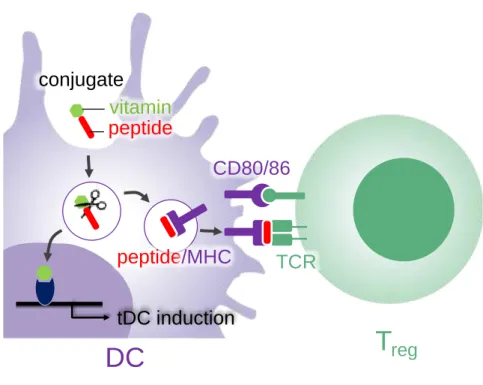
Here we proposed peptide-vitamin conjugates to achieve co-delivery of vitamins and antigen peptide to DCs for effective induction of tDCs and Treg cells (Fig.2.1). We chose ATRA and vitamin D3 (vD3) [12–15] as inducing molecules of tDC. The
conjugates are taken up by DC and release antigen peptides and vitamins from the conjugates by lysosomal peptidases. Antigen peptide is presented on the surface of DC through intracellular process, and vitamins bind to corresponding nuclear receptors to induce differentiation of tDC thereby facilitating the Treg induction. The antigen peptide and vitamin were connected with three arginine residues (Fig. 2.2) which is a cleavable sequence of an endosomal peptidases such as cathepsin B [16,17]. The three-arginine linker will enhance the solubility of the conjugates containing hydrophobic vitamins. We established general scheme to synthesize the conjugates and investigated whether the scheme shown in Fig. 2.1 was conducted in vitro.
Fig. 2.1. Schematic representation of induction of tDC and Treg by peptide-vitamin conjugates. Conjugate taken up DC releases an antigen peptide and a vitamin by lysosomal peptidases. The resulting vitamin bound to a corresponding nuclear receptor to differentiate DC to tDC. The resulting peptide is presented on a major histocompatibility
T
regCD80/86
peptide/MHC TCR
DC
conjugate vitamin peptide
tDC induction
complex (MHC) protein to stimulate a T cell receptor (TCR) thereby inducing Treg cell.
Fig. 2.2. Structure of peptide-ATRA (1) and peptide-vD3 conjugates (2). Three arginine residues on N-terminus are linker between antigen peptide (SIINFEKL) and vitamins.
2.2. Materials and Methods
2.2.1. Synthesis of lysine residue-protected peptide
Peptide was synthesized on Nova syn TGA resin at a 0.2-mmol scale by Fmoc solid-phase peptide synthesis. 1-Hydroxy-1H-benzotriazole hydrate and O-(1H-benzotriazol-1-yl)- N,N,N',N'-tetramethyluronium hexafluorophosphate were used as the condensing agents.
For the N-terminal amino acid, tert-butoxycarbonyl (Boc)-protected one was used. After the completion of elongation of peptide main chain, 4-methyltrityl (Mtt) protection group of lysine residue was removed by 10% trifluoroacetic acid (TFA) / dichloromethane (DCM). Then, N-(9-fluorenylmethoxycarbonyloxy) (Fmoc) group was modified on the lysine residue by using Fmoc succinimide (Fmoc-OSu). The peptide was then cleaved
from the resin by treatment with TFA containing 2.5% (v/v), triisopropyl silane (TIS) and 2.5% (v/v) water, and purified by reversed-phase high-performance liquid chromatography (HPLC) (Hitachi Chromatomaster, Tokyo, Japan) on an Altantis dC18 OBD column (19x100 mm) (waters corporation, MA, USA) using a mobile phase consisting of acetonitrile/0.1%TFA and H2O/0.1%TFA. The obtained lysine residue- protected peptide was identified by a matrix-assisted laser desorption ionization time-of- flight (MALDI-TOF) mass spectrometer (Bruker Daltonics, Billerica, MA, USA).
2.2.2. Synthesis of conjugate 1 and 2
N-Hydroxysuccinimide ester (NHS) of vitamins (ATRA-NHS, vD3-succinic acid-NHS) were synthesized according to literatures [18,19]. The lysine residue-protected peptide (1 mM) and vitamin-NHS (10 mM) were dissolved in dimethylformamide (DMF). N,N- Diisopropylethylamine (DIEA) (10 mM) was added to the solution and reacted at room temperature overnight under nitrogen. After the reaction, Fmoc group of lysine residue was removed with 20% piperidine (PPD)/DMF and the resulting conjugate was purified by reversed-phase HPLC. The obtained conjugates were identified by a MALDI-TOF mass spectrometer (Fig.2.3).
2.2.3. Cell line and culture
RAW 264.7 macrophages transfected with secreted alkaline phosphatase (SEAP) as a reporter gene under the transcriptional control of a nuclear factor-kappa B (NF-κB) response element (Novusbio) were maintained in Dulbecco’s Modified Eagle’s Medium (DMEM) supplemented with 10% (v/v) fetal bovine serum (FBS), 100 U/mL penicillin, 100 µg/mL streptomycin, 0.25 µg/mL amphotericin B (Invitrogen, grand island, NY, USA) and 500 μg/mL G418 (only for the RAW 264.7 macrophages transfected with SEAP). DC2.4 cells were kindly provided by Dr. Kenneth Rock (University of
Massachusetts Medical Center, Worcester, MA, USA) [20]. The cells were maintained in Roswell Park Memorial Institute media (RPMI1640) medium supplemented with 10%
(v/v) FBS, 100 U/mL penicillin, 100 µg/mL streptomycin, and 0.25 µg/mL amphotericin B. CD8OVA1.3 T hybridoma cell were kindly provided by Dr. Clifford V. Harding (Case Western Reserve University, Cleveland, OH) [21]. The cells were maintained in RPMI1640 medium supplemented with 10% (v/v) FBS, 100 U/mL penicillin, 100 µg/mL streptomycin, 0.25 µg/mL amphotericin B, and 2-mercaptoethanol. Cells were incubated in a humidified atmosphere containing 5% CO2 at 37°C.
2.2.4. Cell viability
DC2.4 cells (2×10⁵ cell/well) were seeded in 48-well plates, grown for 24 h then exposed for 24 h at 37°C to conjugate 1 or 2 in culture media. Propidium iodide (PI) was added a few minutes before analyzing the cells by using a Tali® image-based cytometer (Thermo Fisher Scientific). Excitation and emission wavelengths were 488 and 620 nm respectively.
2.2.5. Alkaline phosphatase assay
RAW 264.7 macrophages transfected with SEAP (4×10⁴ cells/well) were cultured in a 48-well plate with complete DMEM. After incubation overnight, the medium was replaced with complete DMEM containing conjugate 1 or 2. The supernatant were discarded and the cells were treated with lipopolysaccharide (LPS) (Sigma-Aldrich) (10 ng/mL) for 24h. After incubation, the supernatants were mixed with an equal volume of alkaline phosphatase substrate (1 mg/mL p-nitrophenylphosphate) and incubated at room temperature for 1.5 h and then 3 N NaOH was added to stop the reaction. The optical density of the solution was measured at 405 nm with a microplate reader (Wallac ARVO.SX 1420 Multilabel counter).
2.2.6. Quantitative real-time-PCR assays (qRT-PCR)
DC2.4 cells (2×10⁵ cell/well) were plated in 24-well plates with RPMI1640. After incubation overnight, the medium was replaced with complete RPMI1640 containing conjugate 1. After 24 h, LPS (final conc. 10 ng/mL) was added and the cells were incubated for 24 h. The total RNA from the cells was prepared using isoPlus reagent (Takara). The samples were reverse-transcribed using the SuperScript III First-Strand Synthesis System (Invitrogen) and the synthesized cDNA was used as a template in qPCR experiments performed with a LightCycler 1.5 (Roche Diagnostics, Germany) and analyzed with LightCycler Manager software 3.5 (Roche Diagnostics, Germany). The relative expression level was calculated by the ΔΔCt method using Gapdh as a reference gene. All primers were purchased from Takara-Clontech Laboratories (Japan) (Table 2.1).
2.2.7. Flow cytometry
MHC-II, CD80, and CD86 expression levels were measured after incubating DC2.4 cells with conjugate 1 or ATRA (1 µM) for overnight. Cells were washed with DPBS and re- suspended in FACS buffer (2 wt% BSA containing PBS) and stained with APC-anti mouse CD80 mAb (BioLegend), APC-anti mouse CD86 mAb (BioLegend) and PE-anti mouse MHC-II mAb (BioLegend) by incubating in the dark at 4 °C for 15 min. Cells were washed with FACS buffer and analyzed by a cytoflex flow cytometer (Backman coulter).
2.2.8. Antigen presentation assay
DC2.4 cells (2×10⁵ cell/well) were plated in 24-well plates with RPMI1640 and incubated 24 h. Conjugate 1 or free peptide (0.1 µM) were added to each well and incubated for 6 h. After discarding supernatant, suspension of CD8OVA1.3 T cells (2×10⁵ cell/well) was
added in each well, then the plates were centrifuged at 450 g to initiate contact between the cells. The cells were incubated together at 37 °C. Then, the response of CD8OVA1.3 T cells was determined by measuring IL-2 levels in the supernatants with enzyme-linked immunosorbent assay (ELISA) (mouse IL-2, Invitrogen).
2.3. Results and Discussion
2.3.1. Synthesis of vitamin-peptide conjugate
Here we used peptide sequence (SIINFEKL) derived from ovalbumin 257-264, which is widely used as a model antigen and can be presented on an MHC class I. We designed conjugate 1 and 2 with a vitamin on N-terminus of the peptide through a linker of three arginine residues (Fig. 2.2). We failed to synthesize the conjugates on a resin because ATRA and vD3 did not tolerate to strongly acidic condition which was used for cleavage of peptide from a resin. Thus we decided to modify the vitamins on the peptide in solution phase and established general scheme to synthesize conjugates which is applicable to peptides containing lysine residue (Scheme 2.1). After the synthesis of the peptide with N-terminus Boc protection on the resin, the protecting group of the lysine residue was converted from Mtt to acid-tolerable Fmoc, then the peptide was cleaved from the resin and purified. To the peptide with a protected lysine, NHS ester of ATRA was modified on N-terminus and finally Fmoc group was removed by weakly basic condition to which ATRA was tolerable. Conjugate 2 with vD3 was prepared by the same scheme with conjugate 1, except for NHS ester of succinated vD3 was used for conjugation.
Scheme 2.1. Synthesis of 1
Fig. 2.3. MALDI-TOF mass spectra and chromatogram of conjugate 1 and 2.
2.3.2. Cytotoxicity of conjugates
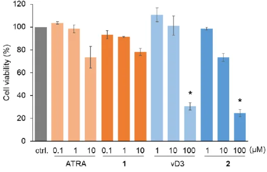
We compared cytotoxicity of the conjugates with free vitamins on a mouse cell line derived from DC (DC2.4). The cell viability was evaluated by PI staining after 24h. As shown in Fig.2.4, cytotoxicity of free vitamins and the corresponding conjugates was almost same. ATRA and conjugate 1 did not show significant cytotoxicity up to the examined concentration range. vD3 and conjugate 2 showed significant toxicity at 100 μM. The same cytotoxic profiles of free vitamins and the corresponding conjugates indicates that the conjugates which have amphiphilic nature due to the hydrophilic peptide
and hydrophobic vitamins does not perturb cell membrane to induce cytotoxicity.
Fig. 2.4. Viability of DC2.4 cells after treatment with conjugates or free vitamins for 24 h. Dead cells were stained with PI and counted using image cytometer. Data represent the mean ± SD (n = 3). * P < 0.05, as compared with non-treated group (ctrl).
2.3.3. Alkaline phosphatase assay
We investigated release of vitamins from the conjugates using mouse macrophage cell line. Here we used RAW 264.7 macrophage transfected with SEAP as a reporter gene induced by the NF-κB transcription factor. If the conjugate shows anti-inflammatory response, free ATRA or vD3 is released from the conjugate. The cells were incubated with the conjugate or free vitamins then stimulated with LPS. As shown in Fig.2.5, free ATRA and vD3 showed suppression of macrophage activation at concentrations of 0.1 and 10 μM, respectively. The requirement of free vD3 for higher concentration will result from the requirement of conversion of vD3 to its active form by intracellular enzymes for binding to a nuclear receptor [22]. As for the conjugates, vD3 conjugate 2 did not show
suppressive effect, while ATRA conjugate 1 showed suppressive effect from the concentration of 1 μM. Although conjugate 1 required higher concentrations than free ATRA, the suppressive effect of 1 showed the release of free ATRA from 1. The requirement of higher concentration of 1 than free ATRA may result from lower efficacy of cellular uptake as well as requirement of enzymatic process to release free ATRA.
Fig. 2.5. Suppressive effect of conjugates and free vitamins on the LPS-induced NF-κB activation in RAW264.7 macrophages. After 24 h from the addition of each conjugate or free vitamin to macrophages, LPS (10 ng/mL) was added to the medium and incubated for a further 24 h. Absorbance resulting from substrate reacted with SEAP in supernatant was measured at 405 nm. Data are the mean ± SD (n = 3). *P < 0.05, **P < 0.01.
2.3.4. Suppression of LPS-induced inflammatory response of DC by ATRA conjugate
Next, we investigated the effects of ATRA conjugate 1 on the gene expression of cytokines in DC2.4 cells stimulated with LPS. As shown in Fig.2.6, both free ATRA and
conjugate 1 suppressed the expression of inflammatory cytokines, IL-6 and TNF-.
However, the expression of anti-inflammatory cytokines (IL-10, TGF-) was not affected by both ATRA and conjugate 1. These results were consistent with previous reports about the response of bone marrow derived DCs to ATRA [23–25]. Therefore, free ATRA would be released from 1 in DC2.4 cells to induce suppressive effect against LPS-induced inflammatory response.
Table 2.1. Primer sequences used for RT-qPCR.
Transcript Forward primer Reverse primer
Il6 5’-CCACTTCACAAGTCGGAGGCTTA-3’ 5’-CCAGTTTGGTAGCATCCATCATTTC-3’
Tnf 5’-ACCCTCACACTCAGATCATCTTC-3’ 5’-TGGTGGTTTGCTACGACGT-3’
Gapdh 5’-CCCAGCAAGGACACTGAGCAAG-3’ 5’-GGTCTGGGATGGAAATTGTGAGGG-3’
Tgf 5’-GTGTGGAGCAACATGTGGAACTCTA-3’ 5’-CGCTGAATCGAAAGCCCTGTA-3’
Il-10 5’-TGCCTTCAGCCAGGTGAAGACTTTC-3’ 5’-CTTGATTTCTGGGCCATGCTTCTCTG-3’
Fig. 2.6. Suppressive effect of conjugates on the gene expression of cytokines in DC2.4 cells. After addition of conjugates to dendritic cells for 24 h, LPS (10 ng/mL) was added and incubated for a further 24 h. And gene expression was evaluated by qRT-PCR. Data are the mean ± SD (n = 3). *P < 0.05, **P < 0.01, ***P < 0.001.
2.3.5. Expression of ligand on surface of DC2.4 cells
We compared effect of conjugate 1 with free ATRA on the ligand expression of DC2.4 cells. As shown in Fig.2.7, conjugate 1 showed similar effect on the ligand expression;
reduction of MHC class II and no effect on costimulatory ligands CD80 and CD86. The effect of ATRA on the ligands expression of DC is controversial because it seems to differ up to the experimental setup. However, the reduction of MHC class II has been observed for many reports [26–29], and this phenotype is typical one in tDC [30].
Fig. 2.7 Comparison of effect of conjugate 1 with free ATRA on the ligand expression of DC2.4 cells. After 24 h from the addition of conjugate or ATRA (0.1 μM) to DC2.4 cells, expression of each protein was analyzed by flow cytometry.
2.3.6. Conjugate induction of antigen-specific immune responses
Presentation of peptide from conjugate 1 on DC2.4 was evaluated by stimulation of CD8+ T cell hybridoma. DC2.4 cells were pulsed with conjugate 1 and to this was added CD8OVA 1.3 T cell hybridoma (which can be activated by OVA257-264/Kb complex) [21]. Stimulation of the T cell hybridoma by DC2.4 was evaluated by secretion of IL-2.
As shown in Fig.2.8, conjugate 1 induced production of IL-2 though the induction efficacy was lower than free peptide (SIIFEKL). This result indicated that peptide was cleaved from the conjugate in DC2.4 cells and presented on the cell surface.
Fig. 2.8. Evaluation of antigen presentation on DC2.4 from conjugate 1 to CD8+ T cell hybridoma. DC2.4 cells were pulsed with conjugate 1 or free peptide (0.1 μM) for 6 h and then co-cultured with CD8OVA1.3 T cells for 18 h. IL-2 production in supernatant was measured by ELISA. Data are the mean ±SD (n = 3). *P < 0.05, ***P < 0.001, as compared with ctrl group (non-treated DC + hybridoma).
2.4. Conclusions
We established synthetic scheme of peptide-vitamin conjugates. ATRA conjugate was edited in both macrophage and DCs to show the anti-inflammatory effect derived from ATRA. ATRA conjugate was also edited in DCs to present peptide/MHC class I complex to stimulate T cell hybridoma. The conjugates developed here will be useful to facilitate the induction of antigen-specific immunotolerance for the therapy of allergic and autoimmune diseases.
2.5. References
[1] E.N. Grant, M. Robin Wagner, K.B. Weiss, Observations on emerging patterns of asthma in our society, J. Allergy Clin. Immunol. 104 (1999) 1–9.
https://doi.org/10.1016/S0091-6749(99)70268-X.
[2] W.-J. Song, M.-G. Kang, Y.-S. Chang, S.-H. Cho, Epidemiology of adult asthma in Asia: toward a better understanding, Asia Pac. Allergy. 4 (2014) 75.
https://doi.org/10.5415/apallergy.2014.4.2.75.
[3] M. Miyara, K. Wing, S. Sakaguchi, Therapeutic approaches to allergy and autoimmunity based on FoxP3+ regulatory T-cell activation and expansion, J.
Allergy Clin. Immunol. 123 (2009) 749–755.
https://doi.org/10.1016/j.jaci.2009.03.001.
[4] A. Schmidt, N. Oberle, P.H. Krammer, Molecular mechanisms oftreg-mediatedt cell suppression, Front. Immunol. 3 (2012) 1–20.
https://doi.org/10.3389/fimmu.2012.00051.
[5] M. Caridade, L. Graca, R.M. Ribeiro, Mechanisms underlying CD4+ Treg immune regulation in the adult: From experiments to models, Front. Immunol. 4 (2013) 1–
9. https://doi.org/10.3389/fimmu.2013.00378.
[6] Y. Yu, X. Ma, R. Gong, J. Zhu, L. Wei, J. Yao, Recent advances in CD8+
regulatory t cell research (Review), Oncol. Lett. 15 (2018) 8187–8194.
https://doi.org/10.3892/ol.2018.8378.
[7] S. Bézie, I. Anegon, C. Guillonneau, Advances on CD8+ Treg Cells and Their Potential in Transplantation, Transplantation. 102 (2018) 1467–1478.
https://doi.org/10.1097/TP.0000000000002258.
[8] A. Schmidt, M. Eriksson, M.M. Shang, H. Weyd, J. Tegnér, Comparative analysis of protocols to induce human CD4+Foxp3+ regulatory T cells by combinations of IL-2, TGF-beta, retinoic acid, rapamycin and butyrate, PLoS One. 11 (2016) 1–31.
https://doi.org/10.1371/journal.pone.0148474.
[9] J.R. Gordon, Y. Ma, L. Churchman, S.A. Gordon, W. Dawicki, Regulatory dendritic cells for immunotherapy in immunologic diseases, Front. Immunol. 5 (2014) 1–19. https://doi.org/10.3389/fimmu.2014.00007.
[10] R.A. Maldonado, R.A. LaMothe, J.D. Ferrari, A.H. Zhang, R.J. Rossi, P.N. Kolte, A.P. Griset, C. O’Neil, D.H. Altreuter, E. Browning, L. Johnston, O.C. Farokhzad, R. Langer, D.W. Scott, U.H. Von Andrian, T.K. Kishimoto, Polymeric synthetic nanoparticles for the induction of antigen-specific immunological tolerance, Proc.
Natl. Acad. Sci. U. S. A. 112 (2015) E156–E165.
https://doi.org/10.1073/pnas.1408686111.
[11] K. Zai, K. Yuzuriha, A. Kishimura, T. Mori, Y. Katayama, Preparation of complexes between ovalbumin nanoparticles and retinoic acid for efficient induction of Tolerogenic dendritic cells, Anal. Sci. 34 (2018) 1243–1248.
https://doi.org/10.2116/analsci.18P252.
[12] T. Nikolic, B.O. Roep, Regulatory multitasking of tolerogenic dendritic cells - lessons taken from vitamin D3-treated tolerogenic dendritic cells, Front. Immunol.
4 (2013) 1–13. https://doi.org/10.3389/fimmu.2013.00113.
[13] G.B. Ferreira, C.A. Gysemans, J. Demengeot, J.P.M.C.M. da Cunha, A.-S.
Vanherwegen, L. Overbergh, T.L. Van Belle, F. Pauwels, A. Verstuyf, H. Korf, C.
Mathieu, 1,25-Dihydroxyvitamin D 3 Promotes Tolerogenic Dendritic Cells with Functional Migratory Properties in NOD Mice , J. Immunol. 192 (2014) 4210–4220. https://doi.org/10.4049/jimmunol.1302350.
[14] S. Agrawal, S. Ganguly, A. Tran, P. Sundaram, A. Agrawal, Retinoic acid treated human dendritic cells induce T regulatory cells via the expression of CD141 and GARP which is impaired with age, Aging (Albany. NY). 8 (2016) 1223–1235.
https://doi.org/10.18632/aging.100973.
[15] G. Bakdash, L.T.C. Vogelpoel, T.M.M. Van Capel, M.L. Kapsenberg, E.C. De Jong, Retinoic acid primes human dendritic cells to induce gut-homing, IL-10- producing regulatory T cells, Mucosal Immunol. 8 (2015) 265–278.
https://doi.org/10.1038/mi.2014.64.
[16] S.S. Cotrin, L. Puzer, W.A. De Souza Judice, L. Juliano, A.K. Carmona, M.A.
Juliano, Positional-scanning combinatorial libraries of fluorescence resonance energy transfer peptides to define substrate specificity of carboxydipeptidases:
Assays with human cathepsin B, Anal. Biochem. 335 (2004) 244–252.
https://doi.org/10.1016/j.ab.2004.09.012.
[17] G. Hook, J.S. Jacobsen, K. Grabstein, M. Kindy, V. Hook, Cathepsin B is a new drug target for traumatic brain injury therapeutics: Evidence for E64d as a promising lead drug candidate, Front. Neurol. 6 (2015) 1–27.
https://doi.org/10.3389/fneur.2015.00178.
[18] G. Magoulas, D. Papaioannou, E. Papadimou, D. Drainas, Preparation of spermine conjugates with acidic retinoids with potent ribonuclease P inhibitory activity, Eur.
J. Med. Chem. 44 (2009) 2689–2695.
https://doi.org/10.1016/j.ejmech.2009.01.001.
[19] S. Patil, S. Gawali, S. Patil, S. Basu, Synthesis, characterization and in vitro evaluation of novel vitamin D3 nanoparticles as a versatile platform for drug delivery in cancer therapy, J. Mater. Chem. B. 1 (2013) 5742–5750.
https://doi.org/10.1039/c3tb21176b.
[20] Z. Shen, G. Reznikoff, G. Dranoff, K.L. Rock, Cloned dendritic cells can present exogenous antigens on both MHC class I and class II molecules., J. Immunol. 158 (1997) 2723–30. http://www.ncbi.nlm.nih.gov/pubmed/9058806.
[21] J.D. Pfeifer, M.J. Wick, R.L. Roberts, K. Findlay, S.J. Normark, C. V. Harding, Phagocytic processing of bacterial antigens for class I MHC presentation to T cells,
Nature. 361 (1993) 359–362. https://doi.org/10.1038/361359a0.
[22] H. Sigmundsdottir, J. Pan, G.F. Debes, C. Alt, A. Habtezion, D. Soler, E.C.
Butcher, DCs metabolize sunlight-induced vitamin D3 to “program” T cell attraction to the epidermal chemokine CCL27, Nat. Immunol. 8 (2007) 285–293.
https://doi.org/10.1038/ni1433.
[23] W. Dawicki, C. Li, J. Town, X. Zhang, J.R. Gordon, Therapeutic reversal of food allergen sensitivity by mature retinoic acid–differentiated dendritic cell induction of LAG3+CD49b−Foxp3− regulatory T cells, J. Allergy Clin. Immunol. 139 (2017) 1608-1620.e3. https://doi.org/10.1016/j.jaci.2016.07.042.
[24] K.A. Wojtal, L. Wolfram, I. Frey-Wagner, S. Lang, M. Scharl, S.R. Vavricka, G.
Rogler, The effects of vitamin A on cells of innate immunity in vitro, Toxicol. Vitr.
27 (2013) 1525–1532. https://doi.org/10.1016/j.tiv.2013.03.013.
[25] Y. Qiang, J. Xu, C. Yan, H. Jin, T. Xiao, N. Yan, L. Zhou, H. An, X. Zhou, Q.
Shao, S. Xia, Butyrate and retinoic acid imprint mucosal-like dendritic cell development synergistically from bone marrow cells, Clin. Exp. Immunol. 189 (2017) 290–297. https://doi.org/10.1111/cei.12990.
[26] T. Feng, Y. Cong, H. Qin, E.N. Benveniste, C.O. Elson, Generation of Mucosal Dendritic Cells from Bone Marrow Reveals a Critical Role of Retinoic Acid, J.
Immunol. 185 (2010) 5915–5925. https://doi.org/10.4049/jimmunol.1001233.
[27] L. Saurer, K.C. McCullough, A. Summerfield, In Vitro Induction of Mucosa- Type Dendritic Cells by All- Trans Retinoic Acid , J. Immunol. 179 (2007) 3504–
3514. https://doi.org/10.4049/jimmunol.179.6.3504.
[28] D. Liang, A. Zuo, H. Shao, W.K. Born, R.L. O’Brien, H.J. Kaplan, D. Sun, Retinoic acid inhibits CD25+ dendritic cell expansion and γδ T-cell activation in experimental autoimmune uveitis., Invest. Ophthalmol. Vis. Sci. 54 (2013) 3493–
3503. https://doi.org/10.1167/iovs.12-11432.
[29] L. Abdelhamid, H. Hussein, M. Ghanem, N. Eissa, Retinoic acid-mediated anti- inflammatory responses in equine immune cells stimulated by LPS and allogeneic mesenchymal stem cells, Res. Vet. Sci. 114 (2017) 225–232.
https://doi.org/10.1016/j.rvsc.2017.05.006.
[30] H. Torres-Aguilar, M. Blank, L.J. Jara, Y. Shoenfeld, Tolerogenic dendritic cells in autoimmune diseases. Crucial players in induction and prevention of
autoimmunity, Autoimmun. Rev. 10 (2010) 8–17.
https://doi.org/10.1016/j.autrev.2010.07.015.
CHAPTER 3
Protection of gut microbiome from antibiotics:
development of a vancomycin-specific adsorbent with high adsorption capacity
3.1. Introduction
Antibiotics are used extensively for the treatment of infectious diseases. However, during antibiotic treatment, the non-absorbed fraction of orally administered antibiotics, along with the fraction excreted into the upper intestine via bile of both orally and parenterally administered antibiotics, reaches the large intestine where it affects the gut microbiome to induce dysbiosis [1]. Dysbiosis in the gut microbiome results in various consequences in the short and long term, including Clostridioides difficile infection (CDI) [2], the generation of antibiotic-resistant bacteria [3], allergies, and metabolic syndromes [4].
To avoid gut dysbiosis induced by antibiotics, strategies to protect the gut microbiome during antibiotic treatment need to be developed. Two strategies have been reported to date. One of these strategies is the oral administration of β-lactamase to degrade residual β-lactam antibiotics that reach the large intestine [5–8]. It has been reported recently that a clinical trial of this therapy for the suppression of antibiotic- induced CDI was successful [9]. However, this therapy is only applicable to β-lactam antibiotics. The other reported strategy to protect the gut microbiome is using activated charcoal (AC) as an adsorbent of antibiotics in the large intestine [10,11]. Utilizing nonspecificity in adsorption of hydrophobic molecules, AC is potentially applicable to a variety of antibiotics. However, the nonspecific characteristics of AC pose the risk of side
effects; they may affect the metabolism of host animals by the nonspecific adsorption of biological compounds in the intestinal fluid, such as bile acids and essential micronutrients. In the treatment of patients with renal failure, orally administered AC nonspecifically adsorbed bile acids [12], which is suspected to be a risk factor for cardiovascular disease [13]. It was reported that AC did not change the amount of necessary trace metal ions such as iron, zinc and manganese in the digestive tract in animal study [14]. Thus, as for metal ions, such AC’s adverse effect may not be a concern.
To avoid the nonspecific adsorption of biological compounds in the upper intestine, colon-specific AC has been developed in which AC was coated with pectin, which is degradable by colonic microflora [15]. A phase I clinical trial of this AC formulation showed that the fecal free-antibiotic level was reduced to a negligible level, without affecting the blood concentration of some biological compounds [10]. Capturing orally administered antibiotics by AC before reaching to intestine, where antibiotics is adsorbed, should be avoided. Thus, giving enough interval between the administration of antibiotics and AC would work to avoid this issue. AC administration at least 2 hours before or 1 hour after the oral administration of antibiotics would avoid the removal of antibiotics in upper gastrointestinal tract.
Here, we sought to develop specific adsorbents for antibiotics, free from the risks associated with nonspecific adsorbents (Fig. 1). As a target antibiotic, we selected vancomycin (VCM), which is used widely against Gram-positive bacteria. VCM treatment via both oral and parenteral administration causes gut dysbiosis [16,17], as well as generating VCM-resistant bacteria colonies in the rectum [18]. VCM specifically binds to the carboxyl-terminus of peptidoglycan precursors terminating in the sequence D-Ala- D-Ala-OH to block the cross-linking reaction in the growing bacterial cell wall [19]. We used this peptide as a ligand to adsorb VCM. It has been reported that cell-wall mimetic peptides, such as N,N’-diacetyl-L-Lys-D-Ala-D-Ala-OH have a relatively high binding
affinity to VCM (dissociation constant Kd = 106 M-1) [20]. To immobilize the peptide ligand, we used a crosslinked polymeric support for the solid phase peptide synthesis because it enables high loading of the ligand peptide. A high ligand content is important to achieve adsorption of VCM in the large intestine using a practical dosage. To raise the loading of the ligand in the microparticles (MPs), the ligand was attached to the MPs via a linker of dendritic D-lysine [21] (Fig. 1b). Using the obtained ligand-modified MPs, we investigated the adsorption of VCM in vivo, and the suppression of CDI in a mouse model.
Fig. 3.1. (a) Mechanism of action of the VCM-specific adsorbent MPs in the large intestine. Pre-administered adsorbent specifically captures VCM and protects the gut microbiome and minimizes the effect on the concentrations of bioactive molecules in the intestinal fluid. (b) Schematic drawing of a MP. The MP is a spherical hydrogel of crosslinked polyethyleneglycol with the VCM-specific ligand (D-Ala-D-Ala-OH) immobilized via D-lysine dendrons. R indicates the ligand.
3.2. Materials and Methods
3.2.1. Preparation of MPs
D-Lysine dendrons terminated with the ligand were synthesized on NovaPEG amino resin (0.2 mmol of amine group) by Fmoc solid-phase peptide synthesis (SPPS) and tert- butyl SPPS for the D-lysine dendron and ligand, respectively. First, the D-lysine dendron was synthesized using Fmoc-D-Lys(Fmoc)-OH and N-[1-(cyano-2-ethoxy-2- oxoethylideneaminooxy) dimethylamino(morpholino)] uronium hexafluorophosphate (COMU) as the carboxyl-group activating agent. Then, succinic anhydride was attached to the lysine α and ɛ-amine groups using DMAP followed by addition of Fmoc-L-Lys- OH•HCl using COMU. After removing the Fmoc group, the α-amine group of L-lysine was acetylated with acetic anhydride. The amount of cleaved Fmoc groups were used to quantify the number of modified L-lysine resides. H-D-Ala-OtBu was attached to the α- carboxyl group of lysine using COMU. After removing the tert-butyl group using trifluoroacetic acid/dichloromethane = 9:1, addition of H-D-Ala-OtBu and removal of the tert-butyl group were repeated. After completion of the reaction, the resin was washed with tetrahydrofuran MeOH, water, and citrate buffer (pH 3.0) and dried in a desiccator.
3.2.2. Field emission scanning electron microscope observations
Dried MPs were placed on carbon sheets and their morphology was observed by Field Emission Scanning Electron Microscope (SEM) (Hitachi SU8000, Japan) operated at 10 kV.
3.2.3. Adsorption of VCM to MPs
VCM (1.0–50 µg/mL), and swelled MPs (2.0 mg) obtained by incubation for 24 h at 37°C in distilled water were mixed in 500 µL of phosphate buffered salts (PBS) (pH 7.4) and
shaken (200 rpm) in a shaker for 24 h at r.t. Then the concentration of VCM in the aqueous phase was determined by high performance liquid chromatography (HPLC). VCM was quantified by a HPLC validated method using an Elite Lachrom L2455 diode array detector (Hitachi, Japan) on a COSMOSIL 5C4 column (5 µm, 10 × 150 mm). An isocratic mobile phase consisting of 10% methanol and 90% milli-Q water was used with a flow rate of 1.0 mL/min and a 50 µL injection volume. VCM was detected at a wavelength of 225 nm. The adsorption isotherm was fitted based on the Langmuir equation as follows using Prism 8 (GraphPad Software):
Ce/Qe = Kd/Qm + Ce/Qm
where Ce (M) is the equilibrium concentration of VCM in solution, Qe (mol-VCM/g-MPs) is the amount of VCM adsorbed at equilibrium, Qm (mol-VCM/g-MPs) is the adsorption capacity of the MPs, and Kd (M) is the dissociation constant.
3.2.4. Evaluation of the minimal inhibitory concentration 50 value
Swelled MPs in MD12 medium (500 µL) were added into the top compartment of transwells (3 µm porosity; BD Biosciences) in 6-well plates (Corning). Staphylococcus lentus isolate in the logarithmic growth period was added to MD12 medium and incubated at 37°C for 24 h. Then, the 50-µL suspensions were mixed with the VCM-containing MD12 medium and the mixtures (1.5 mL) were applied gently into the bottom compartment of the transwells. Then, the plates were incubated at 37°C for 24 h in a constant temperature incubator. The minimal inhibitory concentration (MIC) value was determined as the lowest VCM concentration to show no growth of turbidity with the plate reader at a wavelength of 600 nm. The MIC50 value was defined as the lowest concentration of VCM at which 50% of the bacteria were inhibited.
3.2.5. Determination of the dose of MPs in vivo
CB57/BL6 mice (male; 8 weeks-old) were purchased from Kyudo Co., Ltd. (Saga, Japan). Animal experiments were performed according to the guidelines for animal care and use committee, Kyushu University. Mice were fed with 30 kGy γ-irradiated CE-2 (CLEA Japan Inc.), with access to bedding and tap water. Mice had a cycle of 12 h of light and 12 h of darkness. For in vivo capacity testing, mice (n = 3) were administered MP-G4 (3.0 or 10.0 mg/100 µL Dulbecco's phosphate-buffered saline (DPBS)), and after 1 h, VCM (300 µg/100 µL sterile water) was administered by oral gavage. The feces were collected over 23 h after administration. Distilled water was then added to 1.0 g of a feces sample and the dispersion was vortex mixed. The dispersion was centrifuged at 8,000 rpm for 10 min at 4°C. The supernatant was then filtered through a 0.45 µm filter and diluted with mobile phase before being analyzed using HPLC. The HPLC analysis procedure was the same as that described in Section 2.2.3. To examine the effect of repeated daily treatments, mice were divided into four groups (n = 3 per group) and administered either DPBS or MP-G4 (10.0 mg/100 µL DPBS), and after 1 h, either sterile water or VCM (300 µg/100 µL sterile water) by oral gavage. The treatment was conducted once a day for 7 days and body weight was measured daily.
3.2.6. C.difficile spore preparation
C. difficile VPI10463 (ATCC 43255) spores were prepared as follows. C. difficile VPI10463 was grown overnight in 5 mL of Columbia Broth at 37ºC under anaerobic conditions. The next day, 40 mL of Clospore media was added to the inoculum. The culture media was anaerobically incubated at 37ºC for 7 days. Spores were harvested by centrifugation and washed with cold water three times. Spore stocks were stored at 4ºC in sterile water. Viable spores were enumerated by plating for colony-forming unit (CFU)/mL on taurocholate, cefoxitin, cycloserine, fructose agar (TCCFA) to determine
the challenge dose.
3.2.7. VCM and MP administration and challenge with C. difficile
C57BL/6J WT mice (female; 5-weeks old) were purchased from CLEA Japan Inc.
Mice were fed with 30 kGy γ-irradiated CE-2 (CLEA Japan Inc.), with access to bedding and tap water. Mice had a cycle of 12 h of light and 12 h of darkness. The procedure for the animal experiment is shown in Figure S1. The C57BL/6J WT mice (female; 5-weeks old) were divided into four groups (n = 6 per group) and administered DPBS(-) or MP- G4 (10.0 mg/100 µL DPBS), and after 1 h, either sterile water or vancomycin (300 µg/100 µL sterile water) was administered by oral gavage for 5 days (= treatment period).
Following the treatment period, mice were allowed to drink normal water for 2 days, and the next day, mice were challenged with C. difficile spores 1 x 10⁴ CFU by oral gavage.
These animal experiments were conducted using protocols approved by Keio University Ethics Committee for Animal Experiments.
3.2.8. 16s ribosomal DNA analysis
Mouse fecal samples were collected and preserved at –80ºC. Bacterial DNA was extracted complying with the E.Z.N.A. Stool DNA Kit Pathogen Detection protocol (OMEGA). The V3-V4 region of the 16S rDNA gene was amplified following DNA extraction using universal primers (Table S1). The PCR reaction mixture consisted of 5 ng/µL of DNA extraction mix, 2.5 µL; 2 × CAPA HiFi mix (Illumina), 12.5 µL; and 1 µM of each primer, 5 µL. The cycling conditions were: 95ºC (3 min), 25 cycles of 95ºC (30 s), 55ºC (30 s), and 72ºC (30 s) followed by a final elongation step at 72ºC (5 min).
The amplicon DNA were purified using AMPure XP beads (Beckman Coulter) followed by a second PCR reaction with a mixture consisting of purified DNA, 5 µL; 2 × CAPA HiFi mix (Illumina), 25 µL; distilled water 10 µL; and 1 µM of each primer, Nextera XT
index kit (Illumina), 5 µL. The cycling conditions were: 95ºC (3 min), 8 cycles of 95ºC (30 s), 55ºC (30 s), and 72ºC (30 s) followed by a final elongation step at 72ºC (5 min).
Likewise, tagged DNA were purified with AMPure XP beads, and diluted in 10 mM Tris- HCl buffer (pH 8.5) to 12 pM and all samples were pooled. The completed library was sequenced on an Illumina Miseq 600 cycle V3-V4 kit (Illumina).
3.2.9. Evaluation of fecal CFU
C. difficile CFUs were determined using fecal samples collected on day 1 after infection. Fecal samples were weighed and suspended in 1 mL of DPBS (-) and homogenized in the anaerobic chamber. An aliquot (50 µL) of the suspensions were plated 50 µL on TCCFA and incubated at 37ºC under anaerobic conditions for 3 days. Then, the number of colonies were enumerated.
3.2.10. ELISA of fecal lipocalin-2
Feces collected on day 1 after infection were weighed and stored at –80ºC. The fecal samples were suspended in DPBS (-) containing 0.1% Tween-20 (100 mg/mL), and vortexed for 20 min. The suspension was centrifuged at 12,000 rpm, 4ºC, for 10 min1. The ELISA was conducted according to the protocol of the ELISA kit (R&D Systems, DY1857). Samples were diluted in the kit-recommended reagent diluent [1% BSA in DPBS (-)].
3.2.11. Statistical analysis
Statistical analyses were performed using R studio (Version 1.1.456). The survival rate and significance between the four groups were tested by the Kaplan-Meier test using R Survival package. Bartlett’s test was used to determine significance of variance between the four groups. If the differences were significant (p < 0.05), Scheffe’s test was used for
comparisons of discontinuous variables between the four groups. The Tukey HSD test was used for comparisons between the four groups with similar variance. The diversity of the gut microbiota was analyzed by QIIME13 and R using R vegan and multcomp packages. Intra- and inter-group β-diversity analysis was analyzed by R using R multcomp package. * p < 0.05, ** p < 0.01, *** p < 0.001.

![Fig. 1.6. Effect of microbiome-modulated metabolites on human health. Altered levels of microbiome-modulated metabolites have been associated with immune-mediated and immune-associated disease risk [29]](https://thumb-ap.123doks.com/thumbv2/123deta/9786843.1870023/15.892.126.782.305.766/microbiome-modulated-metabolites-microbiome-modulated-metabolites-associated-associated.webp)
![Fig. 1.7. Dysbiosis caused by medication and therapy [20].](https://thumb-ap.123doks.com/thumbv2/123deta/9786843.1870023/17.892.152.741.162.902/fig-dysbiosis-caused-medication-therapy.webp)