INTRODUCTION
Amino acids and other nutrients are supplied to the inside of cells via transporters on the cell membrane.
The incorporation of required nutrients is increased in cells that proliferate abundantly, such as cancer cells.
Original
Physiological Role of L-type Amino Acid Transporter 1 in Two Lines of Cultured Choriocarcinoma Cells,
JAR and JEG-3
Tatsuko Tochigi, M.D., Ph.D.1,2), Asuka Morita, Ph.D.1), Motoshi Ouchi, M.D., Ph.D.1), Keitaro Hayashi, Ph.D.1), Tomoe Fujita, M.D., Ph.D.1), Ichio Fukazawa, M.D., Ph.D.2)
and Naohiko Anzai, M.D., Ph.D.1,3)
1) Department of Pharmacology and Toxicology, Dokkyo Medical University School of Medicine, Tochigi, Japan
2) Department of Gynecology, Dokkyo Medical University School of Medicine, Tochigi, Japan
3) Department of Pharmacology, Chiba University Graduate School of Medicine, Chiba, Japan
SUMMARY
In cells that proliferate abundantly, such as cancer cells, the expressions of many nutrient transporters increase in order to drive the uptake of required nutrients. The system L transporters are the main sodi- um-independent neutral amino acid transporters. They have four isoforms:L-type amino acid transporter
(LAT) 1, 2, 3, and 4. The expression of LAT1 has been reported in lines of cultured choriocarcinoma cells.
However, the role of LAT1 in choriocarcinomas has not been adequately clarified. This study examined the role of LAT1 in these cell lines JAR and JEG-3.
Based on an examination of mRNA and protein expression, it was determined that both JAR and JEG-3 cells express LAT1. The uptake of leucine, a substrate of LATs, by JAR and JEG-3 cells showed that there are sodium-independent leucine transport. To examine whether leucine transport is mediated by LAT1, a leucine uptake was examined with loading of JPH203, a LAT1 specific inhibitor, and transport inhibition was clearly seen. Cell counts were measured to examine whether cell proliferation was decreased by JPH203 treatment. Inhibition of JEG-3 cell proliferation was seen on day 4 of the treatment. No inhibi- tion of JAR cell proliferation was seen by low concentrations of JPH203, however inhibition was seen on a higher concentration of JPH203. JPH203 decreased cell activity concentration-dependently in JEG-3 cells.
JAR cell, however, showed an increased activity following JPH203 treatment at low concentrations. Thus, the study found that LAT1-mediated amino acid transport occurs in these two different lines of cultured choriocarcinoma cells. Moreover, it showed that the two types of cells responded differently with respect to inhibition of cell proliferation and changes in cell activity as a result of inhibition of LAT1-mediated amino acid transport. The results showed that LAT1-mediated amino acid transport in choriocarcinoma is com- plex and has a variety of functions.
Key Words:cancer, transporter, amino acids, leucine, choriocarcinoma
Received December 1, 2017;accepted December 14, 2017 Reprint requests to:Naohiko Anzai
Department of Pharmacology, Chiba Universi- ty Graduate School of Medicine 1-8-1 Inoha- na, Chuo-ku, Chiba, Chiba 260-8670, Japan
The expression of transporters of many nutrients has been reported to be increased in cancer cells1). The main sodium-independent neutral amino acid trans- porters are the system L2). System L has four iso- forms:L-type amino acid transporter (LAT) 1, 2, 3, and 4. LAT1 was discovered in 19983,4), and LAT1 and LAT2 form heterodimers with the ancillary sub- unit 4F2hc (CD98) when transporting amino acids through the cell membrane3,5). LAT3 and LAT4, on the other hand, transport amino acids independently.
However, LAT1 and LAT2 have a greater capacity for transport2,6,7).
JPH203 (KYT0353) is a tyrosine analog that selec- tively inhibits amino acid transport by LAT18). The specific inhibition of LAT1 has been reported to potently inhibit the proliferation of various cancer cell culture lines, particularly human enterocytes, in both in vitro and in vivo models8). JPH203 has recently been found to inhibit leucine uptake and cell prolifera- tion and thereby exhibit anticancer activity in human oral cancer cells9) and leukemia cells10). JPH203 has shown little toxicity for normal mouse thymocytes and human peripheral blood cells10), so, cancer thera- pies using JPH203 are therefore anticipated. Conse- quently, clinical trials are currently being conducted using JPH203 as an anticancer drug.
Choriocarcinomas are tumors that occur as a result of malignant conversion of trophoblastic cells, and BeWo cells, a line of cultured choriocarcinoma cells, have been found to express LAT111). Treatment of BeWo cells with 2-aminobicyclo-(2,2,1)-heptane- 2-carboxylic acid (BCH), which is an inhibitor of LATs, has been found to induce apoptosis and autoph- agy12). However, the role of LAT1 in individual cho- riocarcinoma cells has not been adequately clarified.
Moreover, although chemotherapy is often used to treat choriocarcinomas, the prognosis is poor for che- moresistant patients13). Consequently, it is important to develop an anticancer agent that is effective against choriocarcinomas, and examining whether JPH203 inhibits choriocarcinoma cell proliferation may yield findings of value for the treatment of choriocarcino- mas in the future.
As a preliminary step in investigating the role of LAT1 in choriocarcinomas, this study examined the physiological role of LAT1 in JAR and JEG-3 cells,
which are lines of cultured choriocarcinoma cells.
METHODS Reagents
JPH203 and anti-LAT1 antibodies were provided by J-Pharma Co., Ltd (Tokyo, Japan). JPH203 was dissolved in 10% DMSO/EtOH for use in the tests.
Culture cells
Human gestational choriocarcinoma JAR cells
(HTB-144) and JEG-3 cells (HTB-36) were pur- chased from American Type Culture Collection
(ATCC, Manassas, VA, USA). The JAR cells were grown in RPMI-1640 medium (Sigma-Aldrich, St.
Louis, MO, USA) supplemented with 2 3 . 8 mM NaHCO3 and 10% fetal bovine serum (FBS). The JEG-3 cells were grown in Minimum Essential Medi- um Eagle ( Sigma-Aldrich) supplemented with 26.2 mM NaHCO3 and 10% FBS. The cell lines were cultured in an incubator at 37℃ under 5% CO2.
Reverse transcription polymerase chain reaction
(RT-PCR), quantitative RT-PCR
The RNeasy Mini kit (QIAGEN, Hilden, Germany)
was used to extract mRNA from the JAR and JEG-3 cells. The experimental method is described in the package insert. Genomic removal was applied to the extracted mRNA (2 µg), and cDNA synthesis was performed using reverse transcriptase. The genomic removal and cDNA synthesis were performed as described in the package insert using the PrimeScript RT Reagent Kit with gDNA Eraser (Takara Bio Inc., Shiga, Japan).
RT-PCR was performed using a Thermal Cycler Dice Touch (Takara Bio Inc.) with PCR Master Mix
(Promega, Madison, WI, USA). Primer was added to a final concentration of 0.5 µM in the reaction solution.
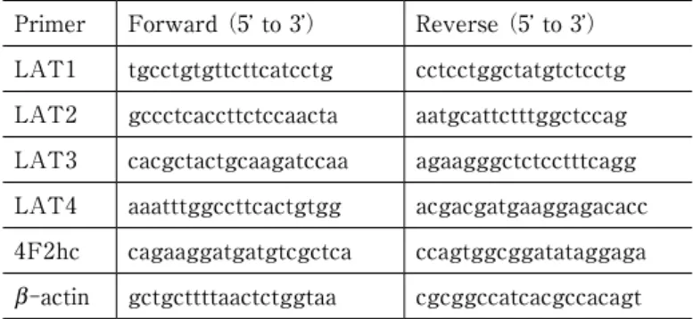
The primer sequences used are shown in Table 1.
The PCR conditions consisted of initial denaturation for 2 min at 95℃, followed by three steps that were repeated for 30 rounds:30 s at 95℃, 30 s at 55℃, and 30 s at 72℃. Finally, the reaction was incubated for 5 min at 72℃. Beta-actin was used as the internal control.
The quantitative RT-PCR reaction was performed using SYBR® Fast qPCR Mix (Takara Bio Inc.) with
the Thermal Cycler Dice Real Time System II (Taka- ra Bio Inc.). Primer was added to a final concentration of 0 . 4 µM in the reaction solution. The primer sequences used are shown in Table 2. The PCR condi- tions consisted of initial denaturation for 30 s at 95℃, followed by incubation of the reaction for 40 cycles, with each cycle consisting of 5 s at 95℃ and 10 s at 60℃. Using beta-actin as the internal control, relative quantitation was performed using the standard curve method. The level of beta-actin expression was assumed to be the same in JAR and JEG-3 cells, and the relative ratio of gene expression in JEG-3 cells was determined with gene expression in JAR cells assigned at a value of 1.
Western blot analysis
The JAR and JEG-3 cells were seeded on 24-well plates (2×10⁵ cells/well) and dissolved 2 days later by adding 1×SDS loading buffer. The cell lysate was boiled for 5 min at 95℃, then electrophoresed through Mini-PROTEAN® TGXTM Precast Gels (Bio-Rad Lab- oratories, Hercules, CA, USA), and transcription was performed on a polyvinylidene difluoride membrane.
Blocking was performed using 5% skim milk, and the membrane was soaked in primary antibody diluted
with blocking buffer and incubated overnight at 4℃.
Anti-LAT1 antibodies were diluted 1,000-fold, and anti-beta-actin, clone C4 antibody (EMD Millipore, Billerica, MA, USA) was diluted 3,000-fold for use.
Anti-mouse IgG-HRP antibody (GE Healthcare, Chi- cago, IL, USA) was diluted 3,000-fold and used as the secondary antibody. The membrane was developed with SuperSignal West Pico Chemiluminescent Sub- strate (Thermo Fisher Scientific, Inc., Waltham, MA, USA), and images were obtained with an ImageQuant LAS 4000 imager (Fuji Film, Tokyo, Japan). A solu- tion of cells that persistently expressed LAT 1
(S2-LAT1) was used as the positive control for LAT18). A solution of cells that persistently expressed LAT2 (S2-LAT2) was used as the negative control8).
[14C]Leucine uptake test
JAR and JEG-3 cells were seeded (3×10⁵ cells/
well) on 24-well plates coated with poly-L-lysine
(Corning Incorporated, Corning, NY, USA) and grown for 2 days. Hank’s balanced salt solution (HBSS)
[125 mM NaCl, 4.8 mM KCl, 25 mM HEPES, 1.2 mM KH2PO4, 1.2 mM MgSO4, 1.3 mM CaCl2, and 5.6 mM glucose (pH 7.4)] or sodium-free HBSS with 125 mM C5H14ClNO added instead of NaCl was used in the Table 1 Sequence of primers used for RT-PCR
Primer Forward (5’ to 3’) Reverse (5’ to 3’)
LAT1 tgcctgtgttcttcatcctg cctcctggctatgtctcctg LAT2 gccctcaccttctccaacta aatgcattctttggctccag LAT3 cacgctactgcaagatccaa agaagggctctcctttcagg LAT4 aaatttggccttcactgtgg acgacgatgaaggagacacc 4F2hc cagaaggatgatgtcgctca ccagtggcggatataggaga b-actin gctgcttttaactctggtaa cgcggccatcacgccacagt
Table 2 The primer sequences used for quantitative RT-PCR Primer Forward (5’ to 3’) Reverse (5’ to 3’)
LAT1 actcaggaccagatgtccgtctc cgctgtgaagtctgtccatgtg LAT2 tgctggacagatagtccttcg ggaacagcaggttgatcttg LAT3 tgtgttcgccttgcttcag gagtgagaatagcaggaggc LAT4 ctgaaggagtgtgaagacgc Tgatcttctggatctgccg 4F2hc atgagattggcctggatgc aagctggactcatcccacag b-actin ctggcatcgtgatggactccgg gtggatgccacaggactccatg
uptake test. The cells were washed twice in 1 mL of HBSS warmed to 37℃ and preincubated for 10 min.
HBSS supplemented with 1 µM of [14C]-leucine
(Moravek, Brea, CA, USA) was then added, and incu- bated for 1, 2, 5, 10, and 30 min. After incubation, the cells were washed twice with cold HBSS and dis- solved with 0.1N NaOH. The radioactivity in the cell lysate was measured using an LSC-7200 liquid scintil- lation counter (Hitachi Aloka Medical America, Inc./
Hitachi Ltd., Tokyo, Japan). In addition, the protein content of the cell lysate was measured using the Pierce BCA Protein Assay Kit (Thermo Fisher Scien- tific Inc.), and protein normalization was performed.
In the test of leucine uptake inhibition by JPH203, JPH203 (0.001, 0.01, 0.1, 1, and 10 µM) was added to 1 µM [14C]-leucine solution using sodium-free HBSS, and the leucine uptake in 1 min was examined. A solu- tion of 0.1% DMSO/1% EtOH was added as the con- trol.
Cell proliferation test
To evaluate the proliferation-inhibiting effect of JPH203, cell proliferation over time was evaluated by measuring cell counts. JAR cells (1×10⁴ cells/well)
and JEG-3 cells (2×10⁴ cells/well) were seeded on 24-well plates, and culture solutions containing 10, 30, and 100 µM JPH203 were substituted the following day. A culture solution containing 0.1% DMSO/1%
EtOH was used as the control. Cell counts were mea- sured using a TC20 Automatic Cell Counter (Bio-Rad Laboratories) immediately after, and also on 2 and 4 days after culture solution replacement.
Cell activity measurement
To evaluate cell activity after the addition of JPH203, cell activity was measured by the fluores- cence method using Alamar Blue (Bio-Rad Laborato- ries). JAR cells (1×10³ cells/well) and JEG-3 cells (2
×103 cells/well) were seeded on 96-well plates, and 100 µL/well of JPH203 at different concentrations (10, 30, and 100 µM) and a control culture solution con- taining 0.1% DMSO/1% EtOH were substituted the following day. Ten microliters per well of Alamar Blue were added immediately after (0 days) and also on 2 and 4 days after culture solution replacement, the cells were incubated under 5% CO2 at 37℃ for 3
h, and fluorescence intensity was measured (measure- ment wavelengths used:544 nm excitation wave- length, 590 nm fluorescence emission wavelength).
Measurements were also performed only for wells with culture solution that had JPH203 added, and the value yielded by subtracting the fluorescence intensity of wells with culture solution alone from the fluores- cence intensity of the test wells was evaluated.
Statistical analysis
In the quantitative RT-PCR analysis, Student’s t-test was used for JAR cell expression. A multigroup analysis of the results of the cell proliferation test and cell activity measurements was performed by analysis of variance (ANOVA) using SPSS Statistics version 24 (IBM Corp., Armonk, NY, USA), and the groups were compared using the Bonferroni method. The means and standard errors were shown for the data.
A p-value <0.05 was considered significant.
RESULTS
Expressions of LATs and 4F2hc in cultured choriocar- cinoma cells
RT-PCR was performed using mRNA extracted from JAR and JEG-3 cells to examine whether LATs were present in these lines of cultured choriocarcino- ma cells using the expressions of LATs and 4F2hc in lines of cultured choriocarcinoma cells (Fig. 1A). RT- PCR of JAR cells showed clear bands for LAT1, LAT4, and 4F2hc. A weak band was seen for LAT2.
With JEG-3 cell RT-PCR, clear bands were detected for LAT1, LAT2, LAT4, and 4F2hc. In addition, quantitative RT-PCR was performed using the same mRNA to assess whether there were differences between the cells in the expression of LAT mRNA
(Fig. 1B). With the expression of LATs in JAR cells assigned at a value of 1, LAT1 expression in JEG-3 cells was 0.8-fold, which was significantly lower. The level of LAT2 expression in JEG-3 cells was 3.5-fold higher. The expressions of LAT3 and LAT4 were lower in JEG-3 cells than in JAR cells (LAT3:0.8- fold, LAT4:0.4-fold). No difference in 4F2hc expres- sion were seen between JAR and JEG-3 cells. LAT1 expression in JAR and JEG-3 cells was also examined by Western blot analysis. Although the homology between LAT1 and LAT2 is 50%5), it was deter-
mined that the LAT1 antibody used in this study rec- ognized only LAT1. Consequently, LAT1 protein expression was determined for both JAR and JEG-3 cells (Fig. 2).
Leucine uptake and its inhibition by JPH203 in a line of cultured choriocarcinoma cells
To examine whether LAT-mediated amino acid uptake occurs in JAR and JEG-3 cells, leucine uptake tests were performed using HBSS containing sodium and sodium-free HBSS, and whether sodium-indepen- dent amino acid transport occurs in these cells was examined. In both JAR and JEG-3 cells, no difference in leucine uptake was seen as a result of the differ- ence in HBSS (Fig. 3A). That is, sodium-independent leucine transport was shown to exist in JAR and
Figure 1 Expression of LATs and 4F2hc in JAR and JEG-3 cells
A:RT-PCR of LATs. RT-PCR of LATs was performed using collected mRNA from JAR and JEG-3 cells. In RT (+) side lane, synthesized cDNA was added to the reac- tion mixture. In RT (−) side lane, mRNA was added. Primers’ sequence was shown in Table 1. B:Quantitative PCR of LATs.
mRNA expression of LATs and 4F2hc was quantified by realtime PCR. Expression level was indicated as expression relative to JAR.
Error bars represent±standard error (SE)
for triplicate experiments.
Figure 2 Western blotting of LAT1
Whole cell proteins were extracted from non-treated JAR and JEG- 3 cells, LAT 1 expressing cells
(S2-LAT1), LAT2 expressing cells (S2-LAT2) and mock cells (S2-mock). Lysate of LAT1 expressing cells was considered a positive control. Blotting for b-actin serves as loading controls.
JEG-3 cells.
Next, a leucine uptake test with JPH203 loaded was performed to examine whether the sodium-indepen- dent leucine transport in JAR and JEG-3 cells is mediated by LAT1. JPH203-concentration-dependent inhibition of leucine uptake was seen in both JAR and JEG-3 cells (Fig. 3B). The IC50 was calculated to be 0.408 µM for JAR cells and 0.475 µM for JEG-3 cells.
These results showed that LAT1-mediated leucine transport exists in JAR and JEG-3 cells.
Proliferation-inhibiting effect of JPH203
To examine whether JAR and JEG-3 cell prolifera-
tion is inhibited by the inhibition of LAT1-mediated amino acid transport, cell counts were measured after JPH203 loading (Fig. 4). No difference in the JAR cell count was seen immediately after culture solution replacement, but the cell count was significantly lower in the 100 µM group than in the control group 2 days after replacement. The cell count was also significant- ly lower in the 100 µM group than in the control group 4 days after replacement. However, cell counts in the 10 and 30 µM groups were nearly identical to those in the control group. With JEG-3 cells, no changes in the cell count were seen due to the addi- tion of JPH203 immediately after or 2 days after cul- Figure 3 Leucine uptake of JAR and JEG-3 cells and inhibition by JPH203
A:Time courses of leucine uptake were examined with HBSS containing Na (Normal HBSS) and Na free HBSS. One µM [14C]-Leucine uptake by JAR and JEG-3 cells was measured during a 30 minutes incubation. B:Inhibition of leucine uptake by JPH203 in JAR and JEG-3 cells. JAR and JEG-3 cells were incubated with [14C]-Leucine and 0.001-10 µM JPH203 for 1 minute. Data are shown as means±SE.
ture solution replacement. However, a JPH203 concen- tration-dependent decrease in cell count was seen 4 days after replacement.
Changes in cell activity due to JPH203
Cell activity following treatment with JPH203 was examined over time to determine whether amino acid transport by LAT1 is involved in the activity of JAR and JEG-3 cells (Fig. 5). The activity of the JAR cells in the groups with 10 and 30 µM added increased on
all of the measurement days. In the group with 100 µM added, cell activity decreased immediately after culture solution replacement (3 h after), but it did not differ significantly compared with the control from 2 days after replacement onward. The activity of JEG-3 cells decreased significantly in the group with 10 µM added immediately after and 2 days after culture solu- tion replacement. The activity of cells in the groups with 30 and 100 µM added decreased significantly immediately after and 2 and 4 days after replacement.
Figure 4 Growth inhibitory effect of JPH203
The cells were treated with 0, 10, 30 and 100µM JPH203 for 0, 2 and 4 days. After incubation, cells were trypsinized and counted by auto cell counter. Data are shown as means±SE. (*:p<0.05, #:p<0.01 compared to the control group)
Figure 5 Metabolic activities of JPH203-treated JAR and JEG-3 cells
JAR and JEG-3 cells were treated with 0, 10, 30 and 100 µM JPH203 for 0, 2 and 4 days. The metabolic activities were determined by Alamar Blue solution. Data are shown as means±SE. (*:p<0.05, #:p<0.01 compared to the control group)
DISCUSSION
In this study, it was found that amino acids are transported by LAT1 in two kinds of choriocarcinoma cell lines, JAR cells and JEG-3 cells. Moreover, these cell lines showed different responses of cytostasis and cell activity changes to LAT1 inhibition.
The quantitative RT-PCR showed that the expres- sions of LAT1, 3 and 4 were slightly but significantly higher in the JAR cells than in the JEG-3 cells. On the contrary, the expression of LAT2 was higher in the JEG-3 cells than in the JAR cells. It is generally known that LAT2 and 4 are expressed in normal cells, while the expressions of LAT 1 and 3 are increased in cancer cells2). Furthermore, it has been reported that the JAR cells proliferate faster and pro- duce more human chorionic gonadotropin than the JEG-3 cells14). From these, it may be said that the JAR cells maintain the characteristics of choriocarci- noma in comparison with the JEG-3 cells. In the Western blotting, the LAT1 protein could be identi- fied in the JEG-3 cells as well as the JAR cells. The LAT1/b-actin ratios were comparable between the two cell lines.
The leucine transport of the JAR cells or the JEG-3 cells was not affected by the existence or the absence of Na in HBSS. It is suggested that the leucine trans- port by LATs plays a substantial role in the amino acid transport by cultured choriocarcinoma cell lines.
The IC50 levels of the leucine uptake were reportedly 0.06 µM in the human intestinal tract cancer cell line
(HT29)9) and 0.79 µM in the human oral cancer cell line ( YD- 3 8)8). In this study, the IC5 0 levels of JPH203 for the leucine uptake inhibition were lower in both the JAR cells and in the JEG-3 cells compared to the YD-38 cells. Thus, it is indicated that the inhib- itory action of JPH203 on leucine uptake is higher in the choriocarcinoma cell lines than in the YD-38 cells.
In addition to the HT-29 cells, JPH203 is known to inhibit the proliferation of many other neoplastic cells8〜10). For example, it has been reported that the 100 µM JPH203 reduced the number of HT-29 cells to 10% of the control after four days8) and also reduced the number of YD-38 cells to 40% of the control9). However, more than half of the JAR and the JEG-3 cells had lived after four days giving 100 µM
JPH203 as compared with the control (Figure 4), indicating that the suppressant effect of JPH203 is less potent in the JAR and the JEG-3 cells than in the HT-29 or the YD-38 cells. The role of LAT1 in cell proliferation may be different between these cell lines
(HT-29 and YD-38) and the choriocarcinoma cell lines. It is known that amino acids are not only used as constituents or nutrients in cancer cells, but also work as a secondary messengers which promotes the proliferation of cancer cells2). A more detailed exami- nations are necessary to clarify whether the difference in the effect of JPH203 on cell proliferation is caused by the depletion of nutrients or by the signal trans- duction disturbance in the cancer cells.
The IC50 levels of JPH203 calculated by the leucine uptake experiment were comparable between the JAR cells and the JEG-3 cells. On the other hand, the cytostatic effect of JPH203 was concentration-depen- dent in the JEG-3 cells, while the effect was only seen by a high-concentration of JPH203 in the JAR cells
(Figure 4). Considering that the JAR cells have high- er proliferative capacity than the JEG-3 cells, it is imagined that the amino acid level necessary for cell proliferation is more in the JAR cells than in the JEG-3 cells (Figure 3A). However, because the amino acid uptake level was higher in the JEG-3 cells than in the JAR cells, it is suggested that these two cell lines have different demand characteristics for amino acids. The higher expression of LAT2 in the JEG-3 cells than in the JAR cells supports a higher demand of amino acids in the JEG-3 cells (Figure 1B). In addition, because JPH203 was added to the culture medium and the RPMI-1640 medium used for the JAR cells contains more amino acids than the MEM medium used for the JEG-3 cells, it is possible that the higher amino acid levels in the medium caused an increased amino acid uptake by the JAR cells than the JEG-3 cells even under the existence of JPH203.
The experiments were performed in the environment that the cells are easy to multiply, but the examina- tion under the condition of same amino acid availabili- ty for each cell lines may provide different results.
The cell activity evaluation using Alamar Blue reflecting the reduction power of viable cells is a quantitative measurement of the cell proliferation and is often performed in a purpose same as cell count15).
However, we examined the cell count and the cell activity measurement separately to evaluate the effects of amino acid transport by LAT1 on cell activi- ty in this study. Interestingly, the concentration- dependent decreasing effect of JPH203 was seen with- in a day in the JEG-3 cells. Whereas, the cell activity started to increase following the addition of JPH203 at concentrations of 10 and 30 µM on the day 0 in the JAR cell (Figure 5). It is thought that the JEG-3 cells could not maintain the metabolism of the cells when the amino acid transport was inhibited by JPH203, which resulted in the decrease of cell activity. On the other hand, the amino acid transport inhibition by JPH203 may have induced the autophagy mechanism which possibly increased the JAR cell activity. It is reported that the cell activity of the human cell line rises with the induction of autophagy mechanism16), and it is speculated that the autophagy mechanism is easier to be induced in the JAR cells than in the JEG-3 cells. However, the decrease in cell count by 100 µM JPH203 in comparison with the control start- ed to be seen on the 2nd day in the JAR cells, which was earlier than the occurrence of cytostatic effect by JPH203 in the JEG-3 cells. Thus, the high concentra- tion JPH203 was shown to restrain cell proliferation irrespective of the kind of choriocarcinoma cell line. It became clear in this study that the cell activities of the JAR cells and the JEG-3 cells respond differently to the low concentration JPH203, but the high concen- tration JPH203 can restrain the cell proliferation of these two kinds of cultured choriocarcinoma cells.
CONCLUSION
The present study demonstrated that amino acid transport by LAT1 functions in two kinds of chorio- carcinoma cell lines. In addition, highly-concentrated JPH203 showed an inhibitory effect on the prolifera- tion of choriocarcinoma cell lines. Furthermore, it was suggested that the amino acid transport by LAT1 has complicated and various roles in the activity of chorio- carcinoma cells.
Acknowledgements The authors thank Ms. S Naka- date, Department of Pharmacology and Toxicology, Dokkyo Medical University School of Medicine, for technical assistance and Dr. H. Endou, J-Pharma, for
providing JPH203 and anti-LAT1 antibody. This study was supported in part by grants from the Japan Society for the Promotion of Science (JSPS KAKENHI 26461258 (N.A.)), Strategic Research Foundation Grant-aided Project for Private Universi- ties (S1412001), the Science Research Promotion Fund of the Japan Private School Promotion Founda- tion, Gout Research Foundation of Japan, The Shimabara Science Promotion Foundation, Dokkyo Medical University, Young Investigator Award (T.T.), Investigator-Initiated Research Grant (M.O.), and Research of Seki Minato Foundation of Seki Minato Memorial Awards (N.A.).
Conflict of Interest No COI for all authors.
REFERENCE
1) Ganapathy V, Thangaraju M, Prasad PD:Nutrient transporters in cancer:relevance to Warburg hypothesis and beyond. Pharmacol Ther 121:29-40, 2009.
2) Qian Wang, Jeff Holst:L-type amino acid transport and cancer:targeting the mTORC1 pathway to inhibit neoplasia. Am J Cancer Res 5:1281-1294, 2015.
3) Kanai Y, Segawa H, Miyamoto Ki, et al:Expression cloning and characterization of a transporter for large neutral amino acids activated by the heavy chain of 4F2 antigen (CD98). J Biol Chem 273:
23629-23632, 1998.
4) Mastroberardino L, Spindler B, Pfeiffer R, et al:Ami- no-acid transport by heterodimers of 4F2hc/CD98 and members of a permease family. Nature 395:
288-291, 1998.
5) Segawa H, Fukasawa Y, Miyamoto K, et al:Identifi- cation and functional characterization of a Na+-inde- pendent neutral amino acid transporter with broad substrate selectivity. J Biol Chem 274:19745-19751, 1999.
6) Babu E, Kanai Y, Chairoungdua A, et al:Identifica- tion of a novel system L amino acid transporter structurally distinct from heterodimeric amino acid transporters. J Biol Chem 278:43838-43845, 2003.
7) Bodoy S, Martin L, Zorzano A, et al:Identification of LAT4, a novel amino acid transporter with system L
activity. J Biol Chem 280:12002-12011, 2005.
8) Oda K, Hosoda N, Endo H, et al:L-type amino acid transporter 1 inhibitors inhibit tumor cell growth.
Cancer Sci 101:173e179, 2010.
9) Yun DW, Lee SA, Park MG, et al:JPH 2 0 3 , an L-type amino acid transporter 1-selective compound, induces apoptosis of YD-38 human oral cancer cells.
J Pharmacol Sci 124:208–217, 2014.
10) Rosilio C, Nebout M, Imbert V, et al:L-type amino- acid transporter 1 (LAT1):a therapeutic target supporting growth and survival of T-cell lymphoblas- tic lymphoma/Tcell acute lymphoblastic leukemia.
Leukemia 29:1253–1266, 2015.
11) Ritchie JW, Taylor PM:Role of the System L perme- ase LAT1 in amino acid and iodothyronine transport in placenta. Biochem J 356:719-725, 2001.
12) He B, Zhang N, Zhao R et al:Dexamethasone Down- regulates SLC7A5 Expression and Promotes Cell Cycle Arrest, Autophagy and Apoptosis in BeWo
Cells. J Cell Physiol 231:233-242, 2016.
13) Bower M, Newlands ES, Holden L, et al:EMA/CO for high-risk gestational trophoblastic tumors:
results from a cohort of 272 patients. J Clin Oncol 15:2636-2643, 1997.
14) Mauschitz R, Cervar M, Hahn T, et al:Self-regula- tion of the endothelin receptor system in choriocarci- noma cells. Biochim Biophys Acta 1502:224-234, 2000.
15) Larson EM, Doughman DJ, Gregerson DS, et al:A new, simple, nonradioactive, nontoxic in vitro assay to monitor corneal endothelial cell viability. Invest Ophthalmol Vis Sci 38:1929-1933, 1997.
16) Kapuy O, Vinod PK, Bánhegyi G:mTOR inhibition increases cell viability via autophagy induction dur- ing endoplasmic reticulum stress-An experimental and modeling study. FEBS Open Bio 4:704-713, 2014.