nervous system’ 1). Neuropathic pain has many serious effects on quality of life and is associated with a high economic burden for both individuals and society, and makes from a heterogeneous group disorders that affect the peripheral and central somatosensory ner- vous systems2). It is now regarded as a distinct clini- cal entity despite a large variety of causes. People with neuropathic pain may experience altered pain sensation, areas of numbness or burning, and continu- ous or intermittent evoked or spontaneous pain. Thus, neuropathic pain is an unpleasant sensory and emo-
INTRODUCTION
Neuropathic pain is now defined by the Internation- al Association for the Study of Pain(IASP)as ‘pain caused by a lesion or disease of the somatosensory
Original
Pre and Post Synaptic Mechanisms about the Peripheral Nerve-injured Plasticity of Nociceptive Transmission in
the Superficial Dorsal Horn of Mouse Spinal Cord
Yuya Sato
1), Takaaki Ajima
1), Toshifumi Takasusuki
1), Eiko Kato
2), Yuuichi Hori
2), Shigeki Yamaguchi
1)Departments of Anesthesiology1) and Physiology2), Dokkyo Medical University School of Medicine
SUMMARY
Long-term potentiation(LTP)and long-term depression(LTD)in synaptic transmission have not only been observed in the hippocampus, but also in the superficial dorsal horn. LTP and LTD in the superficial dorsal horn play an important role of central sensitization, leading to hyperalgesia and allodynia in neuro- pathic pain. In order to clarify its mechanisms, we evaluated evoked excitatory postsynaptic currents(eEP SCs)and asynchronous EPSCs(aEPSCs)in the superficial dorsal horn of a sciatic nerve-ligated neuropath- ic pain mice(nerve-ligated mice)using whole cell patch clamp recording.
Three types of neuron in the superficial dorsal horn of mouse spinal cord were observed after tetanus stimulation which were:eEPSC amplitude-increased(LTP neuron), decreased(LTD neuron)and unchanged neurons. After tetanus stimulation, cumulative probability of aEPSCs amplitude did not change in LTP neuron, but shifted to the left in LTD neuron. In nerve-ligated mice, the ratio of LTP neuron was high. On the other hand, the ratio of both LTD neuron and unchanged neuron was high in sham-operated control mice.
These findings suggest both pre- and post-synaptic mechanisms of central sensitization in the superficial dorsal horn may contribute to neuropathic pain and LTP or LTD may be important role of pre- or post- synaptic action, respectively.
Key Words: neuropathic pain, central sensitization, neuroplasticity, long-term potentiation, long-term depression
Received November 30, 2020;accepted December 14, 2020 Reprint requests to:Shigeki Yamaguichi
Dokkyo Medical University, School of Medicine, Departments of Anesthesiology, 880 Kitako- bayashi, Mibu, Tochigi 321-0293, Japan
tional experience.
For establishing suitable treatments of neuropathic pains, such as pharmacological approach, it is impor- tant to know about the detail mechanisms of neuro- pathic pain. However, its mechanisms are complicated and still have been unclear. Central sensitization is considered as one of it mechanisms, and may lead to hyperalgesia and allodynia. Recent reports suggested a relationship between central sensitization and neuro- plasticity in the spinal cord dorsal horn3).
Long-lasting changes in the efficacy of synaptic transmission, such as long-term potentiation(LTP)
and long-term depression(LTD)play an important role of neuroplasticity and is observed in the hippo- campus and cerebellum4). Both LTP and LTD are also observed in the superficial dorsal horn, which is an important area for transmission and regulation on nociception5〜10). However, the detail mechanisms of neuroplasticity, involved in the pathogenesis of neuro- pathic pain, are not still clear.
In the present study, to clarify mechanism of neuro- plasticity on neuropathic pain, we evaluated evoked excitatory postsynaptic currents(eEPSCs)and asyn- chronous EPSCs(aEPSCs)in the superficial dorsal horn of peripheral nerve-ligated neuropathic pain mice using whole cell patch clamp recording.
MATERIALS AND METHODS
1. AnimalsAll animal experiments were approved by the Insti- tutional Animal Care and Use Committee of Dokkyo Medical University. The care and use of the animals were in accordance with the National Institutes of Health guidelines on animal care and with the guide- lines of the IASP. Experiments were performed on male ICR mice aged 6-8 weeks. Mice were main- tained in a temperature-controlled room on a 12- h light-dark cycle with food and tap water available ad libitum.
2. Partial ligation of the sciatic nerve
All mice were anesthetized with sevoflurane for the sciatic nerve-ligation surgery and for the sham opera- tion. The left sciatic nerve was partially ligated according to the protocol described by Seltzer et al.(nerve-ligated mice)11). In sham-operated control
mice, the sciatic nerve was exposed, but not ligated
(sham-operated mice).
3. Behavioral assessment
To assess the effects of sciatic nerve ligation on the nociceptive behavior, the withdrawal threshold on mechanical stimulation was determined by using an electronic von Frey device(Model 1601;IITC Life Science, Woodland Hills, CA, USA). The use of the electronic von Frey device has been validated for investigations on nociceptive behaviors(Moller et al.12), 1998;Cunha et al., 200413)). The probe of the electronic von Frey device was manually applied with the force increasing at a rate of 1.6-8.1 g/s. Each trial of stimuli was composed of 10 applications of mechani- cal stimulation at approximately 10-s intervals;each trial was repeated three times at approximately 3-min intervals. Behavioral assessments were per- formed from 3 days prior to the surgery till 10 days after surgery13).
4. Electrophysiological experiments 1)Preparation of spinal cord slices
Transverse spinal cord slices were obtained on post-surgical day 10. Briefly, segments of the lumbo- sacral(L4-S1)spinal cord were removed under ket- amine/xylazine anesthesia. Slices(450 µm thick)were cut on a vibratome(Dosaka EM, Japan)in Krebs solu- tion at 4℃. The Krebs’ solution was equilibrated with 95% O2 and 5% CO2. The solution contained the fol- lowing ingredients(in mM):NaCl, 113;KCl, 3;
NaHCO3, 25;NaH2PO4, 1;CaCl2, 2;MgCl2, 1;d-glu- cose, 11. Before electrophysiological experiments, spi- nal cord slices were incubated in Krebs solution at 37
℃ for 60 minutes.
2)Tight-seal whole-cell recordings
The spinal slices were mounted in a recording chamber on a microscope stage(Axioskop FS-II, Zeiss), and continuously perfused with Krebs’ solution.
Conventional tight-seal whole-cell recordings were obtained from neurons located in the superficial dorsal horn(lamina II)under visual control using infrared- differential interference contrast optics and a CCD video camera(IR-CCD 2741;Hamamatsu Photonics), as described previously14,15)The pipette were filled with a solution of the following composition(in
mM):K gluconate, 123;KCl, 14;Na gluconate, 2;
EGTA, 1;HEPES, 10;and the pH of the solution was neutralized to 7.4 with KOH. The currents were recorded in the voltage-clamp mode at a holding potential of -70 mV, using an Axopatch 200B patch- clamp amplifier(Axon Instruments). The data were sampled using a Digidata 1440 interface(Axon Instru- ments). A PCLAMP 10(Axon Instruments)and Mini Analysis 6.0.3(SynaptoSoft)were used to analyze the data.
3)Electrical stimulation-evoked EPSCs(eEPSCs)
To record eEPSCs, the external solution routinely contained strychnine(Sigma, 2-5 µM)and bicuculline
(Sigma, 10 µM). Electrical stimulation was applied using a glass pipette filled with 1 M NaCl with its tip
(diameter, ca 3 µm)placed at the dorsolateral margin of the spinal cord, 100-200 µm away from the record- ed neuron. With a square pulse of 0.1 ms duration, stimulus intensity was adjusted so that an EPSC of similar amplitude was evoked in each experiment.
4)LTP and LTD
For induction of LTP or LTD, tetanus stimulation
(100 Hz, 5s)was applied at the dorsolateral margin of the spinal cord. The external solution routinely con- tained strychnine(Sigma, 2-5 µM)and bicuculline
(Sigma, 10 µM). Electrical stimulation was applied using a glass pipette filled with 1 M NaCl with its tip
(diameter, ca 3 µm)placed at the dorsolateral margin of the spinal cord, 100-200 µm away from the record- ed neuron.
5)Asynchronous EPSCs(aEPSCs)
aEPSCs was recorded under prefusion of Krebs’
solution, replaced CaCl2 with SrCl2(2 mM), which can effectively substitute for calcium in driving synaptic transmission16).
5. Statistical analysis
All data are presented as the mean±Standard Error of the Mean(SEM). Statistical analyses of the data were performed by one-way or two-way analy- sis of variance(ANOVA)followed by post hoc multi- ple comparison(Tukey test). To compare frequencies of LTP and LTD between ligated and control mouse, chi-square test was performed using 3×2 contingency table. Cumulative frequency distribution of aEPSCs amplitude was evaluated by Kolmogorov-Smirnov
test. The level of significance was set at p<0.05.
RESULTS
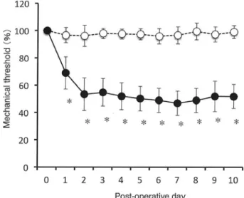
1. Behavioral assessment of mechanical allodynia induced by partial ligation of the sciatic nerve Mechanical withdrawal thresholds of the paw ipsi- lateral to the sciatic nerve ligation were determined using von Frey filaments. Partial sciatic nerve ligation elicited a significant decrease in the threshold for evoking hindpaw withdrawal(Fig. 1, filled circles). In nerve-ligated mice, the decrease in withdrawal thresholds appeared within 1 day after sciatic nerve- ligation and lasted throughout the period of investiga- tion up to 10 days after sciatic nerve-ligation. On the other hand, in sham-operated mice, no significant alterations in withdrawal thresholds were observed
(Fig. 1A, open circles).
Figure 1 Mechanical allodynia induced by the partial ligation of the sciatic nerve.
Mechanical withdrawal thresholds of the hindpaw ipsilat- eral to the sciatic nerve ligation, expressed as percentage to baseline values, are plotted as a function of postopera- tive days. Filled circles represent sciatic nerve-ligated mice(n=13), and open circles represent sham-operated control mice(n=14). Data are expressed as the mean±
SEM. *Indicates a significant decrease in the withdrawal threshold compared with the values before the surgical procedure.
2. Tight-seal whole-cell recordings
1) Various patterns of eEPSCs after tetanus stimulation
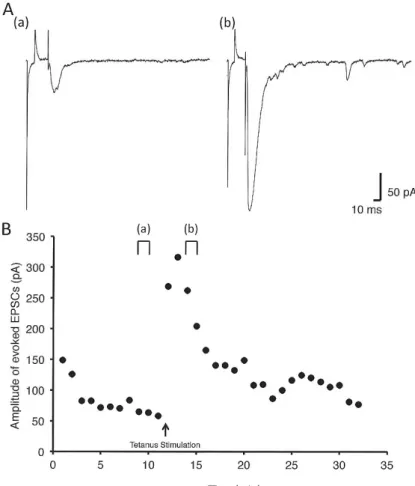
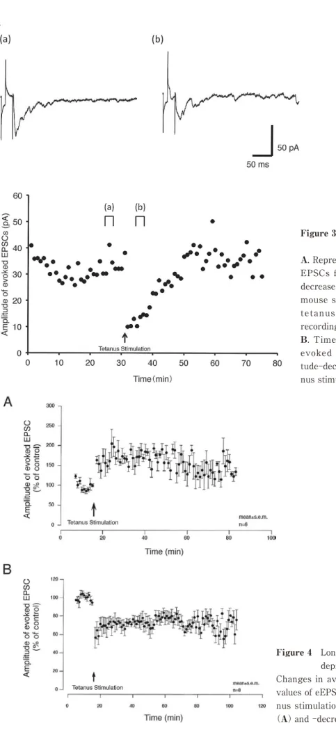
Three types of neuron in the superficial dorsal horn of mouse spinal cord, including eEPSC amplitude- increased(LTP neuron, Fig. 2A, before;a, after;b), decreased(LTD neuron, Fig. 3A, before;a, after;b)
and unchanged neurons were observed after tetanus stimulation. Fig. 4A and B show changes in averages of percentage to baseline values of eEPSC amplitude before and after tetanus stimulation in both LTP and LTD neurons, respectively.
2) Effects of sciatic nerve ligation on the incidence of various patterns of eEPSCs
In both nerve-ligated and sham-operated mice, all types of neurons were observed(Table). However, the ratio of LTP neuron in nerve-ligated mice(74%)
was significantly higher than that in sham-operated mice(29%). On the other hand, the ratio of both LTD and unchanged neurons in nerve-ligated mice(17%, 9%, respectively)was significantly lower than that in
sham-operated mice(32% and 39% , respectively).
3)Effect of tetanus stimulation on aEPSCs.
Figs. 5A and 6A show effect of tetanus stimulation on aEPSCs in LTP and LTD neurons, respectively. In LTP neuron, after tetanus stimulation, cumulative probability of aEPSCs amplitude did not changed, resulted in no change in aEPSCs amplitude due to tet- anus stimulation(Fig. 5B). However, in LTD neuron, after tetanus stimulation, cumulative probability of aEPSCs amplitude shifted to the left, resulted in decrease in aEPSCs amplitude due to tetanus stimula- tion(Fig. 5B).
DISCUSSION
In the present study, before electrophysiological experiment, we evaluated the threshold for evoking hindpaw withdrawal in both mice with sciatic nerve- ligation by Seltzer method10)and sham operation. The threshold in nerve-ligated mice significantly decreased after sciatic nerve-ligation and lasted for ten days, but not in sham-operated mice. It is well
Figure 2 A sample recoding in neuron with long term potentiation
A. Representative recordings showing evoked EPSCs from an evoked EPSC amplitude-increased neuron in superficial dorsal horn of mouse spinal cord before(a)and after(b)tetanus stimulation.
The time points of recording(a and b)are shown in B.
B. Time course of change in amplitude of evoked EPSCs in an evoked EPSC amplitude-increased neuron before and after tetanus stimulation.
Figure 3 A sample recoding in neuron with long term depression
A. Representative recordings showing evoked EPSCs from an evoked EPSC amplitude- decreased neuron in superficial dorsal horn of mouse spinal cord before(a)and after(b)
tetanus stimulation. The time points of recording(a and b)are shown in B.
B. Time course of change in amplitude of evoked EPSCs in an evoked EPSC ampli- tude-decreased neuron before and after teta- nus stimulation.
Figure 4 Long term potentiation and long term depression
Changes in averages of percentage to baseline values of eEPSC amplitude before and after teta- nus stimulation in eEPSC amplitude-increased
(A)and -decreased neurons(B).
Table
LTP LTD no change
sciatic nerve-ligated mice
(n=23 cells) 17(74%) 4(17%) 2(9%)
sham operated mice
(n=31 cells) 9(29%) 10(32%) 12(39%)
LTP:long term potentiation(evoked EPSC amplitude-increased neuron)
LTD:long term depression(evoked EPSC amplitude-decreased neuron)
The ratio of evoked EPSC amplitude-increased, -decreased and unchanged neurons in sciatic-ligated and sham-operated mice. The ratio of eEPSC amplitude-increased neuron in sciatic nerve ligation mice was significantly more than that in sham controlled mice. On the other hand, the ratios of both eEPSC amplitude -decreased and -unchanged neuron in sciatic nerve ligation mice were significantly less than those in sham-controlled mice.
Figure 5 Amplitude of aEPSCs in the superficial dorsal horn with long term potentiation
A. Representative recordings showing asynchronous EPSCs from an evoked EPSC amplitude-increased neuron in superficial dorsal horn of mouse spinal cord before(a)
and after(b)tetanus stimulation. Inset traces show examples of asynchronous EPSCs recorded at the time indicated by
(a)before and(b)after tetanus stimulation.
B. Cumulative probability of asynchronous EPSCs amplitude. There was no difference between before and after tetanus stimula- tion.
known that neuroplasticity in the superficial dorsal horn contributes to hyperalgesia and allodynia. LTP, evoked by high frequency pre-synaptic stimulation, is one of mechanism of neuroplasticity. It is observed not only in the hippocampus, but also in the superfi- cial dorsal horn5,17).
Therefore, we performed whole cell patch clump recording on neurons in the superficial dorsal horn of spinal cord slices in both mice after sciatic nerve-liga- tion or sham operation. Then, three types of neuron in the superficial dorsal horn of mouse spinal cord, including LTP(eEPSC amplitude-increased), LTD
(eEPSC amplitude-decreased)and unchanged neu- rons after tetanus stimulation were recorded in both mice. These results were consistent with previous reports18〜19). However, there was a significant differ- ence in the ratio of these neurons between both mice.
The sciatic nerve-ligation changed the ratio of LTP and LTD neurons in the superficial dorsal horn. Our
results suggest that increase in LTP of excitatory synaptic transmission in the superficial dorsal horn may play an important role in neuropathic pain.
In the previous reports18,20), LTP and LTD were observed in spinal thalamic tract neurons and GAB Aergic neurons, respectively. In our results, the ratio of eEPSC amplitude-increased neuron was significant- ly increased after sciatic nerve-ligation. It means that nociceptive synaptic transmission is sensitized and inhibitory synaptic transmission is depressed. Our results were consistent with the previous reports20〜22).
Furthermore, to confirm pre- or post- synaptic changes in LTP and LTD, we evaluated aEPSC in the superficial dorsal horn23,24). In LTD neuron, amplitude of aEPSC was decreased after initiation of LTD. It may suggest that decrease in sensitivity of post syn- aptic neurons contributes expression of LTD. On the other hand, amplitude of aEPSC was not changed in LTP neuron. It may suggest that increase in sensitivi-
Figure 6 Amplitude of aEPSCs in the superficial dorsal horn with long term depression A. Representative recordings showing asynchronous EPSCs from an evoked EPSC amplitude-decreased neuron in superficial dorsal horn of mouse spinal cord before(a)and after(b)tetanus stimulation.
Inset traces show examples of asynchronous EPSCs recorded at the time indicated by(a)before and(b)
after tetanus stimulation.
B. Cumulative probability of asynchronous EPSCs amplitude. There was no difference between before and after tetanus stimulation. Cumulative probability of aEPSCs amplitude shifted to the left, resulted in decrease in aEPSCs amplitude due to tetanus stimu- lation.
ty of pre synaptic neurons contributes to the expres- sion of LTP in the superficial dorsal horn.
Limitations in this study is that we only investigat- ed pre and post-synaptic electro physiological mecha- nisms in the superficial dorsal horn using neuropathic pain model mouse. Therefore, in the next study, to confirm the theory and to establish suitable pharma- cotherapy for neuropathic pain, another study using presynaptic ligand, Ca2+ channel a2 ligands, such as pregabalin, must be performed.
CONCLUSIONS
Our results, such as no change in aEPSCs after LTP and decrease in aEPSCs after LTD, suggest that both pre synaptic mechanism for LTP and post syn- aptic mechanism for LTD may play an important role in central sensitization in the superficial dorsal horn after peripheral nerve injury.
REFERENCES
1) International Association for the Study of Pain:
IASP Taxonomy. Pain terms. Neuropathic pain Updated 2017 Dec 14. www.iasp-pain.org/Taxonomy
#Neuropathicpain
2) Sumitani M, Sakai T, Matsuda Y, et al:Executive summary of the Clinical Guidelines of Pharmacother- apy for Neuropathic Pain:second edition by the Jap- anese Society of Pain Clinicians. J Anesth 32:463- 478, 2018.
3) Ji RR, Nackley A, Huh Y, et al:Neuroinflammation and Central Sensitization in Chronic and Widespread Pain. Anesthesiology 129:343-366, 2018.
4) Martin SJ, Grimwood PD, Morris RG:Synaptic plas- ticity and memory:an evaluation of the hypothesis.
Annu Rev Neurosci 23:649-711, 2000.
5) Randić M, Jiang MC, Cerne R:Long-term potentia- tion and long-term depression of primary afferent neurotransmission in the rat spinal cord. J Neurosci 13:5228-5241, 1993.
6) Drdla R, Gassner M, Gingl E, et al:Induction of syn- aptic long-term potentiation after opioid withdrawal.
Science 325:207-210, 2009.
7) Costigan M, Scholz J, Woolf CJ:Neuropathic pain:a maladaptive response of the nervous system to dam- age. Annu Rev Neurosci 32:1-32, 2009.
8) Latremoliere A, Woolf CJ:Central sensitization:a
generator of pain hypersensitivity by central neural plasticity. J Pain 10:895-926, 2009,
9) Treede RD:Gain control mechanisms in the nocicep- tive system. Pain 157:1199-1204, 2016.
10) West SJ, Bannister K, Dickenson AH, et al:Circuitry and plasticity of the dorsal horn--toward a better understanding of neuropathic pain. Neuroscience 300:254-275, 2015.
11) Seltzer Z, Dubner R, Shir Y:A novel behavioral model of neuropathic pain disorders produced in rats by partial sciatic nerve injury. Pain 43:205-218, 1990.
12) Möller KA, Johansson B, Berge OG:Assessing mechanical allodynia in the rat paw with a new elec- tronic algometer. J Neurosci Methods 84:41-47, 1998.
13) Cunha TM, Verri Jr WA, Vivancos GG, et al:An electronic pressure-meter nociception paw test for mice. Braz J Med Biol Res 37:401-407, 2004.
14) Blanton MG, Lo Turco JJ, Kriegstein AR:Whole cell recording from neurons in slices of reptilian and mammalian cerebral cortex. J Neurosci Methods 30:
203-210, 1989.
15) Coleman PA, Miller RF:Measurement of passive membrane parameters with whole-cell recording from neurons in the intact amphibian retina. J Neuro- physiol 61:218-230, 1989.
16) Miledi R:Strontium as a substitute for calcium in the process of transmitter release at the neuromus- cular junction. Nature 212:1233-1234, 1966.
17) Ikeda H, Heinke B, Ruscheweyh R, et al:Synaptic plasticity in spinal lamina I projection neurons that mediate hyperalgesia. Science 299:1237-1240, 2003.
18) Kim HY, Jun J, Wang J, et al:Induction of long- term potentiation and long-term depression is cell- type specific in the spinal cord. Pain 156:618-625, 2015.
19) Pockett S, Figurov A:Long-term potentiation and depression in the ventral horn of rat spinal cord in vitro. Neuroreport 4:97-99 1993.
20) Moore KA, Kohno T, Karchewski LA, et al:Partial peripheral nerve injury promotes a selective loss of GABAergic inhibition in the superficial dorsal horn of the spinal cord. J Neurosci 22:6724-6731, 2002.
21) Castro-Lopes JM, Tavares I, Coimbra A:GABA decreases in the spinal cord dorsal horn after periph-
eral neurectomy. Brain Res 620:287-291, 1993.
22) Janssen SP, Truin M, Van Kleef M, et al:Differential GABAergic disinhibition during the development of painful peripheral neuropathy. Neuroscience 184:
183-194, 2011.
23) Dodge FA Jr, Miledi R, Rahamimoff R:Strontium
and quantal release of transmitter at the neuromus- cular junction. J Physiol 200:267-283, 1969.
24) Oliet SH, Malenka RC, Nicoll RA:Bidirectional con- trol of quantal size by synaptic activity in the hippo- campus. Science 271:1294-1297, 1996.