Attempt to Change the Virulence of RH and Beverley Strains of Toxoplasma gondii by Drug Treatment
Akira MlYATA
Department of Epidemiology, Institute for Tropical Medicine, Nagasaki University
ABSTRACT: In the present study, the author attempted 1) to change the virulence of RH strain of Toxoplasma gondii to that of Beverley type by the treatment of antitoxoplasma drugs and 2) to change the virulence of Beverley strain of T. gondii to that of RH type by the treatment of cortison or Kenacort (triamcinolone). The following results were obtained.
1) Mice inoculated RH strain died within 4•`5 days in control without treatment, and mice treated with spiramycin or sulfathiazole-N^4 died of an acute infection, but survival periods were slightly longer than those of non-treated control mice. Mice treated orally with sulfadiazine or Policydal (sulfamethopyrazine) for 10•`20 days survived for a long period, and from few of the mice, cysts were found in their brains. Proliferative forms were also found in several organs of the survival mice including brain. As a result, in the RH strain modi- fication of the virulence by drugs did not succeed. 2) To enhance the virulence of Beverley strain, mice were treated with cortison or Kenacort just before the inoculation of parasite. Five days later a fluid which was taken by washing with a physiological saline of the peritoneal cavity of each mouse was inoculated to each fresh mouse which was treated with cortison or Kenacort. After the same treatment and inoculation were repeated up to five passages of mice, the washing fluid of last passage mouse of each series was inoculated to fresh mouse without the treatment. After stop of the treatment, some mice died of an acute infection, within 4•`5 days and even in the non-treated control series, one case became a virulent type for several passages. But, if once the virulence of Beverley strain enhanced like RH type, often the parasites of "virulent" Beverley type turn again to avirulent Beverley type (cyst-cyst type) after serial passages.
There are well known laboratory strains of Toxoplasma gondii of which virulence is considerably different from each other, RH and Beverley strains. RH strain is highly virulent. Intraperitoneal inoculation of the strain results in death of the mice within 5-8 days in accordance with the number of parasites inoculated, and the infected mice develop 1-2 ml of serous ascites in which about 104-105 banana-shaped parasites per mm3 are counted microscopically. While the case of an avirulent Beverley strain, inoculated mice can survive for a month or more, but in their brains, spherical cysts surrounded by the definite walls
C ontribution No. 767 from the Institute for Tropical Medicine, Nagasaki University
Received for publication, July 31, 1976
are observed showing characteristic of chronic infection. The parasite-stages in those infections in mice are called as "extra-intestinal stages" , and in all host animals except Felidae, only such stages are reported. In the experimental infections of mice, the virulence of each strain, RH or Beverley, becomes stable respectively and their virulences do not change without the intervention of experimental procedures.
The treatments of RH infected mice or other animals with several anti-toxoplasma drugs have been reported by many workers, for example, spiramycin or acetyl-spiramycin by Nakayama and Matsubayashi (1963) and Aoki (1969), sulfa-drugs by Frenkel (1953), Eyles and Coleman (1953) and Nakayama and Matsubayashi (1961), SDDS by Oshima et al. (1967) and Oshima and Kumata (1974), and pyrimethamine by Summers (1953).
Jacob (1973) also has reviewed recent works on treatments. After such treatments, sometimes mice, which survived for a long period, still had toxoplasmas in their tissues especially in their brains as reported by Frenkel (1953), and Nakayama and Matsubayashi (1961 and 1963). According to Nakayama (1964), mice which were previously infected with a non-virulent strain were challenged with the virulent RH strain 6 weeks later, and their brains proved positive of the RH for up to 7 weeks after the challenge. Those parasites were usually within cysts, but in some cases the parasites might be present in tissues or cells of mice without the cyst as pointed by Frenkel (1953).
In the present study, the author intended to change the virulence of each strain by treatment with several chemicals. Although the virulence of RH or Beverley strain could not be changed permanently, some interesting informations obtained have been discussed.
MATERIALS ANDMETHODS
1. The following strains of toxoplasma were used:Beverley, RH, N-130, and N-108.
The last two strains were isolated from pigs in Nagasaki by Nakabayashi et al. (1969).
Mice (DDK) weighing approximately 20-25 g were used for inoculation.
2. To obtain chronic infection, the mice inoculated with the RH strain were treatedwith orally or intraperitoneally with the following anti-toxoplasma drugs : Spiramycin-Kyowa,
Theradiazine (sulfadiazine ; 2 , (p-aminobenzon-sulfonamido)-pyrimidine) , Sulzol S (sulfathi- azole-N4 ; sodium dextrosesulfonate) , SDDS (2-sulfamoyl-4, 4/-diaminodiphenylsulfone) , and Policydal (sulfamethopyrazine). The first time treatment was done at same time of the inoculation and the treatments were repeated daily or every the other day as shown in Table 1.
The extension of survival period (days) of the treated mice, and the detection of cysts in their brains were recorded. The inoculation of the emulsion of the brains was further attempted to fresh mice to examine whether the chronic infection of mice can be continued again in the second passage or turn to acute infection.
3. To reduce resistant ability of inoculated mice against the infection with Beverley strain,
the following substances were given intramusclarly to the mice just before the inoculation :
Cortison (cortison acetate), and Kenacort-A (Squibb triamcinolone acetonide aqueous
suspension , 9a-fluoro-16o:-hydroxyprednisolone acetonide).
Terminology of each stages: Before going to report results, the author must describe his opinion on terminology for extra-intestinal stages. The life cycle of Toxoplasma gondii (Nicolle and Manceaux, 1908) , had been known almost completely in the enteroepithelial cells of cats (Hutchinson et aL, 1968, 1970, and 1971). The enteroepithelial stages such as schi- zogony, gametogony, and oocyst formation are observed only in animals belonged to Felidae, from which the following species were reported as true host, Felis felis, F, yagouarounde, F. pardalis, F, bengalensis, F. concolor, and Lynx rufus (see Jacob, 1973, and Frenkel, 1974). While both extra-intestinal and enteroepithelial stages were reported in Felidae, only extra-intestinal stages were known in other animals. Hoare (1972) has suggested to use the following terms for the extra-intestinal stages of toxoplasma ; endozoite for the rapidly multiplying forms within pseudocysts, and cystozoite for those which develop within cysts, because of existence of morphological differences between endozoites and cystozoites. They are
"distingushable by the position of the nucleus, which is typically central in the endozoites but terminal in the cystozoites" (Zypen and Piekarski, 1967a and b). Frenkel (1973) has also proposed the two terms as follows : tachyzoite for the rapidly multiplying extra-intestinal forms of the acute infection, which reproduces by endodyogeny and eventually destroy their host cells ; and bradyzoite for the more slowly multiplying (by endodyogeny) encysted forms, characteristic of the chronic infection. Both of the authors used the term "cyst" in the same meaning as commonly used. According to Jacob (1973), pseudocyst ("group" in the term by Frenkel) means the aggregation of rapidly dividing tissue forms (tachyzoites or endozoites) within a host cell. The terminology may not be conclusive up to now, and the present author can not identify whether each banana shaped parasite found from tissues or ascite is tachyzoite (endozoite) or excysted bradyzoite (cystozoite) if parasites are freed cyst. Then, in the present paper, he used proliferative form for banana shaped parasites which were found outside of cyst, and cyst in the same sense of Hoare and Frenkel.
RESULTS
1. Attempt to change the virulence ofRH strain to Beverley type by the treatment of anti- toxoplasma drugs
Approximately 5 X 10⁴ parasites which were obtained from ascites of RH inoculated mice were injected to each mouse intraperitoneally. Before the inoculation, each mouse was pre- treated with one of anti-toxoplasma drugs, then, the treatment was repeated every day or every other day after inoculation of RH strain. The drugs, its dosage per day, the method of treatment, and the daily observation of the mice were summarized in Table 1.
The mice treated with the daily dosage of 5 mg or 10 mg of spiramycin died of an acute
infection, and the survival periods of the mice were slightly longer than those of non-treated
control mice. In the case of sulfathiazole-N4, the result obtained was similar to those of
spiramycin, and the survival periods were also slightly longer, but all mice died of the
acute infection within 10 days. With the oral treatments of 4 mg SDDSper day for 7 days,
mice died of the acute infection ; with intraperitoneal treatment, however, mice survived for
up to 30 days in chronic infection.
In mice treated orally with 2.5 mg sulfadiazine per day for 10 days, the survival days were markedly prolonged, however, the mice died of a sub-acute infection. Then, in the next attempt, mice were treated orally with 2 mg sulfadiazine per day for 20 days, and almost all the mice survived for up to at least 30 days in the chronic infection as shown in Table 2. Twenty mice, except one which died of an accidental injury to its stomach with syringe, survived for a long period, and cysts were not found in 17 mice, but the emulsion of the brains infected to fresh mice as shown in Table 2. Each emulsion of the brains obtained from mouse No. 6, 10, 18 and 19 in Table 2 was inoculated to fresh mice intraperitoneally. About 10 days later, the mice died of the acute infection. Mouse inoculated with the emulsion of brain taken from mouse No. 20 survived for a long period without any sign of infection. The brain of mouse No. ll from which cysts (Fig. 1) were detected microscopically was inoculated to fresh mice orally or intraperitoneally. All the mice inoculated intraperitoneally died, but in the mice given orally, only two of them died of the acute infection, and the other 8 mice survived for a long period. About one month later, from one of the survival mice, cysts were found in the brain. Again the brain emulsionwas inoculated to a mouse, but the mouse did not infect. All mice, which were inoculated intraperitoneally with the brain emulsion of mouse No. 17, died of the acute infection, but in the mice, which were given the emulsion orally, survived without any sign of the infection
T
able 1. Experimental treatment of RH inoculated mice with several anti-toxoplasma drugs D ru gs D aily D o sag e (m g ) an d D ays
an d M ethod of T reatm en t
S urviv al D ay s of M ice nu
T y pe of In fection an d O ther O b servation s
n (5)* 9 .4
(7) 7 .6
n A cu te in fection A cu te in fe ction S p iram ycin 10 m g , 10 days , orally*
5 m g , 10 day s, orally***
n
S ulfad iazin e 2 .5 m g , 10 days, orally 2 m g , 20 d ays , orally
(7) 24 .1 (20) 30<
Su b ‑acute infection , n o cyst c h ron ic in fection ,
cysts (2/20) , see T able 2
n
S u lfath ia zole‑ N 4 4 m g , 7 day s, orally
u
(8) 3.3 A cu te in fe ction S D D S 4 4 m g , n ag , 7 7 days , days , orally in trap eriton eally (5) ll.4
(5) 30< A cu te in fe ction
C u re or ch ron ic in fection u
P olicyd al
D
2 m g , 14 days , in traperiton eally j (10) 4 m g , 7 day s, in trap eritoneally (10) 30< 22 .7 4 in g , every the oth er d ay for
14 days , in trap eriton eally (10) 30<
6 m g , 7 day s, in trap eritoneally (7)** 21
(2)** 30<
C u re or ch ron ic in fe ction Su b ‑acute infection , no cyst c u re or ch ron ic in fection Su b ‑acute infection 1 gee c h ron ic in fection , ctr ‑4‑^ J f T‑ 11 T able cyst( + )
C on trol n o treatm en t (5) 6 .0 u
A cu te in fection
* (5) 9.4 means average 9.4 survival days in 5 inoculated mice.
** 7 out of 9 mice died within 30 days after inoculation and rest 2 mice survived for longer than 30 days.
*** Some of mice died before finish of the 10 days treatment.
Each mouse used in this experiment was inoculated intraperitoneally with 5x 10⁴ proliferative
forms of RH strain.
T
able 2. Experimental treatment of RH inoculated mice with sulfadiazine (2.5 mg/day for 20 days, orally)
M o u s e N o . S u r v i v a l D a y s C y s t D e t e c t io n R e s u lt o f S u b ‑ i n o c u la t io n w it h B r a i n
N o . 1 . 5 d ie d 2 2 . 5 * N o 3 1 . 5 * N o 3 4 . 5 k il le d N o 3 4 . 5 '/ N o
3 6 . 5 // N o I B r a i n * ‑ > i p . 2 m i c e ( P + , 9 D ) 3 9 . 5 ', N o
3 9 . 5 's N o 4 1 . 」 N o
1 0 4 2 . 5 N o B r a i n ‑^ i p . 2 m i c e ( P + , 6 D & 8 D ) l l 5 3 . 5 *
5 3 . 5 5 3 . 5 H 5 3 . 5 5 3 . 5 5 3 . 5 5 7 . 5 5 7 . 5 5 7 . 5
2 0 5 7 . 5 *
Y e s B r a i n ‑ n p . 1 0 m ic e ( P + ? A v e , * * 8 , 8 D ) N o
N o N o N o N o
Y e s s e e B r a h A N o t e A p . o r a lly , o r a lly , 5 m i c e 2 / 1 0 m i c e 1 / 5 ( P + , m i c e ( P + , ( P + , A v e . 9 D ) , 9 D ) 1 0 D ) 4 / 5 N e g
N o B r a i n > i p . M ic e ( P + , D ) * * * N o B r a i n ^ i p . M i c e ( P + , D ) N o I B r a i n ^ i p . M ic e ( P N e g ) * * * *
N
ote
5 brains from Mouse No. 12 to No. 16 were pooled and emulsified, then inoculated to fresh mice.
/ip. 9/10 mice (P+, llD) & 1/10 mouse (P Neg)
. ^ orally, 9/10 mice (P & cyst Neg) & 1/10 mouse (cyst + in brain) No 12-16 Retreatment with sulfadiazine
* ip. 2/10 mice (cyst+in brain)^Brain^ip. 1 mouse (P+, D) & orally, 1 mouse (Neg)
\ \Brain->orally, 1 mouse (Neg)
* orally, 10/10 mice (Neg)
B
rain of No. 6 was inoculated intraperitoneally to 2 fresh mice, and the mice died after 9 days and proliferative forms (P) were detected mainly from peritoneal cavity of the mice.
Ave. means average survival days of 5 inoculated mice.
D means death of inoculated mice within 10 days.
**** Neg means negative of cyst and proliferative form.
*#
**#
Fig. 1 Cyst of RH strain obtained by treatment with sulfadiazine (No. ll
in Table 2) (diameter about 30/^0.
except one mouse which died of the acute infection. The pooled emulsion of the brains taken from 5 mice (mouse No. 12-16) was given intraperitoneally or orally to each 10 mice respectively. All the mice inoculated intraperitonealy died of the acute infection except one which survived for a long period, and the mice given orally survived without the infection except one from which cysts were detected in the brain. The same emulsion was given orally or intraperitoneally to another fresh mice, and the mice were treated with sulfadiazine just same as the initial treatment. The mice, which were given the emulsion orally, survived for a long period, and from 2 of 10 mice which inoculated intraperitoneally, cysts were observed in their brains. The brain emulsions from these 2 mice were inoculated intraperitoneally to fresh mice, which died of the acute infection, but the mice to which the emulsion was orally survived. As a result, even if mice survived with a chronic infection of RH strain after the treatment of sulfadiazine, the virulence character of RH strain did not change to Beverley type (cyst-cyst type) consistently, namely, after 1~2 passages of the cyst-type RH strain, the strain enhanced to highly virulent nature.
In Policydal, except a group of mice which died of sub-acute infection after the intraperitoneal treatment with 4 mg per day for 7 days, the other groups of mice survived with the chronic infection or without any sign of the infection. The results of a group which was treated intraperitoneally with 6 mg per day for 7 days, were shown in Table 3. Seven out of 9 inoculated mice died, in which the proliferative forms were detected within 20 days in ascites of the mice. From the brains of 3 mice out of 7, the proliferative forms were also observed with the examination of a fluorescent microscope after acridin orange staining (modified from Sakamoto, 1966). Two mice survived for longer than a month, but finally one of them died 43 days after the inoculation, and in its ascites, some proliferative forms were observed. The last mouse was killed to be examined 48 days after the inoculation.
Some proliferative forms were still found in ascite of the mouse. From its brain, too many cysts and few proliferative forms were detected. In the case of Policydal, the treatment
T
able 3. Experimental treatment of RH inoculated mice with Policydal (6 mg/day for 7 days , intraperitoneally)
M o u se N o . s u rv iv a l D ay s Q i r , ! ; in P e r ito n e a l C a v it P ro life ra tiv e F o rm s P r olife ra tiv e C y s ts in B ra in * F o r m s
N o . 1 0 1 10 2 1 9 1 9 d ie d 's Y e s* Y e s* N o * N o * 1 0 3
1 0 4 1 0 5 1 0 6 1 0 7 1 0 8 1 0 9
N o *
P : Y e s* * N o *
P : Y e s* * P : Y e s* *
P : Y e s* * , C : Y e s **
P : Y e s* * , C : Y e s **
* Fresh preparation was examined without staining.
** Acridin orange staining perparation was examined by fluorescent microscope.
P : Proliferative form C : Cyst
with 4 mg or 6 mg per day for 7 days did not give rise to better curative effect than with 2 mg per day for 14 days, and in the latter treatment inoculated mice survived for a long period.
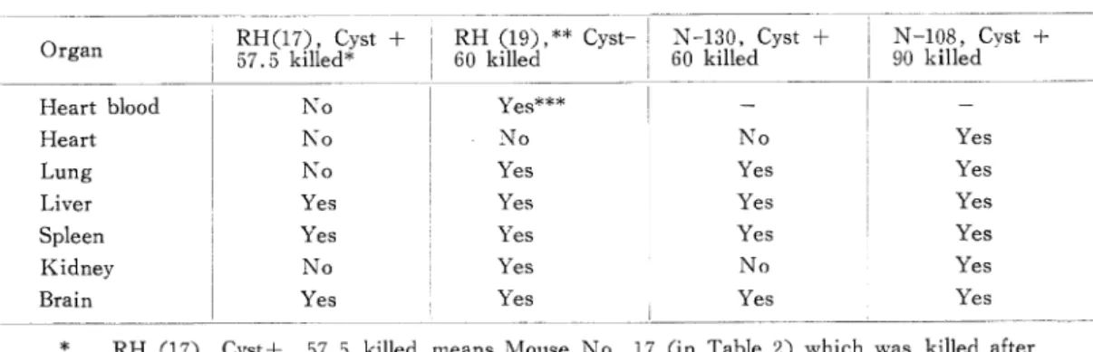
From the present experiments with RH strain, even if the inoculated mice survived with the chronic infection, it was rather difficult to find out cyst in the brain, while the brain emulsions were able to infect fresh mice. Then it seems likely that proliferative from can be present within some tissues or cells of the mice without cyst. Then in the next experiment, such the possibility was examined. Table 4 shows results of examination of the suspect tissues in which the proliferative forms or the cysts might be hiden in the mice of chronic infection. In this experiment, the mice No. 17 and No. 19 in Table 2, andthe mice inoculated with N-130 and N-108 strain respectively, were used, and their tissues were emulsified separately in physiological saline and inoculated intraperitoneally with antibiotics to fresh mice. The parasites still existed even in the heart blood of mouse No. 19 which survived for 57 days in a chronic infection. From brains, livers, and spleens of all four mice examined, the parasites were detected, but from hearts, the parasite was not found except one case which inoculated N-108 strain. Unfortunately, its heart blood was not examined.
2. Attempt to change the virulence ofBeverley strain to that of RH type by the treatment of cortison or Kenacort
To enhance the virulence of Beverley strain, mice were injected with cortison (1.25 mg or 2.5 mg/mouse) or Kenacort (2 mg or 4 mg/mouse) intramusclarly. Then, each mouse was inoculated intraperitoneally with 10^-20 cysts. Five days later, a fluid which was taken by washing with a buffered saline solution of the peritoneal cavity of each mouse was inoculated intraperitoneally to each fresh mouse which was also treated with the same chemical used to the mouse of first passage. After the same treatment and inoculation were repeated up to five passages of mice, washing fluid of each mouse of the last passage was the inoculated to each fresh mouse without the treatment. In the case of Kenacort most of
Table 4. Parasite detection from various organs of chronic infection mice with RH type by sub-inoculated method to fresh mice.
O r g a n R H ( 17 ) , C y st + 5 7 .5 k ille d *
R H ( 1 9 ) ,* * C y st‑ I N ‑ 1 3 0 , C y st 6 0 k ille d I 6 0 k ille d
H
N ‑ 1 0 8 , C y st + 9 0 k ille d
H e a rt b lo o d N o Y e s* * * N o N o
H e a rt N o Y e s
L u n g N o Y e s Y e s u Y e s Y e s Y e s
L iv e r Y e s Y e s
S p le e n Y e s Y e s Y e s Y e s K id n e y N o Y e s N o Y e s B r a in Y e s Y e s u Y e s Y e s
* RH (17), Cyst+, 57.5 killed means Mouse No. 17 (in Table 2) which was killed after 57.5 days from inoculation and cysts were detected in brain.
** RH (19) means Mouse No. 19 in Table 2.
*** Yes means inoculated mouse died of acute infection.
T
able 5. Enhancement of virulence in Beverley strain by using of cortison or Kenacort
D
rug &
Dosage
M
No. ouse
Passages in Treated Mouse
Passages in Untreated Mouse
w
bo
-^
bo
N
o. 201
? 206 207
I 6/7 1-3 passages, Bact. Cont.
1/7 P 2.0-3.7, 5~6D
208 209 210 211 212 213
8D_7D 7D 8D_^7D_
2.8 0.8 0.2 0.4 2.0
7D _^9D _JD _^9D _^8D _
1.0 1.1 0.2 0.7 3.1
6D 6D 7D 7D Bact.
4.6 3.4 0.4 + Cont.
8D 6D 7D 8D 8D_
5.2 0.3 0.3 0.1 0.6
6D Bact. Cont.
7D Bact. Cont.
-4+5D^ 7 Passages<
^-TD^? passages<;
bO a
o
O
bfl
214 215 216 217 218
7D*>_
1.5 C+_
0.06
>C+- 0.02
>C+- 0.04
^C+- 0.02
7D
"0.7 >C+-
P+
C+ C+
0.17 0.02"
,C+
0.02~
^C+
"P +"
,c+ c+
0.05 P-"
C+^C+
0.05 P-"
,9D
,c?
>c+
,c?
.C4-
219 220 221 222 223
lOD^D _
0.01 2.07
C+ .C+_
0.02 P-
c+_^c+_
0.01 0.02
C+^C+_
0.01 0.29
10D 6DU_
0.39 1.38
14D 12D 6D
"l.02 0.35
C+^C+_
0.04 0.12
,C+^C+_
0.07 0.42
,c+^c+
0.01 0.01
22D_^9D
'3.07 1.46
,10D
P+
K^+
.c+
co
o
224 225 226 227 228 I 233
7D
0.2
.»c'+.c+.c+_^c+
*0.1 "^P+ P-~*P-
C+ 8D _^C+^C+^C+
P? 0.03 P- P- P-
llD 7D 8D_JLSD JD_
1.5 1.6 0.4 P- 0.7
10D 5D Bact. Cont.
3.0 P+
C+->C+^C+->C4-->C+
QT^\
2 g : First passage mouse died of an acute infection after 8 days, and the volume of ascite
was2.8ml.
C+ : Cyst positive in the brain of survival mouse
P-f & D : Proliferative form positive and death of an acute infection Bact. Cont. : Bacterial contamination
4passagesP + & D
2 passages P + & D -> Cyst-cyst
1) 2)