Doctoral Dissertation March 2014
Investigation of the germ cell specification in Xenopus embryogenesis
Haru Tada
Graduate School of Life Science, University of Hyogo
Publications
Haru Tada, Makoto Mochii, Hidefumi Orii, Kenji Watanabe
Ectopic formation of primordial germ cells by transplantation of the germ plasm: direct evidence for germ cell determinant in Xenopus.
Developmental Biology 371 (2012) 86–93
Contens
Abstract・・・・・・・・・・・・・・・・・・・・・・・・・・・1
Introduction・・・・・・・・・・・・・・・・・・・・・・・・・2
Materials and Methods・・・・・・・・・・・・・・・・・・・・5
Results・・・・・・・・・・・・・・・・・・・・・・・・・・・ 8
Discussion・・・・・・・・・・・・・・・・・・・・・・・・・ 13
Figures・・・・・・・・・・・・・・・・・・・・・・・・・・・17
Tables・・・・・・・・・・・・・・・・・・・・・・・・・・・ 24
Supplementary data・・・・・・・・・・・・・・・・・・・・・ 25
Acknowledgement・・・・・・・・・・・・・・・・・・・・・・27
References・・・・・・・・・・・・・・・・・・・・・・・・・ 28
1
Abstract
In many animals, the germ line is specified by a distinct cytoplasmic structure called germ plasm (GP). GP is necessary for primordial germ cell (PGC) formation in anuran amphibians including Xenopus. However, it is unclear whether GP is a direct germ cell determinant in vertebrates. Here we demonstrate that GP acts autonomously for germ cell formation in Xenopus.
EGFP-labeled GP from the vegetal pole was transplanted into animal hemisphere of recipient embryos. Cells carrying transplanted GP (T-GP) at the ectopic position showed characteristics similar to the endogenous normal PGCs in subcellular distribution of GP and presence of germ plasm specific molecules. However, T-GP-carrying-cells in the ectopic tissue did not migrate towards the genital ridge. T-GP-carrying cells from gastrula or tailbud embryos were transferred into the endoderm of wild-type hosts. From there, they migrated into the developing gonad. To clarify whether ectopic T-GP-carrying cells can produce functional germ cells, they were identified by changing the recipients, from the wild-type Xenopus to transgenic Xenopus expressing DsRed2. After transferring T-GP carrying cells labeled genetically with DsRed2 into wild-type hosts, I could find chimeric gonads in mature hosts. Furthermore, the spermatozoa and eggs derived from T-GP-carrying cells were fertile. Thus, I have demonstrated that Xenopus germ plasm is sufficient for germ cell determination.
2
Introduction
Most animals are composed of a somatic lineage and a germ cell lineage.
The germ cell lineage is separated from the somatic lineage in early development. Systems of primordial germ cell (PGC) formation are classified into two types. In one system, a unique cytoplasmic structure, the Germ Plasm (GP) is localized in part of the egg and parceled out into a small number of blastomeres, which differentiate into PGCs (Houston and King, 2000b). In the other system, the PGCs are induced from embryonic cells through cell–cell interaction (McLaren, 2003).
GP is contained in oocytes of Xenopus laevis as well as Drosophila melanogaster and Caenorhabditis elegans. In Xenopus, GP contains mRNA such as Xpat (Hudson and Woodland, 1998), Xnanos1 (Mosquera et al., 1993) and Xdazl protein (Houston and King, 2000b). GP also contains abundant mitochondria and endoplasmic reticulum, and aggregates at the vegetal pole of the fertilized egg (Whitington and Dixon, 1975; Taguchi et al., in press).
The blastomeres carrying GP differentiate into PGCs, which are clearly evident at the tailbud stage. Then, PGCs migrate actively towards the most dorsal part of the endoderm and become aligned at the genital ridge of a young tadpole. PGCs differentiate into germ cells in the developing gonad.
GP was discovered in the Rana temporaria egg (Bounoure, 1934) and in various other anuran amphibian eggs. Since then, GP has been believed to be a germ cell determinant. Some experiments were performed to demonstrate the importance of GP in PGC formation. The embryos became sterile or the number of PGCs was reduced when vegetal cytoplasm was physically removed in Xenopus (Buehr and Blackler, 1970), or irradiated with ultraviolet light (UV) in Rana (Smith, 1966). Furthermore, the sterility or reduction of PGCs was restored by injection of vegetal cytoplasm into the vegetal pole region of UV-irradiated embryo in Rana (Smith, 1966 and Wakahara, 1977). These experiments indicated that the vegetal
3
cytoplasm containing GP in anuran amphibian is necessary for PGC formation.
Several investigators have addressed whether GP might be sufficient for germ cell formation. Supernumerary PGCs were induced by injection of extra vegetal cytoplasm in Xenopus (Wakahara, 1978). Xenopus GP was relocated to the ectopic position by injecting GP into the vegetal blastomeres without GP (Ikenishi et al., 1986) or by rotation of young embryo (Cleine and Dixon, 1985). Ikenishi concluded that GP is sufficient for germ cell formation.
However, the investigated blastomeres in these experiments were not enough to be ectopic. Wylie et al. (1985) implanted a labeled probable GP-containing blastomere into the blastocoel of the embryo, traced its progeny, and found no labeled PGCs in the gonad. They supposed that GP is insufficient for PGC formation. However, their labeling methods for tracing the cell lineage might be insufficient.
Drosophila pole plasm, which is synonymous with germ plasm, is localized to the posterior tip of the mature egg. Illmensee and Mahowald (1974) proved definitively that “pole plasm includes the germ cell determinant”.
They injected pole plasm into the anterior tip, which is at an extremely ectopic position. The embryos were chosen as cell donors because of the visible presence of round cells (pole plasm-injected cells) located peripherally to the blastoderm. Then they transferred the pole plasm-injected cells from the anterior tip into the posterior tip of the embryo. The host developed chimeric gonads with functional germ cells derived from pole plasm-injected anterior cells. By utilization of various genetic markers, they were able to verify pole plasm as the germ cell determinant. As there was no method to trace only GP-carrying cells in anuran amphibians, similar tests could not be carried out. Therefore, no experiment has yet provided direct evidence for the hypothesis that “the vertebrate GP includes the germ cell determinant”
in the strict sense.
Recently, Taguchi et al. (2012) have made a transgenic Xenopus line which harbors EGFP gene fused to a mitochondrial transport signal ( Horie et al.,
4
2002), named the Dria line ( Taguchi et al., 2012). Dria line has been shown to be suitable for the observation of GP in early embryos because GP has abundant mitochondria, but the other vegetal cytoplasm contains few mitochondria. In this study, I transplanted the EGFP-labeled GP into an ectopic position, the animal hemisphere of wild-type embryos, and investigated whether or not transplanted GP (T-GP) or T-GP-carrying cells maintain and/or acquire characteristics similar to the endogenous GP or GP-carrying cells. By transferring T-GP-carrying cells from the ectopic position back to the endoderm, I addressed whether T-GP-carrying cells could migrate towards the genital ridge. An important question is whether or not ectopic T-GP-carrying cells can produce functional germ cells, sperm or egg. To answer this question, I utilized another transgenic embryo expressing DsRed2 ubiquitously as a recipient of EGFP-labeled GP. The T-GP-carrying cells genetically labeled with DsRed2 became functional sperm or eggs. My results provide the first direct evidence that “GP includes the germ cell determinant” in vertebrates. Moreover, my study suggests that there is a common mechanism in PGC formation using cytoplasmic determinants among invertebrate and vertebrates.
5
Material and methods
Eggs and embryos
Sexually mature female and male Xenopus laevis were reared at 22 ºC in my laboratory. 50 units of serum gonadotropin (Teikoku Zouki) were injected into the dorsal lymph sac of a female frog, at least two days before induction of ovulation. For induction of full ovulation, the frogs were injected with 700 units of human chorionic gonadotropin (ASKA Pharmaceutical Co., Ltd.) and maintained at 18 ºC. The testes were dissected from a male frog and a fragment was suspended in 0.5 × Marc’s Modified Ringer solution (MMR; 100 mM NaCl/2 mM KCl/2 mM CaCl2/1 mM MgCl2/5 mM HEPES buffer, pH7.5).
Eggs were squeezed manually from the female frog into a dry dish. 500 μl of sperm suspension was sprinkled on the eggs, mixed and kept for five minutes. Sperms were activated by addition of 0.1 × MMR. After twenty minutes, the fertilized eggs were de-jellied with 3% cysteine at pH 7.8 and then washed three times in autoclaved tap water. The fertilized eggs were reared in 0.1 × MMR at 16-22 ºC. In natural mating, males were injected with 50 units of human chorionic gonadotropin, and then they were placed together with the females treated as above in 0.1 × MMR in a dark condition.
Embryos were staged according to Nieuwkoop and Faber (1967).
Transgenic lines
Two transgenic Xenopus laevis was produced according to Kroll and Amaya (1996) with some modifications (Mizuno et al., 2005), using two following plasmids.
Transgenic Xenopus expressing DsRed2 (DsRed Xenopus): A plasmid CAG-DsRed2 encoding DsRed2 sequence under control of CAG promoter (Niwa et al., 1991; Sakamaki et al., 2005) was a gift from Dr. Sakamaki. Dria line: A plasmid RC/CMV-EGFP-OMP25 containing EGFP gene fused to mitochondria outer membrane transport signal (Horie et al., 2002) was a gift
6
from Dr. Sakaguchi. This line has been shown to be suitable for the observation of GP (Taguchi et al., 2012).
Transplantation of germ plasm (GP) and transfer of cells
Dria eight-cell embryo was used as a donor. Wild-type and DsRed Xenopus eight-cell embryos were used as recipients. The vitelline envelope of eight-cell embryos was removed with a pair of tweezers on a 1% agarose gel-coated dish with 100% Steinberg’s solution (58 mM NaCl/0.67 mM KCl/0.34 mM Ca(NO3)24H2O/0.83 mM MgSO47H2O/4.6 mM Tris, pH7.4-7.5).
Under a fluorescence stereomicroscope MZ16 (LEICA), EGFP-labeled GP of Dria eight-cell embryo was taken up and injected into an animal blastomere of the recipient using an injection machine. As a control, cytoplasm of animal blastomere was injected into an animal blastomere of the recipient. After complete wound healing, the embryos were transferred to 10% Steinberg’s solution and cultured. Recipient embryos were reared to stage 11 or stage 28.
A tissue including transplanted GP (T-GP)-carrying cells was cut out from each stage embryo and dissociated into cells, by incubation on a 1% agarose gel-coated dish filled with phosphate-buffered saline without Ca2+ and Mg2+
(PBS(-)) including 0.05% EDTA. EGFP-labeled T-GP-carrying cells were picked up from dissociated cells and transferred into the endoderm of the corresponding host wild-type embryo using a micropipette under a stereomicroscope. As positive control and negative control, I used endogenous PGCs and ectodermal cells from stage 11 or stage 28 Dria embryos.
Immunocytochemistry
Whole-mount specimens were fixed in 2% paraformaldehyde/0.1 M MOPS (pH 7.5)/0.5 M NaCl at 4 ºC for 16 hours, dehydrated through a series of ethanol and hydrated through a series of ethanol-water solutions.
Whole-mount specimens were washed in PBS (-) containing 0.1% Triton X-100 (TPBS (-)), and blocked in TPBS (-) with 10% goat serum at room
7
temperature for 1 or 2 hours. They were reacted with the following antibodies: mouse anti-Xdazl (1:1000; a gift from Dr. Mita (Mita and Yamashita, 2000)) and rabbit anti-GFP (1:100; Clontech) at 4 ºC for 2 days.
Then, they were washed several times in TPBS (-) and reacted with the following second antibodies: Alexa488-conjugated goat anti-rabbit IgG (Molecular Probes) and Alexa546-conjugated goat anti-mouse IgG (Molecular Probes) at 4 ºC overnight. After washing with TPBS (-), they were mounted in 50% glycerol and observed under a fluorescent microscope BX60 (OLYMPUS). The specimens were observed under a stereomicroscope MZ16 (LEICA).
Whole-mount in situ hybridization
Before fixation, I took pictures of the specimens under a fluorescent microscope to record positions of T-GP-carrying cells (EGFP-labeled cells).
Whole-mount in situ hybridization was performed essentially as previously described by Kataoka et al. (2006) using DIG-labeled antisense RNA probes and AP-conjugated anti-DIG antibodies (Roche). BM Purple (Roche) was used as the substrate for the alkaline phosphatase. The specimens were bleached with 10% H2O2/PBS (-) after signal detection.
PCR
Whole ovaries and tadpoles were homogenized, and DNA was purified by standard Proteinase K digestion techniques. Each sample was analyzed by PCR using DsRed2-specific primers
(Fw: CAGGACGGCTGCTTCATCTACAAGGTGAAG and Rv:
CTGGTGGAGTTCAAGTCCATCTACATGGCC).
8
Results
T-GP is localized similarly to endogenous GP
The GP is distributed as numerous patches at peripheral vegetal cytoplasm of the fertilized egg. During the early cleavages, patches of GP are gradually aggregated at the vegetal pole of the four most vegetal blastomeres, and the GP aggregates are maintained at the peripheral cytoplasm through the cleavage stage (Whitington and Dixon, 1975).
To investigate whether T-GP in the animal blastomeres is localized similarly to the normal GP in the cleavage stage, I first observed recipient embryos at early cleavage stages. After transplantation of EGFP-labeled GP, T-GP (Fig. 1A′) was localized beside the cleavage furrow like endogenous GP (Fig. 1B′) at stage 7. These recipient embryos were dissociated into individual cells, and T-GP-carrying cells were observed (Fig. 1D and D′). The T-GP (arrow in Fig. 1D′) was localized as a single aggregate at peripheral cytoplasm like normal GP (arrowheads in Fig. 1E′), while transplanted animal cytoplasm (Fig. 1F′) was not localized but spread all over the cytoplasm.
GP is known to reorganize from peripheral cytoplasm to the perinuclear space around the midblastula transition (MBT) (Whitington and Dixon, 1975; Taguchi et al., in press). To confirm reorganization of T-GP, I observed dissociated cells from the recipient embryo around MBT. T-GP (Fig. 1G′) or normal GP (Fig. 1H′) of dissociated cells at stage 10 was localized to the perinuclear space instead of peripheral cytoplasm. Moreover, the T-GP carrying cell at animal hemisphere was dissociated from late stage 8 embryo and T-GP was observed successively until stage 9. T-GP was found to reorganize from the peripheral cytoplasm to the internal site at stage 9 (Fig.
1J). These observations indicate that T-GP at the ectopic animal hemisphere shows a subcellular localization similar to that of normal GP.
9
T-GP in cells at ectopic area retains GP-specific molecules
The GP contains many maternal products including GP-specific RNAs and proteins, which play important roles in germ cell formation. Xpat (Xenopus primordial germ cell associated transcript) (Hudson and Woodland, 1998), Xnanos1 known as Xcat2 (Mosquera et al., 1993 and Lai et al., 2011) and Xdazl (Xenopus DAZ-like) (Houston et al., 1998) have been identified as GP-specific transcripts. These gene products continue to be in germ line cells during embryogenesis (Hudson and Woodland, 1998; Lai et al., 2011, Houston and King, 2000a and Houston and King, 2000b) and each of them was shown to be essential for GP positioning (Machado et al., 2005), PGC migration and gametogenesis (Houston and King, 2000b).
If T-GP maintains its property in an ectopic position, it would be associated with these specific molecules. At the gastrula stage (stage 11), T-GP-carrying cells (Fig. 2A′, B′ and C′) derived from the blastomere of animal hemisphere retained Xpat mRNA (Fig. 2A and A″), Xnanos1 mRNA (Fig. 2B and B″) and Xdazl protein (Fig. 2C and C″). Even at stage 28, T-GP-carrying cells (Figs.
2D′ and E′) were present in an ectopic position such as head and dorsal ectoderm, and retained Xpat mRNA (Fig. 2D and D″) and Xnanos1 mRNA (Fig. 2E and E″). Most of the EGFP signals by mitochondria were colocalized with the GP-specific molecules.
T-GP-carrying cells do not migrate to the genital ridge
At the tailbud stage, PGCs migrate actively towards dorsal endoderm and reach the most dorsal part of the endoderm by stage 40. Then, PGCs align at the genital ridge (Kataoka et al., 2006). To investigate whether or not T-GP-carrying cells migrate from the ectopic position into the genital ridge, I observed T-GP-carrying cells at the ventral epidermis tissue in tailbud (Fig.
3A-C). However, a mass of T-GP-carrying cells (white arrowheads in Figs.
3A′-C′) that were integrated in the various tissues did not migrate towards the genital ridge and stayed at their ectopic position during the period from stage 28 to stage 39.
10
T-GP-carrying cells express ability to migrate towards the genital ridge Even if T-GP-carrying cells might become PGCs, they might not express migration activity in the ectopic environment. Therefore, T-GP-carrying cells from gastrula- or tailbud-stage embryos were transferred into the endoderm, where endogenous PGCs reside.
First, I transferred four T-GP-carrying cells from donor embryos at gastrula (stage 11) into the blastocoel of a wild-type gastrula (host) (Fig. 4A). I set up positive transfer control using four endogenous PGCs from Dria embryos at the gastrula stage (stage 11), and negative control using four ectodermal cells from Dria embryos at the same stage. About two weeks after transfer, I dissected the gonads from stage 43–46 host tadpoles to find the transferred cells. 27 of 59 tadpoles had one to three transferred T-GP-carrying cells (white arrows in Fig. 4B′), and 33 of 54 tadpoles had one to six transferred endogenous PGCs (positive control) (white arrowheads in Fig. 4C′) in the gonads. In contrast, transferred ectodermal cells (negative control) were not found in the gonads (Table 1).
Second, one or two transferred T-GP-carrying cells from a donor embryo at the tailbud stage (stage 28) were embedded into the endoderm of wild-type embryo and maintained until the tadpole stage (Fig. 4A). I used endogenous PGCs and ectodermal cells from Dria embryo at the tailbud stage (stage 28) as positive control and negative control, respectively. At stages 43–46, eight of the 34 tadpoles had one transferred T-GP-carrying cell and 17 of the 29 tadpoles had transferred endogenous PGCs at the gonads. No transferred ectodermal cells were found in the gonad (Table 1).
As a result, T-GP-carrying cells were shown to retain a critical characteristic of PGC, the ability to migrate towards the genital ridge, even though they were confined at least until stage 28 in the ectopic area differentiating into epidermal or neural cells.
11
T-GP carrying cells can produce functional sperm and eggs
My data demonstrated that T-GP-carrying cells have the characteristics of PGCs. An important remaining question is whether or not ectopic T-GP-carrying cells can produce functional germ cells. To answer this question, GP of the Dria line was transplanted into eight-cell embryos of transgenic Xenopus expressing DsRed2 (DsRed-Xenopus). At stage 11 (gastrula stage), six or seven T-GP-carrying cells of recipient embryos were transferred into a wild-type host embryo at stage 11 (Fig. 5A). If T-GP carrying cells can form functional germ cells, I could obtain germ-line chimera Xenopus with wild-type and DsRed2-expressing germ cells.
I dissected a few host tadpoles, observed the gonads, and found double-fluorescent cells (Fig. 5B). Over five months after the transfer of T-GP-carrying cells, I isolated 17 testes and 11 ovaries from Xenopus hosts, and observed them under a fluorescence stereomicroscope to find chimeric gonads, including DsRed2-expressing germ cells. In seven out of 17 host testes, DsRed2 was expressed in some lobules of the testis (Fig. 5C and C′), but not in the other ten testes. In DsRed-Xenopus as positive control, DsRed2 was expressed in all lobules of the testes (Fig. 5D and D′). DsRed2 fluorescence was not found in the eleven ovaries. Then, I examined the presence of the DsRed2 gene in six ovaries by PCR. Three out of the six ovaries harbored DsRed2 gene, although the level of transgene contribution was very low compared with that of an ovary from DsRed-Xenopus (Fig. 5H).
To determine whether functional sperms harboring DsRed2 gene are present in testes or not, the four males of host Xenopus, seven months after the transfer, were mated with wild-type females. In one couple, I found five DsRed2-expressing offspring in about 2700 wild-type offspring at the larval stage (Fig. 5F and F′). I also obtained 20% of DsRed2-expressing offsprings upon artificial fertilization of wild-type eggs with sperm from a piece of chimeric testis. About 15 months after the transfer of T-GP-carrying cells, a mature host female laid DsRed2-expressing eggs (white arrows in Fig. 5I and I′). These eggs were fertilized and grew apparently normal. Thus,
12
T-GP-carrying cells residing in an extremely ectopic position can become PGCs and produce functional germ cells, sperms and eggs. As a whole, my results provided the first direct evidence for the autonomous function of the GP in vertebrates.
13
Discussion
Germ plasm retains its properties in an extremely ectopic region
I have demonstrated that T-GP in an extremely ectopic position is localized similarly to endogenous GP. By the four- to eight-cell stages, endogenous GP aggregates and forms approximately four patches on the vegetal pole. The aggregation requires polymerized microtubules (Ressom and Dixon, 1988).
In addition, Xklp1 (Xenopus Kinesin-like protein 1) is localized in GP and required for GP aggregation in Xenopus (Robb et al., 1996). Although I always injected GP into the deep cytoplasm of animal blastomeres, T-GP was relocalized at the peripheral cytoplasm about 30 min after transplantation.
Thus there may be a mechanism that actively localizes GP to the peripheral cytoplasm. The microtubules in T-GP might be newly polymerized from inside to the peripheral cytoplasm even in the animal hemisphere. A motor protein such as Xklp1 might carry GP components along microtubules in T-GP. In the ectopic region as well as the vegetal pole, restriction of GP to the peripheral cytoplasm may contribute to yielding a small number of GP-carrying blastomeres in the early embryo.
It is known that there are characteristic events in mid-blastula transition (MBT): initiation of the zygotic transcription and extension of cell cycle.
From MBT onwards, endogenous GP is relocalized from the peripheral cytoplasm to the perinuclear space (Whitington and Dixon, 1975; Taguchi et al., 2012). T-GP in the animal hemisphere was relocalized similarly to endogenous GP in the vegetal hemisphere (Fig. 1). GP and GP-carrying cells might autonomously respond to MBT by an unknown mechanism. The GP-specific molecules (Xpat mRNA, Xnanos1 mRNA and Xdazl protein) could be detected in T-GP at gastrula and tailbud stages (Fig. 2). GP-specific molecules might not have been degraded or were newly expressed in T-GP-carrying cells that were confined in the epidermis (somatic cells).
14
Formation of PGC in ectopic region
T-GP-carrying cells in the extremely ectopic region could not migrate towards the genital ridge (Fig. 3), although they showed characteristics of PGCs (Fig. 1 and Fig. 2). I now consider the case when T-GP-carrying cells were incorporated in the putative epidermal region. The surrounding epidermal cells are tightly connected by tight junction, adherens junction and desmosome. Therefore, even if T-GP-carrying cells become PGCs in the area, they cannot slip out of the epidermal region or they cannot receive adequate migratory signals from the surrounding cells. I found that isolated T-GP-carrying cells from the epidermal region at stage 28 showed reliable motility like isolated endogenous PGCs in vitro, whereas isolated epidermal cells (somatic cells) did not show (data not shown). When the labeled presumptive PGCs were implanted from the posterior halves of endoderm into the anterior halves, the number of PGCs in the genital ridge was decreased (Ikenishi and Tuzaki, 1988). This may be interpreted that even anterior endoderm does not provide the environment necessary for migration of PGCs towards the genital ridge. In my work, ectopic T-GP-carrying cells from stage 11 or stage 28 embryos were transferred into the endoderm region of stage 11 or stage 28 embryos, respectively. Then, they were successful in migrating to the genital ridge (Fig. 4). The migration of PGCs and ectopic T-GP-carrying cells towards the genital ridge would require the endodermal environment.
Germ plasm is a determinant of germ cells in Xenopus
The GP acts as the cell autonomous determinant in Drosophila pole cells (Illmensee and Mahowald, 1974). Also in Xenopus, some investigators intended to prove that GP includes germ cell determinant. Supernumerary PGCs were induced by injection of extra vegetal cytoplasm (Wakahara, 1978).
In addition, Ikenishi et al. (1986) microinjected vegetal cytoplasm containing GP into isolated somatic blastomeres, which are neighbors of GP-carrying blastomeres from 32-cell embryo. The blastomeres were labeled with
15
3H-thymidine and then implanted into the endoderm of unlabeled host neurula. In 7.6% of tadpoles, they found only one 3H-thymidine-labeled cell in the genital ridge. As their implants were vegetal blastomeres, they could not exclude the possibility that the blastomere was already committed and could give rise to PGCs in the intact embryo. Furthermore, they did not show the result of animal blastomere injected with GP. Thus their experiment was insufficient to prove that “GP includes the germ cell determinant”. Wylie et al. (1985) isolated a probable GP-containing blastomere of stage 7 embryo, labeled with TRITC, and implanted into the blastocoel of stage 9 embryo.
They examined the developed embryos at stage 46/47 and found no labeled cell in gonads. They supposed that progeny of GP containing blastomere become somatic cells and that Anuran GP may act only in a permissive role for germ cell formation or require the correct endodermal environment. I have reexamined their experiments, using a blastomere carrying EGFP-labeled GP (of Dria embryo). A single EGFP-labeled GP-carrying blastomere was isolated from stage 7 Dria embryo and was transferred into wild-type host blastocoels at stage 9. Host embryos were allowed to develop to stage 46/47 and dissected gonads were observed. I have found EGFP-labeled PGCs at the gonads in seven of the 17 host tadpoles (Fig. S(C) and Table S, supplementary information). Therefore, the labeling method by Wylie et al. (1985) might be insufficient to trace the PGC fate. In zebrafish, ablation of the cytoplasm at the distal ends of zebrafish cleavage furrows results in a decrease in the number of PGCs (Hashimoto et al., 2004). The localized cytoplasmic factors are required for germ cell formation in zebrafish, but have not been shown to be sufficient for germ cell formation.
My results show that T-GP-carrying cells residing in an extremely ectopic position can become PGCs and produce functional germ cells (Fig. 5).
Therefore, my present results provide the first direct evidence to prove that
“GP includes the germ cell determinant” in vertebrates. The results raise a few important points in terms of germ cell formation. First, there is a conserved mechanism in PGC formation depending on the cytoplasmic
16
determinant among invertebrates and vertebrates. Second, GP-carrying cells can become PGCs in an ectopic position such as putative epidermal or neural area. Determination of germ cell by GP predominates over all specifications of somatic cells by the other cell determinants or cell–cell interaction. Once GP acts on the indifferent nucleus in any area of the early embryo, formation of PGC proceeds cell-autonomously. Third, the environment of the endoderm is not primary for the formation of PGCs, but necessary for migration of PGCs.
What components in GP promote PGC formation? Dazl or Vasa homologue induces germ cells from embryonic stem cells in mouse (Yu et al., 2009) or chicken (Lavial et al., 2009) without GP. So these two molecules are master genes controlling germ cell formation. In Xenopus, Xdazl mRNA and protein were identified as the homologs of Dazl gene products. Functional molecules corresponding to Vasa have not been identified in Xenopus germ-line. They or other factors might play a major role in the formation of germ cell. Many components of GP were identified and their functions have been analyzed in Xenopus. To identify master genes in Xenopus, it is necessary to make PGC from animal cap cells or cultivated cells by introducing putative master genes instead of germ plasm. By such studies, I expect to understand PGC formation by germ plasm by comparison to that by cell–cell interaction.
17
Figures
Fig. 1
Ectopic T-GP in the animal hemisphere was localized like endogenous GP.
(A–C) Lower magnification of stage 7 embryos. T-GP, endogenous GP and transplanted animal cytoplasm in embryonic cells are surrounded by white boxes. (A′–C′) Magnified images of white boxes in A, B and C, respectively.
T-GP (A′) and endogenous GP (B′) were localized beside the cleavage plane.
Transplanted animal cytoplasm spread all over the cytoplasm (C′). Scale bars represent 400 μm in (A-C) and 150 μm in (A′-C′). (D-F′) Blastomeres
18
dissociated from stage 7 embryos. T-GP (D′: arrow) and endogenous GP (E′:
arrowheads) were localized at peripheral cytoplasm. Transplanted animal cytoplasm (F′) spread all over the cell. (D-F) Bright fields under a stereomicroscope. (D′-F′) Fluorescent figures. Scale bars represent 100 μm in (D-F′). (G-I′) Blastomeres dissociated from stage 10 embryo. T-GP carrying blastomere (arrow in G and G′) was contiguous to the animal blastomere (somatic cell). T-GP (G′) or endogenous GP (H′) was localized to perinuclear space in the dissociated blastomeres. Transplanted animal cytoplasm spread all over the cytoplasm (I′). Bright fields (G-I) and fluorescent figures (G′-I′) under upright microscope (OLYMPUS BX60). Nuclei were labeled with Hoechst 33342 (blue) in (G′-I′). Scale bar represents 50 μm in (G-I′). (J) T-GP of the dissociated blastomere was successively observed at stage 9 under a fluorescent stereomicroscope, and found to be reorganized from peripheral cytoplasm to internal site. Scale bar represents 50 μm.
19
Fig. 2
Expression of GP-specific molecules in T-GP-carrying cells. (A and B) Expression of GP-specific molecules in gastrula, viewed from animal side.
(A′-B″) Magnified view of white boxes of (A and B). T-GP-carrying cells (A′
and B′) contain Xpat mRNA (A″) and Xnanos1 mRNA (B″), respectively.
(C-C″) Gastrula doubly stained with anti-GFP (C′) and anti-Xdazl (C″). Scale bars represent 400 μm in A–C. (D and E) Expression of GP-specific molecules in tailbud. (D′-E″) Magnified views of white boxes of (D and E). Almost all T-GP-carrying cells (D′ and E′) contain Xpat mRNA (D″) and Xnanos1 mRNA (E″). In the case of (D), Xpat mRNA was also detected in endogenous PGCs (arrowheads). Scale bar represents 1 mm in (D and E).
20
Fig. 3
T-GP-carrying cells did not migrate from the ectopic area into the genital ridge. The embryo, which had T-GP-carrying cells, was observed successively at stage 28 (A), stage 33/34 (B) and stage 39 (C) under a fluorescent stereomicroscope. (A′-C′) Magnified views of white boxes of (A-C), respectively. A mass of T-GP-carrying cells (white arrowheads) was integrated in the ventral epidermal tissue and did not migrate towards the dorsal genital ridge. Scale bars represent 400 μm in A-C and 100 μm in A′-C′.
21
Fig. 4
After T-GP-carrying cells were transferred at stage 11 or stage 28 into the endoderm, they migrated into the genital ridge. (A) Schematic figure for transfer of T-GP-carrying cells. (B-C′) The genital ridge of the dissected stage 45 tadpoles into which were transferred T-GP-carrying cells or endogenous PGCs. The T-GP-carrying cells (arrows in B′) and the transferred endogenous PGC (arrowheads in C′) resided in the genital ridge. (B and C) Bright fields under a stereomicroscope. (B′ and C′) Fluorescence figures. Scale bars represent 100 μm.
22
Fig. 5 Functional germ cells were formed from T-GP-carrying cells. (A) Scheme to induce functional germ cells from T-GP-carrying cells. (B and B″) After T-GP-carrying cells were transferred, the genital ridge was dissected at stage 43 tadpole (B). (B′ and B″) Magnified view of black box of (B).
EGFP-(B″) and DsRed2-(BB″) positive cells (T-GP-carrying cells) were in the gonad (B′). (C and C′) Testes of mature host Xenopus (C), seven months after transplantation. DsRed2 protein was expressed in part of testis (C′). (D and D′) Testes of DsRed-Xenopus (positive control) six months postnatally (D).
DsRed2 protein was expressed throughout the testes (D′). (E and E′) Testes of wild type (negative control) six months postnatally. (C–E) Bright fields and (C′-E′) DsRed2 fluorescence. Scale bar represents 1 mm. (F and F′) Offspring
23
from male host Xenopus and female wild-type Xenopus. Some offspring carried DsRed2 gene from host Xenopus sperm. DsRed2-expressing offspring are shown by white arrow in (F-F′). (F) Bright field and (F′) DsRed2 fluorescence. Scale bar represents 1 mm. (G) DsRed2 gene was detected in DsRed-positive progeny (+) by PCR but not negative ones (−) derived from sperm in host Xenopus testis. (H) Detection of DsRed2 gene in the ovaries from chimeric Xenopus (host), DsRed-Xenopus and wild type by PCR. Three out of the six host ovaries harbored DsRed2 gene (arrowheads), although the level of transgene was very low compared with that of an ovary from DsRed-Xenopus.(I–I′) Embryos derived from female host Xenopus. DsRed2 expressing eggs are shown by white arrowheads in G and G′. (G) Bright field and (G′) DsRed2 fluorescence. Scale bar represents 1 mm.
24
Table
Table 1 T-GP-carrying cells expressed the ability to migrate towards the genital ridge.
Stage of transfer Donor cells
No. of operated embryos
Survival rate of tadpoles (n)
Proportion of tadpoles with donor cells at genital ridge (n)
T-GP-carrying cells 66 89.4% (59) 45.8% (27)
Gastrula stage (stage 11) Endogenous PGCs 59 91.5% (54) 61.1% (33)
Ectodermal cells 48 95.8% (46) 0% (0)
T-GP-carrying cells 35 97.1% (34) 23.5% (8)
Tailbud stage (stage 28) Endogenous PGCs 31 93.5% (29) 58.6% (17)
Ectodermal cells 17 88.2% (15) 0% (0) Number of transferred donor cells was four in gastrula stage and one or
two in tailbud stage.
25
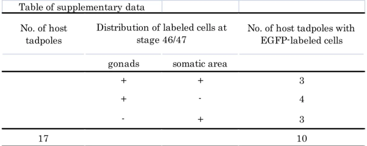
Supplementary data Reexamination of Wylie’s experiment (1985)
(A) Procedure for transfer of EGFP-labeled GP carrying blastomere. Single EGFP-labeled GP carrying blastomere of stage 7 Dria embryo was isolated and was transferred into blastocoel of wild-type host at stage 9. Host embryos were allowed to develop to stage 46/47.
(B) Outside view of jaws of a stage 46 host tadpole. (B’) magnified figure of white box in (B). There were cells (white and yellow arrows) with opaque appearance. (B”) Fluorescent figure of (B’). Most of opaque cells (white and yellow arrows) expressed EGFP. (B”’) Xpat mRNA was detected in some EGFP-labeled cells (yellow arrows) bywhole mount in situ hybridization.
They are probably ectopic PGCs, not somatic cells.
(C, C’) Magnified view of dissected host gonad at stage 46. Several EGFP-labeled cells (white arrows) were found in gonad. A labeled-GP carrying blastomere at stage 7 produced PGCs in gonads. Scale bars represent 1 mm in B and 150 μm in C.
26 No. of host
tadpoles
No. of host tadpoles with EGFP-labeled cells
gonads somatic area
+ + 3
+ - 4
- + 3
17 10
Table of supplementary data
17 tadpoles out of operated embryos showed healthy appearance. Six tadpoles showed EGFP- labeled cells from outside view (at somatic area). Dissected gonads from seven tadpoles possessed EGFP-labeled cells.
Distribution of labeled cells at stage 46/47
27
Acknowledgements
I am very grateful to Dr. M. Yuge (Fukuoka Women’s University) for his technical advice on cytoplasm transplantation. I also thank members of my laboratory for their support.
28
References
Bounoure, L., 1934. Recherches sur la lignée germinale chez la grenouille rousse aux premiers stades du développement. Ann. Sci. Nat. 17, 67-248.
Buehr, M.L., Blackler, A.W., 1970. Sterility and partial sterility in the South African clawed toad following the pricking of the egg. J. Embryol. Exp.
Morph. 23, 375-384.
Cleine, J.H., Dixon, K.E., 1985. The effect of egg rotation on the differentiation of primordial germ cells in Xenopus laevis. J. Embryol. Exp.
Morph. 90, 79-99.
Hashimoto,Y., Maegawa, S., Nagai, T., Yamaha, E., Suzuki, H., Yasuda, K., Inouea, K., 2004. Localized maternal factors are required for zebrafish germ cell formation. Dev. Biol. 268, 152-161.
Horie, C., Suzuki, H., Sakaguchi, M., Mihara, K., 2002. Characterization of Signal That Directs C-Tail–anchored Proteins to Mammalian Mitochondrial Outer Membrane. Mol Biol Cell. 13, 1615-1625.
Houston, D.W., King, M.L., 2000. Germ plasm and molecular determinants of germ cell fate. Curr Top Dev Biol. 50,155-181.
Houston, D.W., King, M.L., 2000. A critical role for Xdazl, a germ plasm-localized RNA, in the differentiation of primordial germ cells in Xenopus. Development. 127, 447-456.
Houston, D.W., Zhang, J., Maines, J.Z., Wasserman, S.A., King, M.L., 1998. A Xenopus DAZ-like gene encodes an RNA component of germ plasm and is a functional homologue of Drosophila boule. Development. 125, 171-180.
29
Hudson, C., Woodland, H.R., 1998. Xpat, a gene expressed specifically in germ plasm and primordial germ cells of Xenopus laevis. Mech. Dev.
73,159-168.
Ikenishi, K., Nakazato, S., Okuda, T., 1986. Direct evidence for the presence of germ cell determinant in vegetal pole cytoplasm of Xenopus laevis and in a subcellular fraction of it. Develop. Growth and Differ. 28, 563-568.
Ikenishi, K., Tsuzaki, Y., 1988. The positional effect of presumptive primordial germ cells (pPGCs) on their differentiation into PGCs in Xenopus.
Development. 102, 527-535.
Illmensee, K., Mahowald, A.P., 1974. Transplantation of posterior polar plasm in Drosophila. Induction of germ cells at the anterior pole of the egg.
Proc. Natl. Acad. Sci. U S A. 71, 1016-1020.
Kataoka, K., Yamaguchi,T., Orii, H., Tazaki, A., Watanabe, K., Mochii, M., 2006. Visualization of the Xenopus primordial germ cells using a green fluorescent protein controlled by cis elements of the 3' untranslated region of the DEADSouth gene. Mech. Dev. 123, 746–760.
Kroll, KL., Amaya, E., 1996. Transgenic Xenopus embryos from sperm nuclear transplantations reveal FGF signaling requirements during gastrulation. Development 122, 3173–3183.
Lai, F., Zhou, Y., Luo, X., Fox, J., King, M.L., 2011. Nanos1 functions as a translational repressor in the Xenopus germline. Mech. Dev.128, 153-163.
Lavial, F., Acloque, H., Bachelard, E., Nieto, M.A., Samarut, J., Pain, B., 2009. Ectopic expression of Cvh (Chicken Vasa homologue) mediates the
30
reprogramming of chicken embryonic stem cells to a germ cell fate. Dev. Biol.
330, 73- 82.
Machado, R.J., Moore, W., Hames, R., Houliston, E., Chang, P., King, M.L., Woodland, H.R., 2005. Xenopus Xpat protein is a major component of germ plasm and may function in its organisation and positioning. Dev. Biol. 287, 289-300.
McLaren, A., 2003. Primordial germ cells in the mouse. Dev. Biol. 262, 1-15
Mita, K., Yamashita, M., 2000. Expression of Xenopus Daz-like protein during gametogenesis and embryogenesis. Mech. Dev. 94, 251-255.
Mizuno, N., Ueda, Y., Kondoh, H., 2005. Requirement for betaB1-crystallin promoter of Xenopus laevis in embryonic lens development and lens regeneration. Dev Growth Differ 47, 131–140.
Mosquera, L., Forristall, C., Zhou, Y., King, M.L., 1993. A mRNA localized to the vegetal cortex of Xenopus oocytes encodes a protein with a nanos-like zinc finger domain. Development 117, 377-386.
Nieuwkoop, P. D., Faber, J., 1967. Normal table of Xenopus laevis (Daudin):
A systematical and Chronological survey of the Development from the fertilized egg till the end of metamorphosis. Garland, New York.
Niwa, H., Yamamura, K., Miyazaki, J., 1991. Efficient selection for high-expression transfectants with a novel eukaryotic vector. Gene 108, 193–199.
Ressom, R.E., Dixon, K.E., 1988. Relocation and reorganization of germ plasm in Xenopus embryos after fertilization. Development. 103, 507-518.
31
Robb, D.L., Heasman, J., Raats, J., Wylie, C., 1996. A Kinesin-like protein is required for germ plasm aggregation in Xenopus. Cell. 87, 823-831.
Sakamaki, K., Takagi, C., Yoshino, J., Yokota, H., Nakamura, S., Kominami, K., Hyodo, A., Takamune, K., Yuge, M., Ueno, N., 2005. Transgenic frogs expressing the highly fluorescent protein venus under the control of a strong mammalian promoter suitable for monitoring living cells. Dev Dyn. 233, 562-569.
Smith, L.D., 1966. The role of a “germinal plasm” in the formation of primordial germ cells in Rana pipiens. Dev. Biol. 14, 330-347.
Taguchi , A., Takii, M., Motoishi, M., Orii, H., Mochii, M., Watanabe, K., 2012.
Analysis of localization and reorganization of germ plasm in Xenopus transgenic line with fluorescence-labeled mitochondria. Dev Growth Differ.
54(8), 767-776.
Wakahara, M., 1977. Partial characterization of ‘primordial germ cell-forming activity' localized in vegetal pole cytoplasm in anuran eggs. J.
Embryol. Exp. Morph. 39, 221-233.
Wakahara, M., 1978. Induction of supernumerary primordial germ cells by injecting vegetal pole cytoplasm into Xenopus eggs. J. Exp.Zool. 203, 159-164.
Whitington. P.M., Dixon. K.E., 1975. Quantitative studies of germ plasm and germ cells during early embryogenesis of Xenopus laevis. J. Embryol. Exp.
Morphol. 33, 57-74.
Wylie, C. C., Heasman, J., Snape, A., O’driscoll, M., Holwill, S., 1985.
32
Primordial germ cells of Xenopus laevis are not irreversibly determined early in development. Dev. Biol. 112, 66-72.
Yu, Z., Ji, P., Cao, J., Zhu, S., Li, Y., Zheng, L., Chen, X., Feng, L., 2009. Dazl Promotes germ cell differentiation from embryonic stem cells. Journal of Molecular Cell Biology. 1, 93–103.