1
顎顔面領域における
MDCT
の有用性Usefulness of multidetector computed tomography for maxillofacial region
日本大学大学院松戸歯学研究科歯学専攻 伊東 浩太郎
(指導 金田 隆 教授)
2 Abstract
Conventional diagnostic imaging in the maxillofacial region was using intraoral radiography,
panoramic radiography and extraoral radiography. The nasopalatine duct in the maxilla contains the
nasopalatine nerve and artery. A nasopalatine duct cyst is a one of nonodontogenic developmental cyst
that arises in the nasopalatine duct. It is the most common type of nonodontogenic cyst, and may arise
from epithelial remnants in the nasopalatine duct. Therefore, differentiation between the normal
nasopalatine duct and duct lesions is very important in clinical situations. However, it is very difficult to
differentiate between nasopalatine duct cyst and normal nasopalatine duct, and to distinction in a
development area with three-dimensional of nasopalatine duct cyst using intraoral radiography and
panoramic radiography. Moreover, it is very difficult to diagnosis of maxillofacial fractures with
three-dimensional deviation, and to evaluate of soft tissue of maxillofacial fractures using panoramic
radiography and extraoral radiography. CT can evaluate three-dimensional structure and soft tissue
condition. Therefore, CT is necessary in recent clinical diagnostic imaging in the maxillofacial region.
MDCT has multidetector in the axis direction, wide range scan with ultra-short time, high quality
reconstructed image and 3D image. Recently, MDCT has been used the diagnosis of many pathologic
conditions such as head and neck, abdominal and any other regions. However, there have been few
reports about MDCT to evaluate maxillofacial region. The purposes of this study were to investigate 1)
image findings of the nasopalatine duct region using MDCT such as sex, form, size and CT value, and 2)
image findings of fractures in maxilla and zygomatic bone using MDCT such as age, sex, cause of injury,
3
fracture location and concomitant damage, and to assessment of usefulness of MDCT for maxillofacial
region.
1) The subjects were consisted of 122 patients who underwent MDCT imaging of the maxilla after
providing informed consent. Anatomic variants of the nasopalatine duct were classified using coronal and
axial slices. Measurements of the duct width diameters and the distances from the nasopalatine foramen
to the incisive foramen as a major axis were performed using sagittal and coronal slices. CT values for the
duct portion with maximum diameter were measured using axial slices.
2) A prospective study was performed in 104 patients with midface fractures and who underwent
MDCT. The midface fracture locations were classified as Le Fort I– III, isolated anterior maxillary sinus,
isolated zygomatic arch, zygomaticomaxillary complex and blowout fractures.
In the nasopalatine duct region, MDCT images showed that the mean width diameter of the duct at
all points using sagittal slices differed significantly between males and females. Furthermore, the
nasopalatine duct was often a single duct, and the mean CT value of males and females was 122.4HU.
MDCT images showed that midface fracture was often a zygomaticomaxillary complex. Furthermore, the
age and sex differences between the falls and violence groups were significant. Image findings of the
nasopalatine duct region such as sex, form, size and CT value and image findings of fractures in maxilla
and zygomatic bone such as age, sex, cause of injury, fracture location and concomitant damage have
been demonstrated using MDCT. These studies suggested that MDCT was a useful examination for
4 maxillofacial region.
5 要 旨
従来から日常臨床において顎顔面領域における画像診断は口内法エックス線検査,パノ
ラマエックス線検査,顔面頭蓋への単純エックス線検査等が施行されてきた。同領域の上
顎骨正中に位置する鼻口蓋管内部には鼻口蓋神経,鼻口蓋動脈が存在している。また,鼻
口蓋管に発生する鼻口蓋管嚢胞は最も一般的な顔裂性嚢胞の一つである。そのため,鼻口
蓋管は他の良性腫瘍や嚢胞等の鑑別も含め,顎顔面領域で重要な解剖構造である。しかし
ながら,従来行われていた口内法エックス線検査,パノラマエックス線検査等では 3次元
的な広がりを持つ鼻口蓋管病変の鑑別や進展範囲の判別は非常に困難であった。また,3次
元的偏位を伴う顎顔面領域の骨折の診断は, パノラマエックス線検査,単純エックス線検査
等の従来の検査では軟組織の状態も含め詳細に評価することは困難であった。CT検査はコ
ンピューターを用いてエックス線情報を処理することにより詳細な断面像が得られ,CT値
の異なる周囲軟組織の情報も評価可能である。よって,近年,顎顔面領域の診断にCT検査
は必要不可欠となっている。MDCTは体軸方向に複数の検出器を有することが最も大きな特
徴である。同装置は,検出器の多列化,画像処理の高速化により,超短時間に広範囲の撮
像が可能になり,より高精細な再構成画像や 3次元画像の作成が可能となった。現在では,
頭頸部のみならず,腹部や全身領域への臨床応用にMDCTは幅広く使用されている。しかし
ながらMDCTを用いた上顎骨や頬骨を含む顎顔面領域への研究や応用は未だ乏しい。本研究
の目的は,1) 鼻口蓋管のMDCTによる性差,形態,大きさおよび内部CT値,2)上顎骨およ
6
び頬骨の骨折のMDCTによる年齢,性別,原因,部位および付随的損傷の検討を行い,顎顔
面領域におけるMDCTの有用性を評価することである。
鼻口蓋管のMDCTによる性差,形態,大きさおよび内部CT値の検討は,2012年4月か
ら2012年8月までの期間に日本大学松戸歯学部付属病院の放射線科において,顎顔面領域
のMDCT検査を施行し,撮像された122名の患者の画像を使用した。鼻口蓋管の形態の分
類において,MDCT前額断像を用い,以下の3つに分類した。(A): single duct, (B): two parallel
ducts, (C): variations of Y type ducts. また,鼻口蓋管の幅径および長径において,MDCT矢状
断像および前額断像を用いて以下の点で計測を行った。(ⅰ)鼻口蓋孔, (ⅱ)鼻口蓋孔と切歯孔
の中間点, (ⅲ)切歯孔, (ⅳ)鼻口蓋孔と切歯孔との距離。さらに,MDCT水平断像を用いて鼻
口蓋管のCT値の計測も行った。次に,上顎骨および頬骨の骨折の MDCTによる年齢,性
別,原因,部位および付随的損傷の検討は, 2006年4月から2014年5月までの期間に日
本大学松戸歯学部付属病院の放射線科においてMDCT検査を施行し,撮像された104名の
患者の画像を使用した。顎顔面領域の骨折部位を,上顎洞前壁の骨折,頬骨弓の骨折,頬
骨上顎複合骨折,吹き抜け骨折,Le Fort Ⅰ-Ⅲ型上顎骨骨折に分類した。また,上顎骨およ
び頬骨の骨折患者の統計分析はFisher’s exact testを用いて以下の項目で行った。ⅰ)損傷の
原因(転倒または暴力),ⅱ)年齢,ⅲ)性別,ⅳ)付随的損傷,ⅴ)骨折の部位。鼻口蓋
管のMDCT特徴像は,幅径および長径の矢状断像において,男性が女性より優位に大きか
った。また,鼻口蓋管の形態はsingle ductが一番多く,CT値は122.4HUだった。顎顔面領
7
域の骨折のMDCT 特徴像は,頬骨上顎複合骨折が一番多かった。さらに,転倒による骨折
は61 歳以上が 60 歳以下より有意に多く,暴力による骨折は男性が女性より有意に多かっ
た。本検討により,MDCT は正常な鼻口蓋管および骨折を詳細に観察することができた。
以上の検討結果より,顎顔面領域においてMDCTは有用であることが示唆された。
Keywords computed tomography maxillofacial region nasopalatine duct fracture
コンピューター断層撮影装置 顎顔面領域 鼻口蓋管 骨折
8 緒 言
従来から日常臨床において顎顔面領域の画像診断は口内法エックス線検査,パノラマエ
ックス線検査,顔面頭蓋への単純エックス線検査等が施行されてきた 1)。同領域の上顎骨
正中に位置する鼻口蓋管(切歯管:以下,鼻口蓋管とする)は,口蓋突起の癒合後に上顎
骨前方で形成され,内部には上顎神経の中隔後鼻枝である鼻口蓋神経,大口蓋動脈と蝶口
蓋動脈の中隔後鼻枝が吻合している鼻口蓋動脈が存在している 2)。また,鼻口蓋管に発生
する鼻口蓋管嚢胞は最も一般的な顔裂性嚢胞の一つである 3)。そのため,鼻口蓋管は他の
良性腫瘍や嚢胞等の鑑別も含め,顎顔面領域で重要な解剖構造である。しかしながら,従
来行われていた口内法エックス線検査,パノラマエックス線検査等では 3次元的な広がり
を持つ鼻口蓋管病変の鑑別や進展範囲の判別は非常に困難であった。
3次元的偏位を伴う顎顔面領域の骨折の診断は,パノラマエックス線検査,単純エックス
線検査等の従来の検査では軟組織の状態も含め詳細に評価することは困難であった。
Computed tomography (以下,CTとする) 装置は1972年に英国EMI社のHounsfieldによって
発明され,臨床医学の発展に寄与してきた。CT検査はコンピューターを用いてエックス線
情報を処理することにより詳細な断面像が得られ,CT値の異なる周囲軟組織の情報も評価
可能である。また,近年,CT検査は,容積画像による 3次元的情報に加え,任意の断面が
抽出可能なMulti-planar reconstruction (以下,MPRとする) を用いることにより多方向の正確
な断層像評価も可能となった。よって,近年,顎顔面領域の診断にCT検査は必要不可欠と
9 なっている。
Multidetector CT (以下,MDCTとする) は体軸方向に複数の検出器を有することが最も大
きな特徴である。同装置は,検出器の多列化,画像処理の高速化により,超短時間に広範
囲の撮像が可能になり,より高精細な再構成画像や 3次元画像の作成が可能となった。現
在では,頭頸部のみならず,腹部や全身領域への臨床応用にMDCTは幅広く使用されている。
しかしながらMDCTを用いた上顎骨や頬骨を含む顎顔面領域への研究や応用は未だ乏しい。
本研究の目的は,1) 鼻口蓋管のMDCTによる性差,形態,大きさおよび内部CT値,2) 上
顎骨および頬骨の骨折のMDCTによる年齢,性別,原因,部位および付随的損傷の検討を行
い,顎顔面領域におけるMDCTの有用性を評価することである。
資料および方法
本研究は,日本大学松戸歯学部倫理委員会(No.EC10-039, No. EC12-009)によって承認を
得ている。
1) 鼻口蓋管のMDCTによる性差,形態,大きさおよび内部CT値
2012年4月から2012年8月までの期間に日本大学松戸歯学部付属病院の放射線科におい
て,顎顔面領域のMDCT (Aquilion 64; Toshiba Medical Systems, Tokyo, Japan) 検査を施行し,
撮像された患者のうち,上顎骨に病変および金属アーチファクトがなく,上顎中切歯が存
在する122名の患者(年齢17-88歳,平均年齢49.8歳の男性61名,女性61名)の画像を
10
使用した。MDCTの撮像条件は管電圧120kV,管電流100mA,撮像領域240×240mm,
ヘリカルピッチ 41,回転時間1.0 秒,再構成スライス厚は 0.3mmであった。画像評価は,
ディスプレイモニター (RadiForce G31; Eizo Nanao, Ishikawa, Japan)を用いて経験年数5年以
上の2 名の歯科放射線科医が各々MDCT 画像を評価し,それぞれの評価に相違が生じたも
のについては討議により合意を得て1つの評価にした。
鼻口蓋管の形態の分類において,MDCT前額断像を用いて行い、以下の3つに分類した。
(A): single duct, (B): two parallel ducts, (C): variations of Y type ducts(Fig. 1)4) . 前額断像およ
び水平断像の各0.3mmの全スライスを確認しながら,(A)鼻中隔が鼻腔底までのもの,(B)
鼻中隔が切歯孔まで及ぶもの,(C)鼻中隔が鼻腔底より低位かつ切歯孔まで及ばないもの
とした。また,鼻口蓋管の幅径(3点)および長径において,MDCT 矢状断像および前額
断像を用いて以下の点で計測を行った4)。(ⅰ)鼻口蓋孔, (ⅱ)鼻口蓋孔と切歯孔の中間点, (ⅲ)
切歯孔, (ⅳ)鼻口蓋孔と切歯孔との距離 (Fig. 2) さらに,鼻口蓋管のCT値計測は,水平
断像を用いて画像上鼻口蓋管の直径が最大径となるスライスにて関心領域(Region of
Interest) を設定し,計測を行った。鼻口蓋管の幅径および長径の男女間における比較検討は
Mann–Whitney U testを用いて行い,P < 0.05を有意差がありとした。
2) 上顎骨および頬骨の骨折のMDCTによる年齢,性別,原因,部位および付随的損傷
2006年4月から2014年5月までの期間に日本大学松戸歯学部付属病院の放射線科におい
て外傷により顎顔面領域のMDCT (Aquilion 64; Toshiba Medical Systems, Tokyo, Japan) 検査
11
を施行し,撮像された104名の患者(年齢12-83歳,平均年齢42.0歳の男性75名,女性29
名)の画像を使用した。MDCT の撮像条件は管電圧 120kV,管電流 100mA,撮像領域
240×240mm,ヘリカルピッチ41,回転時間1.0秒,再構成スライス厚は0.3mmであった。
画像評価は,ディスプレイモニター (RadiForce G31; Eizo Nanao, Ishikawa, Japan)を用いて経
験年数5年以上の2名の歯科放射線科医が各々MDCT 画像を評価し,それぞれの評価に相
違が生じたものについては討議により合意を得て1つの評価にした。
上顎骨および頬骨の骨折部位を,上顎洞前壁の骨折,頬骨弓の骨折,頬骨上顎複合骨折,
吹き抜け骨折,Le Fort Ⅰ-Ⅲ型上顎骨骨折に分類した5) 。上顎骨および頬骨の骨折患者の
統計分析はFisher’s exact testを用い,ⅰ)損傷の原因(転倒または暴力),ⅱ)年齢,ⅲ)性
別,ⅳ)合併症,ⅴ)骨折の部位の項目で行われた。これらはStatistical package IBM SPSS
Statistics, version 20 (IBM Japan, Tokyo, Japan)を用いて行われ,P < 0.05を有意差ありとした。
結 果
1) 鼻口蓋管のMDCTによる性差,形態,大きさおよび内部CT値
前額断像を用いた形態の分類において,122名の鼻口蓋管のうち,69名(56.5%)がsingle
duct に分類され,13名(10.7%)がtwo parallel ductsに分類され,40名(32.8%)がY type
ducts に分類された(Table1)。 全患者 122 名の鼻口蓋管の幅径および長径の平均値におい
て,矢状断像では,鼻口蓋孔部で3.2±1.3mm,中間点で2.7±0.9mm,切歯孔部で3.1±0.9mm,
12
直径は14.0±2.6mmであった。また,そのうち最大値は鼻口蓋孔部で6.3mm,中間点で5.1mm,
切歯孔部で5.8mm,直径は21.4mmであり,最小値は鼻口蓋孔部で0.6mm,中間点で0.7mm,
切歯孔部で0.8mm,直径は5.6mmであった(Table 2)。 全患者122名の鼻口蓋管の幅径お
よび長径の平均値において,前額断像では,鼻口蓋孔部で4.3±1.1mm,中間点で3.6±1.0mm,
切歯孔部で3.8±1.0mm,直径は11.6±2.4mmであった。また,そのうち最大値は鼻口蓋孔部
で6.8mm,中間点で6.4mm,切歯孔部で6.2mm,直径は17.3mmであり,最小値は鼻口蓋孔
部で1.7mm,中間点で1.0mm,切歯孔部で1.1mm,直径は5.4mmであった(Table 3)。 鼻
口蓋管を男女別に観察した結果,鼻口蓋管の幅径および長径の平均値において,矢状断像
では,鼻口蓋孔部で男性3.4±1.1mm 女性2.9±1.1mm (P < 0.05),中間点で男性2.9±0.9mm 女
性2.4±0.8mm (P < 0.05),切歯孔部で男性3.3±0.9mm 女性2.8±0.9mm (P < 0.05),直径は男
性14.5±2.4mm 女性13.4±2.6mm (P < 0.05)であった(Table 4)。 鼻口蓋管を男女別に観察
した結果,鼻口蓋管の幅径および長径の平均値において,前額断像では,鼻口蓋孔部で男
性4.5±1.2mm 女性4.2±1.0mm,中間点で男性3.7±1.1mm 女性3.6±0.8mm,切歯孔部で男性
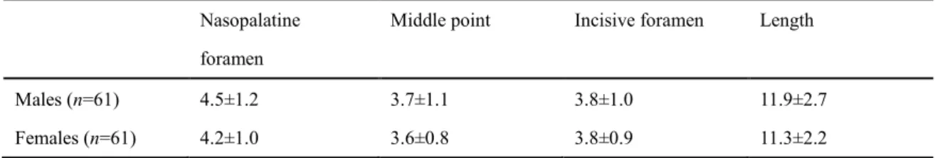
3.8±1.0mm 女性3.8±0.9mm,直径は男性11.9±2.7mm 女性11.3±2.2mmであった(Table 5)。
全患者122名の鼻口蓋管のCT値の平均は122.4HUであった。
2) 上顎骨および頬骨の骨折のMDCTによる年齢,性別,原因,部位および付随的損傷
上顎骨および頬骨の骨折患者の原因の 70.2%は転倒,29.8%は暴力によるものであった。
転倒患者のうち93.8%が61歳以上であった。年齢差と性差は,転倒患者群と暴力患者群と
13
の間において有意に差があった(Table 6)。上顎骨および頬骨の骨折は右側より左側に多く
存在した。左右側いずれも転倒患者群と暴力患者群との間において有意差はみられなかっ
た(Table 7)。付随的損傷は,8名に複視,3名に脳出血,2名に複視および脳出血がみられ
た。付随的損傷において,転倒患者群と暴力患者群との間に有意差はなかった(Table 7)。患
者104名の骨折部位のうち,頬骨上顎複雑骨折に分類されたものが66症例,Le Fort Ⅰ型の
上顎骨骨折に分類されたものが11症例,上顎洞前壁の骨折に分類されたものが16症例,吹
き抜け骨折に分類されたものが7症例であった。頬骨上顎複雑骨折は77.3%が転倒,22.7%
が暴力に起因しており,転倒患者群と暴力患者群との間において有意差がみられた(Fig. 3)。
上顎洞前壁の骨折は43.8%が転倒,56.2%が暴力に起因しており,転倒患者群と暴力患者群
との間において有意差がみられた (Table 8)(Fig. 4)。
考 察
(1) 鼻口蓋管のMDCTによる性差,形態,大きさおよび内部CT値
本検討では56.5%がsingle duct ,10.7%がtwo parallel ducts,32.8%がY type ductsであっ
た。Suterらは,鼻口蓋管の形態は45%がsingle duct に分類され,15%がtwo parallel ducts
に分類され,40%がY type ductsに分類されていたと報告している6) 。
本検討では,矢状断像による鼻口蓋管の幅径および長径は,すべての個所において男性
と女性において有意に差がみられた。Ling らの報告では,鼻口蓋管の長径は有歯顎者と無
14
歯顎者では有意に有歯顎者の鼻口蓋管の長径が長かったとしている。また,鼻口蓋管の幅
径は加齢と共に拡大する傾向があるともしている7)。これらのことから鼻口蓋管の長径およ
び幅径は上顎骨の大きさに影響を受けると考えられる。
鼻口蓋管の中には鼻口蓋神経,鼻口蓋動脈が含まれている 2)。また,扁平上皮や繊毛上皮
も含まれている。よって,発育性の嚢胞が鼻口蓋管中の残留した上皮から発生する3)。しか
しながら,拡大した正常鼻口蓋管と鼻口蓋管嚢胞との鑑別は非常に困難であるといわれて
いる8)。Swansonらの報告によると,鼻口蓋管嚢胞の平均径は17.1mmであり,全鼻口蓋管
嚢胞中75%は20mm未満であり,症状は70%の症例でみられたとしている9)。Suterらの報
告では,CBCT前額断像上で鼻口蓋管の平均径は12.5mmであった。また,最小径は5.5mm
であり,最大径は32.6mmであった6)。さらに,Nicholasらによると,咬合法エックス線写
真上で鼻口蓋管の正常の範囲は 6mm までと言われている 8) 。本検討では正常鼻口蓋管の
最大径が矢状断像では鼻口蓋孔部で 6.3mm,中間点で 5.1mm,切歯孔部で5.8mm,前額断
像では,鼻口蓋孔部で6.8mm,中間点で6.4mm,切歯孔部で6.2mmであった。鼻口蓋管の
幅径がこの数値を超える場合,鼻口蓋管嚢胞を疑う必要があると示唆された。
本検討では鼻口蓋管のCT 値は122.4HUであった。MDCT は高い軟組織の抽出能を有し
ており,腫瘍性病変に対する高い診断能力がある 10)。鼻口蓋管内部は主に神経と血管によ
り構成されている。神経や血管のCT値は80‐90HUであるといわれている11)。しかしなが
ら,Nandalurらによると,線維化や石灰化によって血管のCT値は70‐1200HUの間で変動
15
すると報告されている12)。したがって鼻口蓋管のCT値は神経や血管のCT値80‐90HUよ
り高値になった可能性が考えられる3, 11, 12)。
(2) 上顎骨および頬骨の骨折のMDCTによる年齢,性別,原因,部位および付随的損傷
上顎骨および頬骨の骨折にはいくつかのタイプがあり,最も一般的なもので,Le Fort Ⅰ-
Ⅲ型,頬骨上顎複合骨折,上顎洞前壁の骨折がある。Iidaらの報告では,中顔面の骨折の多
くが頬骨上顎複合骨折であると示されている 13)。本検討では,頬骨上顎複合骨折が一番多
く,Le Fort Ⅰ型,上顎洞前壁の骨折が続いている。
Yamamotoらの報告によると,高齢者において,骨折の原因の多くを転倒が占めるとされ
ている14)。本検討では転倒による骨折の多くは61歳以上の女性だった。Yamamotoらはま
た,頬骨上顎複合骨折の原因の多くは転倒だとしている15)。Salonenらの報告でも,頬骨上
顎複合骨折は転倒した場合に最も引き起こされやすい骨折であるといわれている 16, 17)。本
検討では,頬骨上顎複合骨折の病因は 77.3%が転倒であり,転倒が頬骨上顎複合骨折の特
徴的病因であることを示した。顎顔面領域において,転倒の外力が頬骨,上顎骨の直接的
な骨折を引き起こしていると考えられる。
Salonenらによると,暴力の多くは拳による殴打であるが,金属製の凶器を用いた殴打や
キックにより受けた暴力は,より損傷を増大させるとされている 18)。本検討により,顎顔
面領域の上顎洞前壁の骨折は高エネルギーの暴力に起因していると考えられた。本検討に
おいて,暴力による上顎骨および頬骨の骨折は左側が右側より多かった。ほとんどの人々
16
は右ききであるため暴力関連の骨折は左側に多いと考えられる。
家庭内暴力は女性の顎顔面領域の損傷の原因の一つといわれている19)。 しかし,家庭内
暴力による外傷は,はっきりとした特徴不足のため識別が困難である 20)。本検討では,上
顎洞前壁の骨折の病因は43.8%が転倒,56.2%が暴力であり,上顎洞前壁の骨折の多くが暴
力に起因していることを示した。これらのことから,病因が不明の上顎洞前壁の骨折患者
が家庭内暴力によるものである可能性が考えられた。
本検討において,骨折の付随的損傷として,複視を認めたものが8例,脳出血を認めた
ものが3例,両者を認めたものが2例であった。MDCTは顎顔面領域の骨片の偏位,軟組
織浮腫および出血を検出するのに効果的な検査だと考えられる21- 24)。このことから,頭蓋
顎顔面領域の広範囲な骨折を有する患者や,意識の混濁および消失している患者において,
MDCT検査は有効であると考えられた。
結 語
本研究で,MDCT は正常な鼻口蓋管および骨折を詳細に観察することができた。MDCT
による鼻口蓋管は,幅径および長径の矢状断像において,男性が女性より優位に大きかっ
た。また,形態はsingle ductが一番多く,CT値の平均は122.4HUだった。MDCTによる上
顎骨および頬骨の骨折は,頬骨上顎複合骨折が一番多かった。さらに,転倒による骨折は 61歳以上が60歳以下より有意に多く,暴力による骨折は男性が女性より有意に多かった。
17
以上の検討結果より,顎顔面領域においてMDCTは有用であることが示唆された。
18 文 献
1. Frommer HH, Stabulas-Savage JJ. Radiology for the dental professional. 8th ed. St.Louis:
ELSEVIER; 2005. p.331-333.
2. Naidich TP, Blaser SI, Lien RJ, Mclone DG, Fatterpekar GM, Bauer BS, Embryology and
congenital lesions of the midface. In: Som PM, Curtin HD, editors. Head and neck imaging 5th
ed. St.Louis: ELSEVIER; 2011.p. 110-118.
3. Koenig LJ. Cysts, nonodontogenic/mandible and maxilla. In: Koenig LJ, Tamimi D, Petrikowski
CG, Harnsberger HR, Ruprecht A, Benson BW, Van Dis M, Hatcher D, editors. Diagnostic
imaging oral and maxillofacial. 1st ed. Friesens: AMIRSYS; 2012. p. 66–9.
4. Bornstein MM, Balsiger R, Sendi P, von Arx T. Morphology of the nasopalatine canal and dental
implant surgery: a radiographic analysis of 100 consecutive patients using limited cone-beam
computed tomography. Clin Oral Implants Res. 2011;22:295–301.
5. Hopper RA, Salemy S, Sze RW. Diagnosis of midface fractures with CT: what the surgeon
needs to know. Radiographics. 2006;26:783–93.
6. Suter VG, Sendi P, Reichart PA, Bornstein MM. The nasopalatine duct cyst: an analysis of the
relation between clinical symptoms, cyst dimensions, and involvement of neighboring
anatomical structures using cone beam computed tomography. J Oral Maxillofac Surg.
2011;69:2595–603.
19
7. Liang X, Jacobs R, Martens W, Hu YQ, Adriaensens P, Quirynen M, et al. Macro- and
micro-anatomical, histological and computed tomography scan characterization of the
nasopalatine canal. J Clin Periodontol. 2009;36:598–603.
8. Goodger NM, Hendy CW. Cystic lesions of the jaws/non-odontogenic cysts. In: Andersson L,
Kahnberg KE, Pogrel MA, editors. Oral and maxillofacial surgery. 1st edition. Oxford:
Wiley-Blackwell; 2010. p. 624–5.
9. Swanson KS, Kaugars GE, Gunsolley JC. Nasopalatine duct cyst: an analysis of 334 cases. J
Oral Maxillofac Surg. 1991;49:268–71.
10. Ludlow JB, Mol A. Other imaging modalities/computed tomographic scanning. In: White SC,
Pharoah MJ. Oral radiology principles and interpretation. 7th edition. St. Louis: Elsevier; 2014.
p. 232–3.
11. Matsumoto Y, Krishnan S, Fowler SJ, Saremi F, Kondo T, Ahsan C, et al. Detection of phrenic
nerves and their relation to cardiac anatomy using 64-slice multidetector computed tomography.
Am J Cardiol. 2007;100:133–7.
12. Nandalur KR, Baskurt E, Hagspiel KD, Phillips CD, Kramer CM. Calcified carotid
atherosclerotic plaque is associated less with ischemic symptoms than is noncalcified plaque on
MDCT. AJR Am J Roentgenol. 2005;184:295–8.
13. Iida S, Kogo M, Sugiura T, Mima T, Matsuya T. Retrospective analysis of 1502 patients with
20
facial fractures. Int J Oral Maxillofac Surg. 2001;30:286–90.
14. Yamamoto K, Matsusue Y, Murakami K, Horita S, Sugiura T, Kirita T. Maxillofacial fractures
in older patients. J Oral Maxillofac Surg. 2011;69:2204–10.
15. Yamamoto K, Kuraki M, Kurihara M, Matsusue Y, Murakami K, Horita S, et al. Maxillofacial
fractures resulting from falls. J Oral Maxillofac Surg. 2010;68:1602–7.
16. Salonen EM, Koivikko MP, Koskinen SK. Multidetector computed tomography imaging of
facial trauma in accidental falls from heights. Acta Radiol. 2007;48:449–55.
17. Salonen EM, Koivikko MP, Koskinen SK. Acute facial trauma in falling accidents: MDCT
analysis of 500 patients. Emerg Radiol. 2008;15:241–7.
18. Salonen EM, Koivikko MP, Koskinen SK. Violence-related facial trauma: analysis of
multidetector computed tomography findings of 727 patients. Dentomaxillofac Radiol.
2010;39:107–12.
19. Hashemi HM, Beshkar M. The prevalence of maxillofacial fractures due to domestic violence: a
retrospective study in a hospital in Tehran, Iran. Dent Traumatol. 2011;27:385–8.
20. Perciaccante VJ, Ochs HA, Dodson TB. Head, neck, and facial injuries as markers of domestic
violence in women. J Oral Maxillofac Surg. 1999;57:760–2.
21. Chu ZG, Yang ZG, Dong ZH, Chen TW, Zhu ZY, Deng W, et al. Features of
cranio-maxillofacial trauma in the massive Sichuan earthquake: analysis of 221 cases with
21
multi-detector row CT. J Craniomaxillofac Surg. 2011;39:503–8.
22. Sohns JW, Staab W, Sohns C, Schwarz A, Streit U, Hosseini AS, et al. Current perspective of
multidetector computed tomography (MDCT) in patients after midface and craniofacial trauma.
Clin Imaging. 2013;37:728–33.
23. Ogura I, Sasaki Y, Kaneda T. Analysis of mandibular condylar and glenoid fossa fractures with
computed tomography. Eur Radiol. 2014;24:902–6.
24. Ogura I, Sasaki Y, Kaneda T. Multidetector computed tomography of maxillofacial fractures.
Jpn Dent Sci Rev. 2014;50:86–90.
22
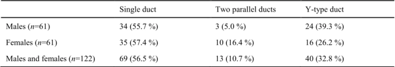
Table 1 Classification of anatomic variations of the nasopalatine duct using reformatted coronal slices from 64-MDCT
Single duct Two parallel ducts Y-type duct
Males (n=61) 34 (55.7 %) 3 (5.0 %) 24 (39.3 %)
Females (n=61) 35 (57.4 %) 10 (16.4 %) 16 (26.2 %)
Males and females (n=122) 69 (56.5 %) 13 (10.7 %) 40 (32.8 %) Data are shown as n (%).
23
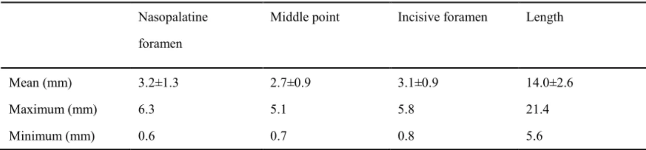
Table 2 Analysis of the nasopalatine duct using reformatted sagittal slices from 64-MDCT imaging (n=122)
Nasopalatine foramen
Middle point Incisive foramen Length
Mean (mm) 3.2±1.3 2.7±0.9 3.1±0.9 14.0±2.6
Maximum (mm) 6.3 5.1 5.8 21.4
Minimum (mm) 0.6 0.7 0.8 5.6
Data are shown as means ± SD.
24
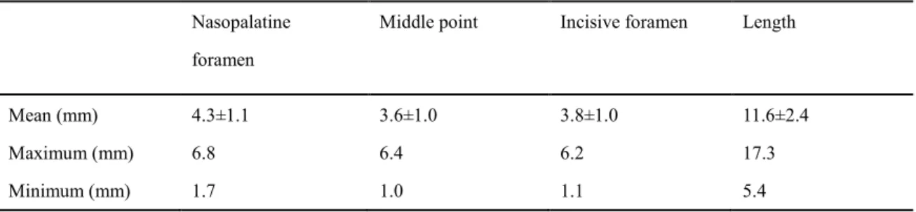
Table 3 Analysis of the nasopalatine duct using reformatted coronal slices from 64-MDCT imaging (n=122)
Nasopalatine foramen
Middle point Incisive foramen Length
Mean (mm) 4.3±1.1 3.6±1.0 3.8±1.0 11.6±2.4
Maximum (mm) 6.8 6.4 6.2 17.3
Minimum (mm) 1.7 1.0 1.1 5.4
Data are shown as means ± SD.
25
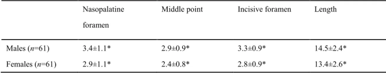
Table 4 Effect of sex on the diameters of the nasopalatine duct using reformatted sagittal slices from 64-MDCT imaging
Nasopalatine foramen
Middle point Incisive foramen Length
Males (n=61) 3.4±1.1* 2.9±0.9* 3.3±0.9* 14.5±2.4*
Females (n=61) 2.9±1.1* 2.4±0.8* 2.8±0.9* 13.4±2.6*
Data are shown as means ± SD.
*P<0.01
26
Table 5 Effect of sex on the diameters of the nasopalatine duct using reformatted coronal slices from 64-MDCT imaging
Nasopalatine foramen
Middle point Incisive foramen Length
Males (n=61) 4.5±1.2 3.7±1.1 3.8±1.0 11.9±2.7
Females (n=61) 4.2±1.0 3.6±0.8 3.8±0.9 11.3±2.2
Data are shown as means ± SD.
27
Table 6 Correlation of midface fracture patients’ characteristics with cause of injury
Parameter No. Cause of injury
Falls Violence
Total patients 104 73 (70.2 %) 31 (29.8 %)
Age (years) *
>60 16 15 (93.8 %) 1 (6.2 %)
≦60 88 58 (65.9 %) 30 (34.1 %)
Sex**
Male 75 46 (61.3 %) 29 (38.7 %)
Female 29 27 (93.1 %) 2 (6.9 %)
*P<0.05, **P<0.01
28
Table 7 Correlation of midface fracture patients’ characteristics with cause of injury
Parameter No. Cause of injury
Falls Violence
Concomitant damage
Eye (diplopia) 8 5 (62.5 %) 3 (37.5 %)
Brain (hemorrhage)
3 3 (100 %) 0 (0 %)
Eye and brain 2 1 (50.0 %) 1 (50.0 %)
Side
Right 39 29 (74.4 %) 10 (25.6 %)
Left 46 32 (69.6 %) 14 (30.4 %)
Bilateral 19 12 (63.2 %) 7 (36.8 %)
29
Table 8 Correlation of midface fracture patients’ characteristics with cause of injury
Parameter No. Cause of injury
Falls Violence
Fracture location Zygomaticomaxillary complex
66 51 (77.3 %)* 15 (22.7 %)*
Le Fort I 17 14 (82.4 %) 3 (17.6 %)
Isolated anterior maxillary sinus
16 7 (43.8 %)* 9 (56.2 %)*
Isolated zygomatic arch
11 6 (54.5 %) 5 (45.5 %)
Blowout fracture 7 3 (42.9 %) 4 (57.1 %)
*P<0.05
30 )LJ
7KHDQDWRPLFYDULDQWVRIWKHQDVRSDODWLQHGXFWZHUHGLIIHUHQWLDWHGLQWRWKUHHJURXSVDVLQJOHGXFWE WZRSDUDOOHOGXFWVDQGFYDULDWLRQVRI<W\SHGXFWVZLWKRQHRUDOSDODWDORSHQLQJDQGWZRRUPRUHQDVDO RSHQLQJV
31 )LJ
7KH IROORZLQJ ODQGPDUNV ZHUH VHOHFWHG IRU DQDO\VLV RI WKH VDJLWWDO DQG FRURQDO 0'&7 LPDJHV LQ D VDJLWWDOVOLFHVDQGEFRURQDOVOLFHVLGLDPHWHURIWKHQDVRSDODWLQHIRUDPHQLLGLDPHWHURIWKHPLGGOH SRLQWRIWKHQDVRSDODWLQHGXFWLLLGLDPHWHURIWKHLQFLVLYHIRUDPHQDQGLYOHQJWKRIWKHQDVRSDODWLQH GXFW$OOPHDVXUHPHQWVZHUHPDGHLQPLOOLPHWHUV
32 Fig. 3
A 59-year-old man had midface fractures resulting from a fall. (a) Axial image shows zygomaticomaxillary complex fractures with soft tissue edema (arrow) and hemorrhage in the maxillary sinus. (b)Axial image shows zygomaticomaxillary complex fractures (arrows) with hemorrhage in the maxillary sinus. (c) Coronal image shows zygomaticomaxillary complex and Le Fort I fractures with soft tissue edema (arrow) and hemorrhage in the maxillary sinus. (d) Coronal image shows zygomaticomaxillary complex (arrow) and Le Fort I (arrowheads) fractures with hemorrhage in the maxillary sinus. (e, f )Three-dimensional images show the zygomaticomaxillary complex (arrow) and Le Fort I (arrowheads) fractures to better advantage.
33 Fig. 4
A 45-year-old man had midface fractures resulting from violence. (a) Axial image shows an isolated anterior maxillary sinus fracture with soft tissue edema (arrow). (b)Axial image shows an isolated anterior maxillary sinus fracture (arrow). (c) Coronal image shows a blowout fracture (arrowhead) and an isolated anterior maxillary sinus fracture with soft tissue edema (arrow). (d) Coronal image shows a blowout fracture (arrowhead) and isolated anterior maxillary sinus fracture (arrow). (e, f) 3D images show the isolated anterior maxillary sinus fracture (arrow) to better advantage.