Tohoku J. Exp. Med., 2018, 245, 269-275
269
Received May 1, 2018; revised and accepted August 16, 2018. Published online August 30, 2018; doi: 10.1620/tjem.245.269.
Correspondence: Kiyoto Shiga, M.D., Ph.D., Department of Head and Neck Surgery, Iwate Medical University, 19-1 Uchimaru, Morioka, Iwate 020-8505, Japan.
e-mail: [email protected]
Treatment with Lactobacillus Retards the Tumor Growth of Head and Neck Squamous Cell Carcinoma Cells Inoculated in Mice
Jun Miyaguchi,
1Kiyoto Shiga,
1Kazumi Ogawa,
2Fumiko Suzuki,
3Katsunori Katagiri,
1Daisuke Saito,
1Aya Ikeda,
1Akira Horii,
2Mika Watanabe
4and Shizunobu Igimi
51Department of Head and Neck Surgery, Iwate Medical University, Morioka, Iwate, Japan
2Department of Molecular Pathology, Tohoku University Graduate School of Medicine, Sendai, Miyagi, Japan
3Department of Otolaryngology-Head and Neck Surgery, Tohoku University Graduate School of Medicine, Sendai, Miyagi, Japan
4Division of Pathology, Tohoku University Hospital, Sendai, Miyagi, Japan
5Department of Agricultural Chemistry, Tokyo University of Agriculture, Tokyo, Japan
Bacteria have been used for more than a century to treat solid tumors. Because solid tumors generate an anaerobic environment, we evaluated the anti-tumor effect of the obligate anaerobe strain KK378, derived from Lactobacillus casei (L. casei), using mice bearing head and neck cancer. Wild-type L. casei is a nonpathogenic bacterium that is commonly used in foods. Moreover, patients with head and neck squamous cell carcinoma often have multiple cancers and cervical lymph node metastasis that can be directly sensed beneath the skin. To establish the animal model bearing head and neck cancer, we inoculated each of human squamous cell carcinoma cell lines, SAS, HSQ89, and HSC2, on the back skin of BALB/cSlc-nu/nu mice. After tumor formation, L. casei KK378 was administered directly into the tumor, and tumor size and serum cytokine levels were analyzed. Mice injected with 108 cfu of L. casei KK378 showed reduction in tumor growth compared with PBS control; especially, the SAS tumor was significantly reduced (p = 0.008). Administered L. casei KK378 was detected in tumor tissues but not in normal tissues (liver, kidney, and lung) of SAS tumor-bearing mice, which was associated with increased blood cytokines (TNF-α, IFN-γ, IL-5, IL-10, and IL-12). Among these cytokines, the serum levels of IFN-γ and TNF-α were significantly increased (p < 0.05). In conclusion, L. casei KK378 infection may suppress tumor growth by inducing the host immune response. Direct injection of Lactobacillus into the tumor could be a potential strategy to treat head and neck squamous cell carcinoma.
Keywords: cytokine; head and neck squamous cell carcinoma; immuno-modulator; Lactobacillus casei; tumor suppression
Tohoku J. Exp. Med., 2018 August, 245(4), 269-275. © 2018 Tohoku University Medical Press Introduction
Bacteria have been used for more than a century to treat solid tumors. Various bacterial strains have been used as treatment strategies for malignant tumors since the 19th century. Coley (1891) described a patient who had recurrent round-celled sarcoma tumors, who was cured by severe erysipelas infection. He attempted to treat patients with malignant tumors by infecting the patient with live Streptococcus pyogenes, although he subsequently abandoned the use of live bacteria in favor of isolated preparations of bacteria toxins (Coley 1906).
It was first shown in 1947 that direct injection of spores of Clostridium histolyticus into a transplantable mouse sarcoma caused oncolysis and tumor regression
(Parker et al. 1947). However, very few animals survived this treatment because of acute toxic effects resulting in death. Kimura and colleagues implanted Ehrlich ascites tumors in mice and injected a suspension of lyophilized Bifidobacteria via the tail vein. The bacteria were highly localized within the tumor, with virtually no bacteria in other organs showing a very high tumor-targeting phenomenon (Kimura et al. 1980).
Salmonella, gram-negative facultative anaerobes, are also known to colonize human tumors (Graham and Coleman 1952; Gill and Holden 1996). Although systemic infection of Salmonella induces septic shock and high mortality in humans if not treated soon enough, Bacon et al.
(1951) showed Salmonella virulence in mice is attenuated in some auxotrophic mutants. In 1997, Pawelek et al.
(1997) reported that Salmonella auxotrophs that were injected into tumor-bearing mice would preferentially replicate within the tumor, indicating this microbe as a tumor-targeting agent.
The use of bacteria for treating tumors clinically causes problems of bacterial pathogenicity and influence on normal tissues. To overcome these problems, we tested the effect of Lactobacillus casei (L. casei), a nonpathogenic bacterium that is commonly used in foods, on the growth of head and neck squamous cell carcinoma. Wild-type L.
casei is a facultative anaerobe that can grow under relatively high oxygen concentrations, which would permit growth both in the hypoxic tumor environment as well as in normal tissues (Brown 1999). Instead, we used anaerobic L. casei KK378, a derivative strain that was mutated to grow under anaerobic conditions. Importantly, L. casei KK378 has low or no growth potential under aerobic conditions, and thus it may not cause severe side effects in normal tissues.
Here we report that direct injection of anaerobic L.
casei into the tumor site retarded the growth of squamous cell carcinoma cells inoculated in mice, which was associated with the increases in serum cytokine levels.
Materials and Methods
Preparation of L. casei
Anaerobic L. casei KK378 was developed by chemical mutagenesis of facultative anaerobic L. casei with 1-Methyl-3-nitro- 1-nitrosoguanidine (MNNG) under anaerobic culture conditions. The obtained L. casei KK378 was cultured in MRS broth at 37°C without oxygen.
Cell lines of human squamous cell carcinoma
SAS is a poorly differentiated squamous cell carcinoma cell line derived from human tongue cancer (Takahashi et al. 1989) and was obtained from Cell Resource Center for Biomedical Research, Institute of Development, Aging and Cancer, Tohoku University, Sendai, Miyagi, Japan. Cells were cultured in RPMI1640 (Sigma R8796, Sigma Aldrich, St. Louis, MO, USA) containing 10% fetal bovine serum (Invitrogen, San Diego, Ca, USA) at 37℃ under atmo- sphere with 5% CO2.
HSC2 is a squamous cell carcinoma cell line derived from human oral cancer (Momose et al. 1989), and HSQ89 is a squamous cell carcinoma cell line derived from human maxillary cancer (Wakamatsu et al. 1993). HSC2 and HSQ89 cell lines were obtained from Riken Bio Resource Center, Tsukuba, Ibaraki, Japan. These cells were cultured in DMEM (Sigma D5546) containing 10% fetal bovine serum (Invitrogen) at 37℃ under atmosphere with 5% CO2. Preparation of tumor-bearing mice
Male five-week-old BALB/cSlc-nu/nu mice (Japan SLC, Inc.
Shizuoka Japan) were used in this study. These mice are immunodeficient as they lack the thymus and are unable to produce T-cells. Aliquots of 3.0 × 10⁶ tumor cells suspended in 200 μl Matrigel (BD Biosciences, Tokyo, Japan) were inoculated on the back skin of the mice. For each tumor, the maximum diameter (A) and diameter at a right angle to A (B) were measured daily using a sliding caliper. Tumor volume was estimated as 0.4 × A × B2. Relative
tumor volume (%) was calculated every day as follows: (tumor vol- ume day X)/(tumor volume on the first day of L. casei injection day 0).
The animal experiments in this study were carried out in accordance with the Guidelines for Animal Experimentation Guidelines of Tohoku University and Iwate Medical University.
Topical injection of L. casei KK378 into tumor
Bacterial cell numbers, shown as colony forming unit (cfu), were calculated from the absorbance of the L. casei culture fluid. The tumor cells-bearing mice were randomly divided into 4 groups (n = 12 for each cancer cell line). When SAS, HSQ89 and HSC2 tumors reached approximately 200 mm3 (6 mm in diameter), a solution containing either 1.0 × 10⁴, 1.0 × 10⁶, or 1.0 × 10⁸ cfu of L. casei KK378 was injected in a volume of 100 µl into the tumor of three groups of mice, and PBS was injected as a control into the tumor of the fourth group. The growing speed of the tumor depended on each cell line. Thus, the day of the L. casei injection varied among these tumor bearing mice: about 10th day after SAS inoculation, 9th day after HSQ89 inoculation, and 30th day after HSC2 inoculation. L.
casei KK378 was administered only once.
Gram staining
Mice were sacrificed under general anesthesia at the indicated day after inoculation of the tumor and their liver, lung, kidney, and tumor were obtained. Two serial 3 µm-thick sections were made from a representative block of each tissue that had been 10%
formalin-fixed and paraffin-embedded. One section was stained with hematoxylin and eosin, and other section was used for Gram staining.
Gram staining was done using Hucker’s crystal violet as the primary stain, and iodine and 0.25% safranin as the counterstain as described previously (Shiga et al. 2001). All reagents were purchased from Muto Pure Chemicals Co. Inc., Tokyo.
Cytokine assay
Mouse serum samples were collected and analyzed for levels of eight cytokines (IL-2, IL-4. IL-5, IL-10, IL-12p70, GM-CSF, IFN-γ and TNF-α) by Bio-Plex cytokine reagent kit with Bio-Plex mouse cytokine Th1/Th2 Panel in the Bio-Plex 200 system (Bio-Rad, Hercules, CA, USA), as directed by the manufacturer.
Statistical analysis
Statistical analyses were carried out using Students’ t-test, and all p values < 0.05 were considered significant.
Results Inhibition of tumor growth
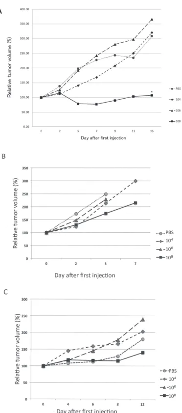
Of the 11 cell lines tested for the growth after inoculation on the back skin of nude mice, we found that three cell lines, HSC2, HSQ89 and SAS, had characteristics of rapid growth and tumor formation. We used these cell lines to test whether L. casei KK378 can suppress tumor growth after injection into the tumor inoculated on the back skin of nude mice. Three dosages of bacteria, 104, 106, and 108 cfu were injected directly into the tumor formed on the mice when tumor volume reached around 200 mm3. Fig. 1 shows the typical result of this experiment. A dose- dependent suppression of tumor growth was seen in SAS tumors infected with L. casei KK378, where 108 Lactobacilli
showed dramatic reduction in tumor size within five days of infection (Fig. 1A). By contrast, only restricted suppression of tumor growth was observed in the HSQ89 tumor (Fig.
1B), despite that 108 Lactobacilli were injected into the tumor. Approximately 50% reduction of the tumor volume was observed on the fifth day after Lactobacilli injection compared with the control group (Fig. 1B), but there was no significant difference. In addition, HSQ89 tumor- bearing mice died during the early days of this experiment, perhaps because HSQ89 tumor cells could induce cachexy in the mouse body. When HSC2 tumor was used, 108 Lactobacilli showed approximately 50% reduction of the tumor volume by the 12th day after Lactobacilli injection compared with the control group (Fig. 1C). However, there was no significant difference between these two groups.
Localization of L. casei KK378 in mice tissues
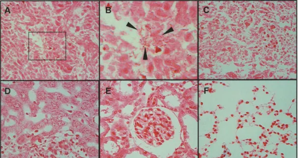
To confirm the localization of Lactobacilli in the injected tumor tissues and absence in other tissues, Gram staining was carried out on tissue sections (Fig. 2). We sacrificed L. casei KK378-injected SAS tumor-bearing mice 21 days after injection. Bacterial colonies were detected only in tumor tissues, and no bacteria were detected in all other normal tissues analyzed (liver, kidneys, and lung). The tumor tissues in which Lactobacilli were injected had necrotic regions inside the tumors and selective localization of L. casei at the boundary between the necrotic region and the tumor region.
Serum concentration of cytokines
To examine the influence of L. casei on the host immune system, we analyzed the serum concentrations of various cytokines in the SAS tumor-bearing nude mice.
Serum concentrations of IL-2, IL-4. IL-5, IL-10, IL-12p70, GM-CSF, IFN-γ, and TNF-α are shown in Fig. 3. Serum levels of IL-5, IL-10, IL-12, TNF-α, and IFN-γ were increased, depending on the number of L. casei KK378 administered compared with the control group. Sera of the mice that received 1.0 × 10⁸ cfu of L. casei KK378 showed the largest increase in the concentrations of these cytokines.
Especially, the IFN-γ and TNF-α levels were significantly increased, compared with those in control mice (Fig. 3).
Discussion
Records of cancer treatment by bacteria have existed for over 100 years. William Coley treated head and neck cancer patients in 1891 using Streptococcus pyogenes (Coley 1891). However, these treatments are usually asso- ciated with problems of bacterial pathogenicity and poor reproducibility, and therefore the main treatment choice for cancer has been radiotherapy and/or surgery. However, treatment of cancer using bacteria has been used so far, although not in a usual matter (Ishii et al. 1976; Peavy et al.
1979; Yasutake et al. 1984; Matsuzaki et al. 1988a).
Activation of tumor immunity and activation of che- motherapy by lactic acid bacteria, which are nonpathogenic
and indigenous, have been reported and used for the treat- ment of various cancers (Asano et al. 1986; Matsuzaki et al.
1988b; Kato et al. 1988; Takagi et al. 1999). Recent studies on the relationship between immune checkpoint inhibitor and intestinal bacterial flora have been attracting attention (Sivan et al. 2015; Routy et al. 2018; Gopalakrishnan et al.
2018). Consequently, these findings are also drawing atten- tion to bacteria for cancer treatments.
This is the first report demonstrating that direct injec- tion of Lactobacillus into the human squamous cell carci- noma established on backs of nude mice suppressed tumor growth. The tumor inhibiting effect was most effective in the group of mice injected with 1.0 × 10⁸ cfu L. casei in all three groups of mice bearing SAS, HSQ89, and HSC2. No tumor inhibiting effect was observed in the group of mice injected with PBS, and little tumor inhibiting effect was observed in 1.0 × 10⁴ or 1.0 × 10⁶ cfu of L. casei compared with PBS. In addition, the tumor suppressing effect was not efficient in HSQ89 and HSC2 tumor-bearing mice by injec- tion of 108 L. casei. Our results indicated that tumor sup- pressing effect of L. casei was dose-dependent and at least 1.0 × 10⁸ cfu of bacteria were required to cause tumor sup- pression in SAS tumor-bearing mice. These results could reflect the characteristic features of the cell lines. Yazawa et al. (2001) have reported the tumor inhibiting effects of intravenously injected Bifidobacterium longum at 5-6 × 10⁶ cfu in mice. These studies suggested that a certain number of bacteria is necessary to obtain noticeable effect of tumor suppression. It is the most interesting and crucial matter that the tumor suppressing effects among these three cell lines were different. However, it is another problem to clar- ify by independent research with phenotype analyses and molecular biological analyses.
We investigated serum cytokines in the SAS tumor- bearing mice that received 1.0 × 10⁴ cfu and 1.0 × 10⁸ cfu of L. casei KK378. IL-5, IL-10, IL-12, IFN-γ and TNF-α showed elevation in the sera of Lactobacillus-injected mice in dose-dependent manner. In particular, serum levels of TNF-α and IFN-γ in mice that received 1.0 × 10⁸ cfu L.
casei KK378 were significantly higher than those in the PBS injected group (TNF-α: p = 0.047; IFN-γ: p = 0.039).
These results indicate that tumor inhibition by L. casei KK378 could be mediated by activation of the host immune response and that L. casei KK378 influenced this response both locally and systematically. Several studies demonstrated that L. casei is phagocytosed and can induce cytokine production (Miettinen et al. 1998; Shida et al.
2006). It is suggested that tumor suppression develops when bacteria accumulate in the tumor. Therefore, route of injection is important in order to accumulate bacteria locally within the tumor.
In the present study, we injected bacteria directly into the tumor. Patients with head and neck squamous cell carcinoma, which we target, often have multiple cancers and cervical lymph node metastasis. Subcutaneous cervical lymph node metastasis or tumor invasion can often be
Fig. 1. Tumor growth curves.
Nude mice were inoculated SAS (A), HSQ89 (B), and HSC2 (C) human squamous cell carcinoma cells, and tumors were established on their backs (n = 12). PBS, 104, 106, or 108 cfu L. casei KK378 were injected in a volume of 100 µl on day 10 after SAS inoculation, on day 9 after HSQ89 inoculation, and on day 30 after HSC2 inoculation when the tumor volume was reached around 200 mm3. Tumor growth curves were calculated and scored by the average of three tumors for each case. Tumor volumes are expressed as 100% on day 0 of L. casei injection. When 108 cfu L. casei KK378 were injected to the SAS tumor, their growth was significantly reduced compared with PBS (p = 0.008) and 106 cfu L. casei KK378 (p = 0.003) injected tumors. Astarisk shows the statistically significant difference (A). When 108 cfu L. casei KK378 were injected to the HSQ89 tumor, their growth was reduced approximately 50%, but no significant reduction was observed compared with PBS (p = 0.21) (B). Similarly, when 108 cfu L. casei KK378 were injected to the HSC2 tumor, their growth was reduced 50%, but no significant reduction was observed compared with PBS (p = 0.41) (C).
A
Fig. 2. Gram staining of tissues of the L. casei KK378-injected mice.
Nude mice were inoculated SAS cells and one tumor was grown on each back. PBS or 108 cfu L. casei were injected when the tumor volume was reached around 200 mm3. Mice were sacrificed under general anesthesia and their organs and tumors were obtained as pathological examined specimens. Gram staining was performed each organs and tumor tissues. (A) tumor tissue injected with 108 cfu L. casei, (B) magnified view of A, the enlarged area is indicated by black- lined rectangle in (A), (C) tumor tissue injected with PBS; (D) liver; (E) kidney; (F) lung. There are irregular shaped tumor cells located in the tumor tissue (A) and Lactobacilli are clearly visible in B. Arrowheads indicate Lactobacilli.
Fig. 3. Serum cytokine concentration in L. casei KK378-injected mice.
Nude mice were inoculated with SAS cells to establish one tumor on each back. These mice were classified into 3 groups and PBS, 104 or 108 cfu L. casei KK378 were injected when the tumor volume was reached around 200 mm3. Mice were sacrificed under general anesthesia and their blood was obtained. Serum cytokine concentrations were ana- lyzed by EIA. Cytokine concentrations are indicated as pg/ml at vertical axis. Serum TNF-α level was significantly higher in mice injected with 108 cfu L. casei than that in PBS injected mice (p = 0.047). Also, serum IFN-γ levels were significantly higher in 108 cfu L. casei-injected mice compared with those in PBS-injected mice (p = 0.039). Apparent- ly, serum levels of IL-2, IL-5, IL-10 and IL-12p70 in mice injected 108 cfu L. casei were higher than those of PBS- injected mice, but there were no significant differences. (A) All eight cytokine levels are shown. (B) Levels of all cyto- kines but TNF-α are shown to clearly depict the cytokine concentration, with the exception of TNF-α that showed much higher concentrations than those of the other cytokines. Asterisks show the statistically significant difference compared with the PBS-injected control.
directly sensed beneath the skin. Local injection is simpler in these cases compared to other solid tumors, as the tumor can be reached easily, especially to cervical lymph nodes.
We think that the control injection must be PBS or saline because some immunoreactive effect was expected using killed or inactivated Lactobacilli (Lee et al. 2015).
However, it is an interesting research to use killed or inacti- vated Lactobacilli so that the experiment will be prepared in the future.
Hypoxia within the tumor is useful for accumulating anaerobic bacteria in the tumor. The system for delivering drugs into tumors has been devised to counter hypoxia in solid tumors (Taniguchi et al. 2010; Fang et al. 2014).
Direct injection of anaerobic L. casei KK378 into the tumor could ensure their proliferation under the hypoxic environ- ment. The antitumor effect of Lactobacillus can be expected when injected directly where the treatment is nec- essary. If anaerobic L. casei shows a significant therapeutic effect in the hypoxic environment within the tumor, new treatments for solid tumors will be possible. This treatment can be used in combination with existing chemotherapy and radiotherapy. Hypoxia in solid tumors has been reported to inhibit radiation therapy and chemotherapy (Ishii et al.
1976; Wan et al. 2012). If bacterial treatment can be com- bined with chemotherapy or radiotherapy, it is not only pos- sible to reduce side effects but also to increase the effective- ness of antitumor treatment. In our study, although microbes were observed in the tumor tissues, no microbes were detected in the normal tissues and tumor suppression was observed. Compared to previous studies that show antitumor effects by intravenous administration, our local injection method is less prone to bacteremia and thus may be a more effective and safe treatment for patients with head and neck squamous cell carcinoma. However, there have been reports of abscess formation and bacteremia in infections with lactic acid bacteria (Cannon et al. 2005).
Even if L. casei is nonpathogenic and injected directly into the tumor site, there is a risk of live bacteria leaking out of the tumor tissue, proliferating in normal tissues, resulting in side effects in normal tissues. Patients with head and neck cancer, particularly those in advanced stages, easily become immunosuppressed by the tumor itself or by chemotherapy and/or radiotherapy. In these cases, viral or bacterial infection can be a life-threatening matter for the patients. Therefore, injection of dead cells of L. casei or injection of supernatant fluid into the tumor seems to be a preferable method, if these methods show the similar tumor suppressing effects as live bacteria. However, the efficacy of these treatments should be examined in the future stud- ies.
Acknowledgments
This work was partially supported by a Grant-in-aid for JSPS KAKENHI Grant Number 23659791, Toshio Kurokawa Cancer Research Foundation, and Yakult Bio-Science Foundation.
We thank “Editage” staffs for their help to prepare this manu-
script by editing our English.
Conflict of Interest
The authors declare no conflict of interest.
References
Asano, M., Karasawa, E. & Takayama, T. (1986) Antitumor activity of Lactobacillus casei (LC 9018) against experimental mouse bladder tumor (MBT-2). J. Urol., 136, 719-721.
Bacon, G.A., Burrows, T.W. & Yates, M. (1951) The effects of biochemical mutation on the virulence of Bacterium typhosum: the loss of virulence of certain mutants. Br. J. Exp.
Pathol., 32, 85-96.
Brown, J.M. (1999) The hypoxic cell: a target for selective cancer therapy – eighteenth Bruce F. Cain Memorial Award lecture.
Cancer Res., 59, 5863-5870.
Cannon, J.P., Lee, T.A., Bolanos, J.T. & Danziger, L.H. (2005) Pathogenic relevance of Lactobacillus: a retrospective review of over 200 cases. Eur. J. Clin. Microbiol. Infect. Dis., 24, 31-40.
Coley, W.B. (1891) II. Contribution to the knowledge of sarcoma.
Ann. Surg., 14, 199-220.
Coley, W.B. (1906) Late results of the treatment of inoperable sarcoma by the mixed toxins of erysipelas and Bacillus prodigiosus. Am. J. Med. Sci., 131, 375-430.
Fang, J., Liao, L., Yin, H., Nakamura, H., Shin, T. & Maeda, H.
(2014) Enhanced bacterial tumor delivery by modulating the EPR effect and therapeutic potential of Lactobacillus casei. J.
Pharm. Sci., 103, 3235-3243.
Gill, G.V. & Holden, A. (1996) A malignant pleural effusion infected with Salmonella enteritidis. Thorax, 51, 104-105.
Gopalakrishnan, V., Spencer, C.N., Nezi, L., Reuben, A., Andrews, M.C., Karpinets, T.V., Prieto, P.A., Vicente, D., Hoffman, K., Wei, S.C., Cogdill, A.P., Zhao, L., Hudgens, C.W., Hutchinson, D.S., Manzo, T., et al. (2018) Gut microbiome modulates response to anti-PD-1 immunotherapy in melanoma patients.
Science, 359, 97-103.
Graham, F.O. & Coleman, P.N. (1952) Infection of a secondary carcinoma by Salmonella montevideo. Br. Med. J., 1, 1116.
Ishii, Y., Yamaoka, H., Toh, K. & Kikuchi, K. (1976) Inhibition of tumor growth in vivo and in vitro by macrophages from rats treated with a streptococcal preparation, OK-432. Gan, 67, 115-119.
Kato, I., Yokokura, T. & Mutai, M. (1988) Correlation between increase in Ia-bearing macrophages and induction of T cell- dependent antitumor activity by Lactobacillus casei in mice.
Cancer Immunol. Immunother., 26, 215-221.
Kimura, N.T., Taniguchi, S., Aoki, K. & Baba, T. (1980) Selective localization and growth of Bilidobacterium bifidum in mouse tumors following intravenous administration. Cancer Res., 40, 2061-2068.
Lee, H.A., Kim, H., Lee, K.W. & Park, K.Y. (2015) Dead nano- sized Lactobacillus plantarum inhibits azoxymethane/dextran sulfate sodium-induced colon cancer in Balb/c mice. J. Med.
Food, 18, 1400-1405.
Matsuzaki, T., Yokokura, T. & Mutai, M. (1988a) Antitumor effect of intrapleural administration of Lactobacillus casei in mice.
Cancer Immunol. Immunother., 26, 209-214.
Matsuzaki, T., Yokokura, T. & Mutai, M. (1988b) The role of lymph node cells in the inhibition of metastasis by subcutaneous injection of Lactobacillus casei in mice. Med. Microbiol.
Immunol., 177, 245-253.
Miettinen, M., Matikainen, S., Vuopio-varkila, J., Pirhonen, J., Varkila, K., Kurimoto, M. & Julkunen, I. (1998) Lactobacilli and Streptococci induce interleukin-12 (IL-12), IL-18, and gamma interferon production in human peripheral blood mononuclear cells. Infect. Immun., 66, 6058-6062.
Momose, F., Araida, T., Negishi, A., Ichijo, H., Shioda, S. &
Sasaki, S. (1989) Variant sublines with different metastatic potentials selected in nude mice from human oral squamous cell carcinomas. J. Oral Pathol. Med., 18, 391-395.
Parker, R.C., Plummer, H.C., Siebenmann, C.O. & Chapman, M.G.
(1947) Effect of histolyticus infection and toxin on transplantable mouse tumors. Proc. Soc. Exp. Biol. Med., 66, 461-467.
Pawelek, J.M., Low, K.B. & Bermudes, D. (1997) Tumor-targeted Salmonella as a novel anticancer vector. Cancer Res., 57, 4537-4544.
Peavy, D.L., Baughn, R.E., Musher, D.M. & Musher, D.M. (1979) Effects of BCG infection on the susceptibility of mouse macrophages to endotoxin. Infect. Immun., 24, 59-64.
Routy, B., Le Chatelier, E., Derosa, L., Duong, C.P.M., Alou, M.T., Daillère, R., Fluckiger, A., Messaoudene, M., Rauber, C., Roberti, M.P., Fidelle, M., Flament, C., Poirier-Colame, V., Opolon, P., Klein, C., et al. (2018) Gut microbiome influences efficacy of PD-1-based immunotherapy against epithelial tumors. Science, 359, 91-97.
Shida, K., Suzuki, T., Kiyoshima-Shibata, J., Shimada, S. &
Nanno, M. (2006) Essential roles of monocytes in stimulating human peripheral blood mononuclear cells with Lactobacillus casei to produce cytokines and augment natural killer cell activity. Clin. Vaccine Immunol., 13, 997-1003.
Shiga, K., Tateda, M., Saijo, S., Hori, T., Sato, I., Tateno, H., Matsuura, K., Takasaka, T. & Miyagi, T. (2001) Presence of Streptococcus infection in extra-oropharyngeal head and neck squamous cell carcinoma and its implication in carcinogenesis.
Oncol. Rep., 8, 245-248.
Sivan, A., Corrales, L., Hubert, N., Williams, J.B., Aquino- Michaels, K., Earley, Z.M., Benyamin, F.W., Lei, Y.M., Jabri, B., Alegre, M.L., Chang, E.B. & Gajewski, T.F. (2015)
Commensal Bifidobacterium promotes antitumor immunity and facilitates anti-PD-L1 efficacy. Science, 350, 1084-1089.
Takagi, A., Matsuzaki, T., Sato, M., Nomoto, K., Morotomi, M. &
Yokokura, T. (1999) Inhibitory effect of oral administration of Lactobacillus casei on 3-methylcholanthrene-induced carcino- genesis in mice. Med. Microbiol. Immunol., 188, 111-116.
Takahashi, K., Kanazawa, H., Akiyama, Y., Tazaki, S., Takahara, M., Muto, T., Tanzawa, H. & Sato, K. (1989) Establishment and characterization of a cell line (SAS) from poorly differen- tiated human squamous cell carcinoma of the tongue. J. Jpn.
Stomatol. Soc., 38, 20-28.
Taniguchi, S., Fujimori, M., Sasaki, T., Tsutsui, H., Shimatani, Y., Seki, K. & Amano, J. (2010) Targeting solid tumors with non- pathogenic obligate anaerobic bacteria. Cancer Sci., 101, 1925-1932.
Wakamatsu, Y., Yamaguchi, Y., Nakagawa, K., Numata, M., Hiromatsu, K., Hasegawa, K. & Aramaki, M. (1993) Estab- lishment of cell line cytotoxic effect of several anticancer drugs on a cultured human squamous carcinoma cell line HSQ-89. Nihon Univ. Dent. J., 67, 457-465.
Wan, X.B., Fan, X.J., Huang, P.Y., Dong, D., Zhang, Y., Chen, M.Y., Xiang, J., Xu, J., Liu, L., Zhou, W.H., Lv, Y.C., Wu, X.Y., Hong, M.H. & Liu, Q. (2012) Aurora-A activation, correlated with hypoxia-inducible factor-1a, promotes radio- chemoresistance and predicts poor outcome for nasopharyn- geal carcinoma. Cancer Sci., 103, 1586-1594.
Yasutake, N., Kato, I., Ohwaki, M., Yokokura, T. & Mutai, M.
(1984) Host-mediated antitumor activity of Lactobacillus casei in mice. Gan, 75, 72-80.
Yazawa, K., Fujimori, M., Nakamura, T., Sasaki, T., Amano, J., Kano, Y. & Taniguchi, S. (2001) Bifidobacterium longum as a delivery system for gene therapy of chemically induced rat mammary tumors. Breast Cancer Res. Treat., 66, 165-170.