The pretreatment neutrophil-to-lymphocyte ratio predicts therapeutic response to radiation
therapy and concurrent chemo-radiation therapy in uterine cervical cancer
(
治療前の好中球
/リンパ球比は子宮頸癌における放射線療法と化学療法同時放射線療法の 治療的反応性を予測する
)申請者 弘前大学大学院医学研究科
腫瘍制御科領域学 婦人科腫瘍学教育研究分野 氏 名 水沼 槙人
指導教授 水沼 英樹
Abstract
Objective: The aim of this study was to investigate the prognostic role of the pretreatment neutrophil-to-lymphocyte ratio (NLR) as a predictive marker prior to treatment of cervical cancer with radiation therapy (RT) alone or concurrent chemoradiation therapy (CCRT).
Methods: Fifty-six patients with squamous cell carcinoma (SCC) of the uterine cervix who underwent RT or CCRT from 2005 to 2013 at this Hospital were retrospectively identified using electronic databases.
Patients were divided into a high NLR group (≥ 2.5) and a low NLR group (< 2.5). The efficacy of RT and CCRT in the two groups was compared.
Result: Of the 56 patients, 35 were in the high NLR group and 21 were in the low NLR group. In comparison to a high NLR, a low NLR was significantly associated with a complete response (P < 0.001).
When cancer was divided into stages I/II and III/IV, patients with a low NLR had a significantly better therapeutic outcome than those with a high NLR (P < 0.05). Multivariate analysis showed that only the NLR was a significant prognostic factor for progression-free survival (PFS). Patients with a high NLR had a significantly shorter PFS and overall survival than those with a low NLR.
Conclusion: Results showed that a low NLR before treatment can predict a good response to RT or CCRT by all stages of uterine cervical cancer. The NLR may be a promising parameter on which to base the choice of a therapeutic strategy to treat SCC of the uterine cervix.
Keywords: NLR, uterine cervical cancer, radiation therapy, concurrent chemoradiation therapy, therapeutic outcome.
INTRODUCTION
Even though the incidence and mortality of invasive uterine cervical cancer have steadily decreased, this disease is still the second most common type of cancer in females worldwide and the leading cause of cancer deaths in women in developing countries [1].
The neutrophil-to-lymphocyte ratio (NLR) has been used as a cost-effective and simple parameter to indicate systemic inflammation or stress in critically ill patients without cancer [2]. The NLR may also relate to the prognosis of many types of cancer, including gastrointestinal tract malignancies [3], hepatocellular cancer [4], pancreatic cancer [5], non-small cell lung cancer [6], and urinary cancer [7].
Furthermore, the pretreatment NLR predicts survival in epithelial ovarian cancer [8] and uterine cervical cancer [9, 10]. However, the NLR was not a predictive factor in patients with uterine cervical cancer treated with neoadjuvant chemotherapy and radical hysterectomy [11]. Thus, the clinical implications of the NLR have yet to be determined.
Despite the improved survival rate for patients who receive concurrent chemo-radiation therapy (CCRT) [12], some patients are resistant to this treatment. The aim of this study was to investigate the prognostic role of the NLR as a pretreatment marker prior to treatment of uterine cervical cancer with radiation therapy (RT) alone or CCRT.
METHODS Patients
Records of patients who had stage IB1 to IV uterine cervical cancer according to the staging system of the International Federation of Gynecology and Obstetrics (FIGO) were retrospectively reviewed using electronic databases. These patients were histologically diagnosed with squamous cell carcinoma and underwent RT or CCRT at Hirosaki University Hospital from 2005 to 2013. The following patients were excluded: 6 patients in whom the differential white blood count was not determined, 5 patients who had to be treated for another inflammatory disease just prior to RT or CCRT, and 1 patient who underwent rounds of RT at another hospital. This study ultimately examined the records of 56 patients.
Treatment
RT: External beam radiation was delivered with anteroposterior and posteroanterior opposed beams generated by an X-ray accelerator with an energy of 10 MV at a distance of 100 cm. Whole pelvis irradiation with a total dose of 50 Gy was delivered with a fraction dose of 2 Gy per day for 5 days a week over a 5-week period. After a dose of approximately 30 Gy, a central shield with a width of 4 cm at the midline was used and intracavitary brachytherapy was performed using a RALS with a Co 60 source.
Intracavitary brachytherapy, with fractions of 6 Gy delivered to point A, was performed once a week for a total of 4 times [13].
Chemotherapy: Thirty mg/m2 of nedaplatin was dissolved in a 500-mL saline solution and infused intravenously over 180 min. The first round was administered on the day that external beam RT started.
The regimen was repeated weekly for a total of 6 times. Nedaplatin infusion was completed 1 hr before irradiation [13]. Nine patients who had not completed the concurrent chemotherapy schedule were also included in this study. Two patients used nedaplatin nor cisplatin, one used irinotecan, the other used paclitaxel and carboplatin.
Outcomes
The primary statistical end point was a tumor response, which was assessed by means of a clinical examination, including histological examination of the uterine cervix and a computed tomography scan from the chest to the pelvis. These examinations were performed about 1 month after completion of RT or CCRT. Patients with no evidence of disease were deemed to have a clinical complete response (CR). If tumors shrank after treatment but cancer cells or tumors were still noted, the patient was not deemed to have a CR.
Progression-free survival (PFS) was defined as the time between the date of the completion of treatment and the date of identification of progressive disease. We identified the case that tumor size were bigger than the size after treatment or new tumors were noted by a computed tomography scan or cancer
cells were noted by histological examination of the uterine cervix as progressive disease. Overall survival (OS) was defined as the time between the date of the completion of treatment and the date of death.
Calculation of NLR and determination of the NLR cut-off level
The NLR was defined as the absolute neutrophil count divided by the absolute lymphocyte count.
Before treatment, the median NLR was 2.4, and patients were divided into a high NLR group (≥ 2.5) and a low NLR group (< 2.5).
Statistical analysis
The following variables were analyzed to predict tumor response and survival: FIGO stage, tumor size, treatment (patients were grouped as RT alone or CCRT), baseline hemoglobin (Hb) level, baseline white blood cell (WBC) and lymphocyte counts, and the NLR. The chi-square test and Student’s t test were used to analyze categorical data, and the Welsh test was used for continuous variables. Receiver opening characteristic plots were conducted to determine the maximum sensitivity and specificity of a threshold value to stratify patients at high risk of recurrence. Relation of NLR to WBC, neutrophil and lymphocyte was calculated using Spearman's rank correlations coefficient.
Multiple logistic regression models were used to identify independent prognostic factors for a response and PFS. The following factors were included in the multivariable models: age, FIGO stage, pretreatment SCC value, tumor size, baseline neutrophil and lymphocyte counts, Hb level, and the NLR.
The OS and PFS were calculated using the Kaplan-Meier method. A log-rank test was used to assess statistical significance. All p values were two-tailed and p <0.05 was considered statistically significant.
All statistical analyses were performed using SPSS (version 21, SPSS Inc., Chicago, IL, USA).
RESULTS
Patient characteristics
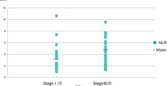
This study analyzed data from 56 patients with a median age of 65.1 years (range, 35-89 years). In terms of the FIGO stage, 13 patients had stage I cancer, 11 had stage II, 17 had stage III, and 15 had stage IV. Of these 56 patients, 35 were in the high NLR group and 21 were in the low NLR group. All pretreatment NLR values according to stage are plotted in Fig.1. Clinical characteristics of patients and tumors by the pretreatment NLR are shown in Table 1. As seen in this Table, patients with ≧ 2.5 had advanced tumor in terms of tumor size, SCC level and lymph node metastasis.
When this analysis was performed, clinical outcomes were recorded for 47 patients (83.9%) and 2 patients (3.6%) who had been followed until recurrence. Cancer recurred in 20 patients (35.7%) and 16 patients (28.6%) died due to the cancer. The mean period to recurrence was 5.3 months (range: 1 to 18 months) and the mean period to disease-related death was 14.2 months (range: 2 to 41 months).
Pretreatment factors and therapeutic response
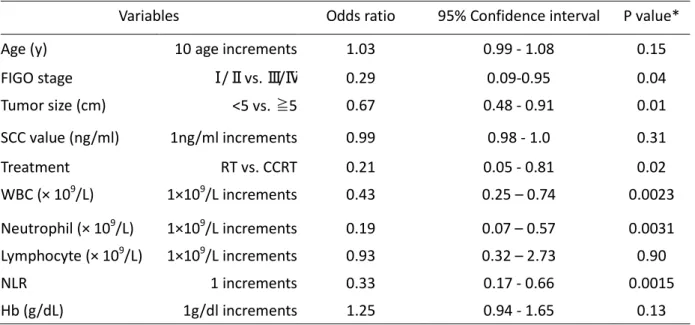
At the completion of treatment, 25 patients (44.6%) had a clinical CR. Table 2 shows relationship between pretreatment factors and complete response obtained from our subjects. As shown in Table 2, univariate logistic regression analysis found a significant association between CR and FIGO stage, tumor size, type of treatment, WBC count, neutrophile count and NLR. In this study, a higher baseline neutrophil count was also a significant predictor of non-response (Table 2). In comparison to a high NLR, a low NLR was significantly associated with a CR (P < 0.00005) (Table 3). When cancer was divided into stages I/II and III/IV, patients with a low NLR had a significantly better therapeutic outcome than those with a high NLR (Table 3).
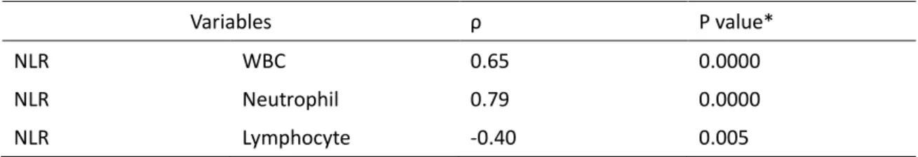
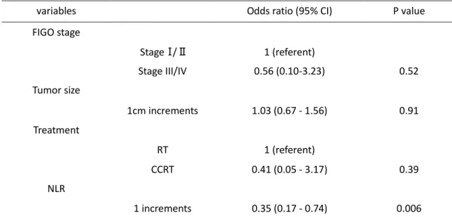
In multivariate logistic regression analysis with regard to CR, variables that were considered to statistically significant factors in univariate regression were selected. These variables were the FIGO stage, tumor size, treatment, and the NLR. The pretreatment NLR was closely correlated with the pretreatment WBC count and neutrophil count, as indicated by Spearman's rank correlation coefficients (Table 4). From among the variables WBC count, neutrophil count, and the NLR, only the NLR was selected as the most significant factor (p=0.0015) because of multi-collinearity. Multivariate logistic regression revealed that the NLR was a significant prognostic factor with an odds ratio of 0.35 (95%
confidence interval; 0.17-0.74, Table 5). Receiver operating characteristic (ROC) curves were derived for age, SCC, tumor size, the NLR, and Hb levels with CR as an explanatory variable. Examination of the parameters that had the largest positive likelihood ratios indicated that a cut-off level of 2.5 for the NLR had the largest positive likelihood ratio. This level was close to the median NLR (Table 6).
The relationship between pretreatment factors and PFS and OS
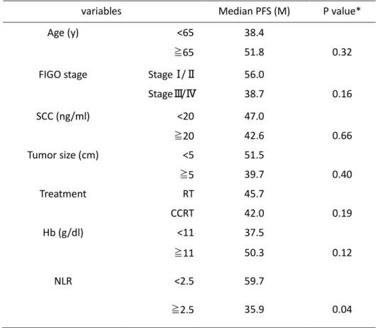
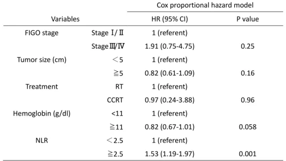
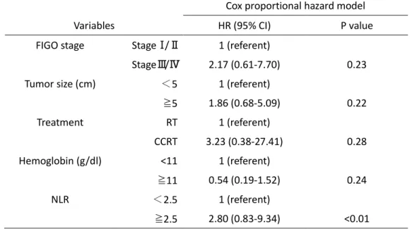
Univariate and multivariate analyses revealed that only the NLR was a significant prognostic factor for PFS (Table 7 and 8). On the other hand, NLR and treatment were associated with OS in univariate analyses, and in multivariate only lower NLR was associated with longer survival(Table 9 and 10).
Patients with a high NLR had a significantly shorter PFS and OS than those with a low NLR (Fig. 2A and B). The pretreatment hemoglobin level was closely associated with PFS (p=0.058) (Table 8).
DISCUSSION
Previous studies [14, 15] have shown that FIGO stage, Hb level, and tumor size are significant prognostic factors of cervical cancer and the results of the present study were compatible as shown in Table 1. In addition, treatment modality (CCRT vs. RT) was also reported to be an impact prognostic factor, as CCRT had a higher rate of success than RT alone [12]. The present study showed that the NLR before treatment was significantly related to the outcomes of radiation for the uterine cervical cancer, and supported results shown by Lee et al. [9] and Zhang et al. [10]. In addition, the present study showed that a low NLR (less than 2.5) was significantly associated with a CR and longer PFS and longer OS in comparison to a high NLR (2.5 and over).
The fact that a high NLR is a factor for a poor prognosis after surgery or after chemotherapy has been reported in relation to various types of carcinomas [3-10]. Looking specifically at cervical cancer, Lee et al. reported that the NLR may be a predictor of prognosis for RT after surgery or definitive RT [9]. The current study involved a choice of only RT or CCRT to treat cervical cancer, but the results were similar to those of Lee et al.
An increase in neutrophils or a decrease in lymphocytes is reported to be associated with a shorter survival time. Why does a high NLR diminish the effectiveness of therapy and why does it increase recurrence and predict a poor prognosis? The specific mechanisms by which it causes these phenomena have yet to be identified. A G-CSF-producing tumor is typically characterized by a rapid progression and obvious leukocytosis, resulting in a poor prognosis. In 4 patients with G-CSF-producing cervical cancer, Matsumoto et al. reported that the cancer recurred in all 4 patients within 6 months and that all of the patients died within 15 months [16]. In addition, the presence of inflammatory foci may increase the risk of cancer progression. Inflammation is known to change a tumor’s microenvironment and promote angiogenesis and metastasis [17]. Inflammatory changes are normally accompanied by an increase in neutrophils. Therefore, one could logically predict that cervical cancer and associated leukocytosis would have a poor prognosis. Recently, a view on the reason for the poor prognosis of cervical cancer and associated leukocytosis has been presented. G-CSF is produced by a tumor and in turn induces the production of myeloid-derived suppressor cells (MDSCs). These MDSCs cause rapid progression of cervical cancer and leukocytosis and radioresistance [18]. Mabuchi et al. depleted MDSCs, which succeeded in inhibiting the progression of cervical cancer and leukocytosis and enhancing radiosensitivity [18]. MDSCs have been found in bone marrow, blood, and the spleen of individuals with tumors. In response to the progression of inflammation and cancer, these cells spread throughout the body as part of the host’s immune response [19]. Although cisplatin is a key drug in CCRT to treat cervical cancer, a study has reported that it induces MDSCs [20]. Meanwhile, gemcitabine is known to be an anticancer drug that reduces the number of MDSCs [21], then gemcitabine may be favorable for chemoradiation therapy of cervical cancer patients with high NLR.
Recent studies have found increased levels of pro-inflammatory cytokines in patients with an increased NLR [22, 23] and increased peri-tumor macrophage infiltration [24]. These results suggest that an increased NLR reflects an up-regulated innate immune response [21]. The immune response has been found to differ depending on the type of cancer. With pancratic cancer, a marked increase in lymphocytes occurs preoperatively and postoperative [25]. The NLR was not associated with the prognosis of pancratic cancer. In addition, a study has reported that combining chemotherapy and polysaccharide K (PSK) after surgery for colon cancer allows the NLR (which is correlated with prognosis) to be kept to a low level, improving the patient’s prognosis [26]. Chemotherapy triggers the apoptosis of peripheral blood T lymphocytes, but immunotherapy with PSK is presumably effective because it controls that apoptosis [27].
RT is known to primarily have little effect in a hypoxic state. A hypoxia state is related to inflammation and anemia, and elements of the innate inflammatory response are associated with the up-regulation of growth factors and angiogenesis [28, 29]. Systemic inflammatory responses are associated with alteration in the circulating WBC count, with neutrophilia and relative lymphopenia [30].
Santin et al. investigated lymphocyte populations including CD3+, CD4+ and CD8+ T-cell subsets, B cells (CD19+), and natural killer cells (CD56+, CD16+, CD3-) before, during, and after RT in patients with locally advanced cervical cancer [31]. While both RT and CCRT with cisplatin significantly decreased the mean absolute number of all lymphocyte subsets compared to pretreatment levels, Santin et al. detected no differences in the characteristics or the magnitude of the lymphopenia induced by the two treatments. Thus, cisplatin does not enhance the magnitude or characteristics of radiation-induced immunosuppression [31].
In conclusion, the current study showed that a low NLR before treatment can predict a good response to RT or CCRT in terms of CT and PFS by all stages of uterine cervical cancer. The NLR may be a promising parameter on which to base the choice of a therapeutic strategy to treat SCC of the uterine cervix. This study involved a relatively small sample size, so further validation is required prior to incorporating the NLR as a prognostic indicator.
CONFLICT OF INTEREST
No potential conflict of interest relevant to this article was reported.
REFERENCES
1. Siegel R, Naishadham D, Jemal A. Cancer statistics. CA Cancer J Clin 2013;63:11-30.
2. Zahorec R. Ratio of neutrophil to lymphocyte counts-rapid and simple parameter of systemic inflammation and stress in the critically ill. Bratisl Lek Listy 2001;102:5-14.
3. Walsh SR, Cook EJ, Goulder F, Justin TA, Keeling NJ. Neutrophil-lymphocyte ratio as a prognostic factor in colorectal cancer. J Surg Oncol 2005;91:181-4.
4. Halazun KJ, Hardy MA, Rana AA, Woodland DCt, Luyten EJ, Mahadev S, et al. Negative impact of neutrophil-lymphocyte ratio on outcome after liver transplantation for hepatocellular carcinoma.
Ann Surg 2009;250:141-51.
5. Bhatti I, Peacock O, Lloyd G, Larvin M, Hall RI. Preoperative hematologic markers as independent predictors of Prognosis in resected pancreatic ductal adenocarcinoma: neutrophil-lymphocyte versus platelet-lymphocyte ratio. Am J Surg 2010;200:197-203.
6. Sarraf KM, Belcher E, Raevsky E, Nicholson AG, Goldstraw P, Lim E. Neutrophil/lympocyte ratio and its association with survival after complete resection in non-small cell lung cancer. J Thorac Cardiovasc Surg 2009;137:425-8.
7. Wei Y, JiangY-Z, Qian W-H. Prognostic Role of NLR in Urinary Cancers: A Meta-Analysis. PLoS one 2014;9:e92079 doi:10.1371/journal.pone.0092079.
8. Cho HB, Hur HW, Kim SW, Kim SH, Kim JH, Kim YT, et al. Pre-treatment neutrophil to lymphocyte ratio is elevated in epithelial ovarian cancer and predicts survival after treatment.
Cancer Immunol Immunother 2009;58:15-23.
9. Lee Y-Y, Choi CH, Kim H-J, Kim T-J, Lee J-W, Lee J-H, et al. Pretreatment Neutrophil:Lymphocyte Ratio as a Prognostic Factor in Cervical Carcinoma. Anticancer Res 2012;32:1555-62.
10. Zhang Y, Wang L, Liu Y, Wang S, Shang P, Gao Y, et al. Preoperative neutrophil-lymphocyte ratio before platelet-lymphocyte ratio predicts clinical outcome in patients with cervical cancer treated with initial radical surgery. Int J Gynecol Cancer 2014;24:1319-25.
11. Wang D, Wu M, Feng F-Z, Huang H-F, Yang J-X, Shen K, et al. Pretreatment neutrophil-to-lymphocyte and platelet-to-lymphocyte ratios do not predict survival in patients with cervical cancer treated with neoadjuvant chemotherapy and radical hysterectomy. Cin Med J (Engl) 2013;126:1464-8.
12. Morris M, Eifel PJ, Lu J, Grigsby PW, Levenback C, Stevens RE, et al. Pelvic radiation with concurrent chemotherapy compared with pelvic and para-aortic radiation for high-risk cervical cancer. N Engl J Med 1999;340:1137-43.
13. Yokoyama Y, Takano T, Nakahara K, Shoji T, Sato H, Yamada H, et al. A phase II multicenter trial of concurrent chemoradiotherapy with weekly nedaplatin in advanced uterine cervical carcinoma:
Tohoku Gynecologic Cancer Unit Study. Oncol Rep 2008;19:1551-6.
14. Noh JM, Park W, Kim YS, Kim JY, Kim HJ, Kim J, et al. Comparison of clinical outcomes of adenocarcinoma and adenosquamous carcinoma in uterine cervical cancer patients receiving surgical resection followed by radiotherapy: a multicenter retrospective study (KROG 13-10).
Gynecol Oncol 2014;132:618-23.
15. Grigiene R, Valuckas KP, Aleknavicius E, Kurtinaitis J, Letautiene SR. The value of prognostic factors for uterine cervical cancer patients treated with irradiation alone. BMC Cancer 2007;7:234 doi:10.1186/1471-2407-7-234.
16. Matsumoto Y, Mabuchi S, Muraji M, Morii E, Kimura T. Squamous cell carcinoma of the uterine cervix producing granulocyte colony-stimulating factor: a report of 4 cases and a review of the literature. Int J Gynecol Cancer 2010;20:417-21.
17. Mantovani A, Allavena P, Sica A, Balkwill F. Cancer-related inflammation. Nature 2008;454:436-44.
18. Mabuchi S, Matsumoto Y, Kawano M, Minami K, Seo Y, Sasano T, et al. Uterine cervical cancer displaying tumor-related leukocytosis: a distinct clinical entity with radioresistant feature. J Natl Cancer Inst 2014;106. pii: dju147. doi: 10.1093/jnci/dju147.
19. Cortez-Retamozo V, Etzrodt M, Newton A, Rauch PJ, Chudnovskiy A, Berger C, et al. Origins of tumor-associated macrophages and neutrophils. Proc Natl Acad Sci USA 2012;109:2491-6.
20. Wang T, Chu Z, Lin H, Jiang J, Zhou X, Liang X. Galectin-3 contributes to cisplatin-induced myeloid derived suppressor cells (MDSCs) recruitment in Lewis lung cancer-bearing mice. Mol Biol Rep 2014;41:4069-76.
21. Najjar YG, Finke JH. Clinical perspectives on targeting of myeloid derived suppressor cells in the treatment of cancer. Front Oncol. 2013;3:49 doi: 10.3389/fonc.2013.00049.
22. Motomura T, Shirabe K, Mano Y, Muto J, Toshima T, Umemoto Y, et al. Neutrophil-lymphocye ratio reflects hepatocellular carcinoma recurrence after liver transplantation via inflammatory microenvironment. J Hepatol 2013;58:58-64.
23. Kantola T, Klintrup K, Väyrynen JP, Vornanen J, Bloigu R, Karhu T, et al. Stage-dependent alterations of the serum cytokine pattern in colorectal carcinoma. Br J cancer 2012;107:1729-36.
24. Romano F, Uggeri F, Crippa S, Di Stefano G, Scotti M, Scaini A, et al. Immunodeficiency in different histotypes of radically operable gastrointestinal cancers. J Exp Clin Cancer Res 2004;23:195-200.
25. Clark EJ, Connor S, Taylor MA, Madhavan KK, Garden OJ, Parks RW. Preoperative lymphocyte count as a prognostic factor in resected pancreatic ductal adenocarcinoma. HPB (Oxford) 2007;9:456-60.
26. Yoshioka S, Kubo M, Yanagisawa K, Hata T, Takiuchi D, Hamano R, et al. Clinical significance of neutrophil to lymphocyte ratio in colorectal cancer patients receiving adjuvant chemotherapy combined with polysaccharide-K. Jpn J Cancer Chemother 2013;40:2086-8.
27. Kono K, Kawaguchi Y, Mizukami Y, Mimura K, Sugai H, Akaike H, et al. Protein-bound polysaccharide K partially prevents apoptosis of circulating T cells induced by anti-cancer drug S-1 in patients with gastric cancer. Oncology 2008;74:143-9.
28. Young CA, Murray LJ, Karakaya E, Thygesen HH, Sen M, Prestwich RJ. The prognostic role of the neutrophil-to-lymphocyte ratio in oropharyngeal carcinoma treated with chemoradiotherapy.
Clin Med Insights Oncol 2014;8:81-6.
29. Roxburgh CS, McMillan DC. Role of systemic inflammatory response in predicting survival in patients with primary operable cancer. Future Oncol 2010;6:149-63.
30. Gabay C, Kushner I. Acute-phase proteins and other systemic responses to inflammation. N Engl J Med 1999;340:448-54.
31. Santin AD, Hermonat PL, Ravaggi A, Bellone S, Roman J, Pecorelli S, et al. Effects of concurrent cisplatinum administration during radiotherapy vs. radiotherapy alone on the immune function of patients with cancer of the uterine cervix. Int J Radiat Oncol Biol Phys 2000;48:997-1006.
Figure legend
Fig. 2. A, Kaplan-Meier survival curves for progression-free survival (PFS) of patients with a high NLR and those with a low NLR. Patients with a high NLR had a significantly shorter PFS than those with a low NLR (log-rank test, P < 0.05). B, Kaplan-Meier survival curves for overall survival (OS) of patients with a high NLR and those with a low NLR. Patients with a high NLR had a significantly shorter OS than those with a low NLR (log-rank test, P < 0.001).
Figure1.
0 2 4 6 8 10 12
0 1 2 3
FIGO stage
● NLR
− Mean
StageⅠ/Ⅱ StageⅢ/Ⅳ
Table 1. The relationship between NLR status and clinical factors (n=56) Clinical factors NLR <2.5
(n=21)
NLR ≧2.5 (n=35)
P value
Mean age (y) 68.8 ± 2.7 62.8 ± 2.4 0.12*
FIGO stage
< 0.05**
Stage Ⅰ/Ⅱ 13 (62%) 11 (31%) Stage Ⅲ/Ⅳ 8 (38%) 24 (69%) Mean SCC value
(ng/ml)
10.2 ± 2.6 41.1 ± 13.2 < 0.001*
Tumor size
<5cm 16 (76%) 14 (41%) < 0.05**
≧5cm 5 (24%) 20 (59%) Lymph node metastasis
< 0.005**
Yes 3 (14%) 18 (51%) No 18 (86%) 17 (49%) Treatment
0.09**
RT alone 10 9
CCRT 11 26
*Student’s t-test
**Chi-square test
Table 2. The relationship between pretreatment factors and complete response
Variables Odds ratio 95% Confidence interval P value*
Age (y) 10 age increments 1.03 0.99 - 1.08 0.15
FIGO stage Tumor size (cm)
Ⅰ/Ⅱ vs. Ⅲ/Ⅳ
<5 vs. ≧5
0.29 0.67
0.09-0.95 0.48 - 0.91
0.04 0.01
SCC value (ng/ml) 1ng/ml increments 0.99 0.98 - 1.0 0.31
Treatment WBC (× 109/L)
RT vs. CCRT 1×10⁹/L increments
0.21 0.43
0.05 - 0.81 0.25 – 0.74
0.02 0.0023 Neutrophil (× 109/L) 1×10⁹/L increments 0.19 0.07 – 0.57 0.0031 Lymphocyte (× 109/L) 1×10⁹/L increments 0.93 0.32 – 2.73 0.90
NLR 1 increments 0.33 0.17 - 0.66 0.0015
Hb (g/dL) 1g/dl increments 1.25 0.94 - 1.65 0.13
*Univariate logistic regression analysis with regard to complete response
Table 3. NLR value and therapeutic outcome
No. of patients Complete response P value*
Cases %
NLR
<2.5 21 17 81.0 <0.00005
≧2.5 35 8 22.9
FIGO stage Ⅰ/Ⅱ
NLR <2.5 13 11 84.6 <0.05
NLR≧2.5 11 4 36.3
FIGO stage Ⅲ/Ⅳ
NLR<2.5 8 6 75.0 <0.005
NLR≧2.5 24 4 16.7
* Chi-square test
Table 4 The correlation of NLR with WBC, neutrophil and lymphocyte
Variables ρ P value*
NLR WBC 0.65 0.0000
NLR Neutrophil 0.79 0.0000
NLR Lymphocyte -0.40 0.005
*Spearman's rank correlation coefficient
Table.5 Multiple logistic regression analysis with regard to complete response
variables Odds ratio (95% CI) P value
FIGO stage
StageⅠ/Ⅱ Stage III/IV
1 (referent)
0.56 (0.10-3.23) 0.52
Tumor size
1cm increments 1.03 (0.67 - 1.56) 0.91
Treatment
RT 1 (referent)
CCRT 0.41 (0.05 - 3.17) 0.39
NLR
1 increments 0.35 (0.17 - 0.74) 0.006
CI: confidence interval
Table 6 Receiver operating characteristic (ROC) analysis
Variables Odds ratio Cut-off value
Age (y) 0.89 53
SCC (ng/ml) 4.29 4.1
Tumor size (cm) 2.86 5.5
NLR 25.9 2.52
Hb (g/dl) 0.75 11.1
Table 7 Uninvariate analysis for progression free survival (PFS)
variables Median PFS (M) P value*
Age (y) <65 38.4
≧65 51.8 0.32
FIGO stage StageⅠ/Ⅱ 56.0
StageⅢ/Ⅳ 38.7 0.16
SCC (ng/ml) <20 47.0
≧20 42.6 0.66
Tumor size (cm) <5 51.5
≧5 39.7 0.40
Treatment RT 45.7
CCRT 42.0 0.19
Hb (g/dl) <11 37.5
≧11 50.3 0.12
NLR <2.5 59.7
≧2.5 35.9 0.04
* logrank test
Table 8 Multivariate analysis for progression free survival
Cox proportional hazard model
Variables HR (95% CI) P value
FIGO stage StageⅠ/Ⅱ 1 (referent)
StageⅢ/Ⅳ 1.91 (0.75-4.75) 0.25
Tumor size (cm)
<
5 1 (referent)≧5 0.82 (0.61-1.09) 0.16
Treatment RT 1 (referent)
CCRT 0.97 (0.24-3.88) 0.96
Hemoglobin (g/dl) <11
≧11
1 (referent)
0.82 (0.67-1.01) 0.058
NLR
<
2.5 1 (referent)≧2.5 1.53 (1.19-1.97) 0.001
HR: hazard ratio, CI: confidence interval
Table 9 Uninvariate analysis for overall survival (OS)
variables Median OS (M) P value*
Age (y) <65 40.9
≧65 66.4 0.11
FIGO stage StageⅠ/Ⅱ 66.8
StageⅢ/Ⅳ 45.6 0.056
SCC (ng/ml) <20 56.3
≧20 53.7 0.69
Tumor size (cm) <5 62.4
≧5 42.5 0.12
Treatment RT 58.7
CCRT 47.8 <0.05
Hb (g/dl) <11 42.4
≧11 62.9 0.12
NLR <2.5 72.4
≧2.5 37.7 <0.01
* logrank test
Table 10 Multivariate analysis for overall survival
Cox proportional hazard model
Variables HR (95% CI) P value
FIGO stage StageⅠ/Ⅱ 1 (referent)
StageⅢ/Ⅳ 2.17 (0.61-7.70) 0.23
Tumor size (cm)
<
5 1 (referent)≧5 1.86 (0.68-5.09) 0.22
Treatment RT 1 (referent)
CCRT 3.23 (0.38-27.41) 0.28
Hemoglobin (g/dl) <11
≧11
1 (referent)
0.54 (0.19-1.52) 0.24
NLR
<
2.5 1 (referent)≧2.5 2.80 (0.83-9.34) <0.01 HR: hazard ratio, CI: confidence interval
No. patients 24 32 Mean 3.29 4.17
0 2 4 6 8 10 12
0 1 2 3
FIGO stage
● NLR
− Mean
StageⅠ/Ⅱ StageⅢ/Ⅳ
0.00 0.20 0.40 0.60 0.80 1.00
0 20 40 60 80 100
Overall survival
Months
0.00 0.20 0.40 0.60 0.80 1.00
0 20 40 60 80 100
Progression free survival
Months (M)