日医大 誌 第62巻 第5号(1995) (439) 27 --原 著
-ヒ ト成人 お よび胎児 の腰部 多裂筋 の解剖
神 経 分 布 形 態 か ら の検 討 進 藤 久 夫 日本医科大学解剖学第2教室Anatomical study of the lumbar multifidus muscle and its innervation in human adults and fetuses
Hisao Shindo
Department of Anatomy, Nippon Medical School
The lumbar multifidus muscle was dissected with particular reference to its innervation in 10 Japanese adults and 10 fetuses from 5 to 10 months old. The results were as follows:
1) The multifidus muscle attached to the spinous process of a lumbar vertebra was segmentally innervated by the medial branch of the dorsal ramus of the lumbar nerve on a level with the spinous process.
2) The motor point of the nerve innervating the multifidus muscle in adults was slightly ventro-caudal to that portion of the lumbar spinous process with muscle attached.
3) The multifidus muscle in fetuses was thinner and flatter than that in adults. The motor point of the nerve innervating this muscle was slightly caudal at the lateral margin of the muscle to that portion of the lumbar spinous process with the muscle attached. These differences can be attributed to the fact that muscles used to oppose gravity are undeveloped in fetuses.
4) There was variation in 5 fascicles in total, in which the medial branch of the dorsal ramus of a lumbar nerve entered the multifidus muscle originating at the spinous process of a lower lumbar vertebra.
Key words: multifidus muscle, lumbar region, fetus, medial branch of dorsal ramus
緒 言 多裂 筋 の 研 究 は,現 在 さ ま ざ ま な方 法 で行 わ れ て い る.そ れ らは い ず れ も生 体 で,組 織 化 学 的 方 法 を 用 い て 本 筋 の筋 線 維 の タ イ プ の 分 類 と,そ の 量 的 割 合 を調 べ た もの1,2),多 裂 筋 の形 状 ・分 節 性 の神 経 支 配 を利 用 し て超 音 波 断 層 装 置 を用 い,多 裂 筋 の 断 面 積 ・断面 の 形 状 を測 定 して 腰 痛 の種 類 お よ び高 位 診 断 を試 み よ う とす る方 法3,4),さ らに 筋 電 図 学 的 検 索5,6)も行 わ れ て い る. しか しな が ら,こ れ らの 研 究 は いず れ も成 人 を 使 用 した もの で あ り,胎 児 を用 い て の報 告 は見 当 らな い. そ こで,今 回著 者 は ヒ ト成 人10体 お よ び ヒ ト胎 児10体 を 用 い,Machintosh7)やBogdukら8,9)の よ う に棘 突 起 を 中 心 に腰 部 多 裂 筋 の 解 剖 を 行 っ た.神 経 分 布 形 態 の観 点 か ら検 索 し,興 味 あ る知 見 を得 た の で報 告 す る. 研 究 材 料 お よび 方 法 材料 は 日本 医科大学解剖学第2教 室所蔵 の もの Correspondence to Hisao Shindo, Department of
Anatomy, Nippon Medical School, 1-1-5 Sendagi, Bunkyo-ku, Tokyo, 113 Japan
- 28 - (440) で,1つ の 棘 突 起 に 収 束 す る 多 裂 筋 を1束 と し て 本 邦 人 成 人 遺 体10体100束(年 齢66∼99歳,平 均 年 齢77歳)(Table1),と 胎 児10体100束 (Table2)を 使 用 し た. Table 1 Materials
Table 2 Materials (Fetal)
検 索 方 法 は,腹 大 動 脈 を露 出切 開 して,第1よ り第4腰 動 脈 の 起 始 口 に 直 径1mmの 持 続 硬 膜 外 カ テ ー テ ル チ ュ ー ブ を加 熱 伸 張 し口径 を細 くし た の を挿 入 し,さ ら に両 側 の総 腸 骨 動 脈 の起 始 口 (内 ・外 腸 骨 動 脈 は結 紮)へ 太 い カ ニ ュ ー レ を挿 入 し,そ れ ぞ れ 着 色 ラテ ッ ク ス ゴ ム を手 動 圧 に て注 入 し,神 経 の 同 定 を容 易 に した.神 経 の走 行,分 布 の観 察 は い ず れ も実 体 顕 微 鏡 下 で 行 っ た. な お,使 用 した胎 児 は,昭 和61年 以 前 に収 集 さ れ 死 体 解 剖 保 存 法 に した が っ て 日本 医 科 大 学 解 剖 学 第2教 室 に保 存 され た もの で あ る. 結 果 多 裂 筋 は成 書 で は横 突 棘 筋 の 一 部 で,横 突 起 か ら起 こ っ て上 行 し,3∼5個 上 位 の 棘 突 起 に停 止 す る筋 で 軸 椎 か ら第5仙 椎 の 間 に あ る と定 義 さ れ,回 旋 筋 や 半 棘 筋 と区 別 され て い る10).この筋 が 最 も強 力 な の は腰 部 で あ り,腰 部 背 筋 群 の 中 で最 大 で あ る8).一 方,棘 突 起 を中 心 に 考 え る と,1つ の 棘 突 起 を停 止 とす る多 裂 筋 は,一 般 に 異 な る3 つ の横 突 起 を起 始 と して お り,こ れ は棘 突 起 と同 高 位 の後 枝 内側 枝 に よっ て 分 節 性 に 神 経 支 配 を受 け る とされ る7∼9).後者 の考 え方 か らみ る と多 裂 筋 は,棘 突 起 か ら斜 下 外 側 方 に拡 が る3つ の重 な り 合 った 大 き な筋 束 か ら な っ て い る.頭 側 部 で これ らの 筋 束 は共 通 腱 とな り,棘 突 起 の 基 部 ・外 側 部 遠位 端 に付 着 して い る.尾 側 部 は分 岐 して任 意 の 棘 突 起 よ り3∼5下 方 の 椎 骨 の乳 頭 突 起 お よび 副 突起,椎 間 関 節 の 関 節 包11),さ らに は腸 骨 稜,仙 骨 へ と別 れ て それ ぞれ 付 着 す る. 1.成 人 の 多 裂 筋 とそ の 神 経 分 布 形 態 多 裂 筋 は腰 部 背 筋 群 の 中 で 最 大 で,最 も内 側 に あ る筋 群 で あ る(Photo1).L1の 棘 突 起 に収 束 す る本 筋 は,L4・L5の 乳 頭 突起 お よび 副 突起, S1の 乳 頭 突 起,L3/4・L4/5・L5/S1の 椎 間 関 節 の 関節 包,上 後 腸 骨 棘 お よび 後 仙 腸 靱 帯 に付 着 す る.同 様 に,L2の 棘 突起 に収 束 す る本 筋 は,L5 の乳 頭 突 起 お よび 副 突 起,S1の 乳 頭 突 起,L4/5・ L5/S1の 椎 間 関節 の 関 節 包,上 後 腸 骨 棘 お よ び 後 仙 腸 靱 帯,腸 骨 稜 に 付 着 す る.L3の 棘 突 起 に収 束 す る本 筋 は,S1の 乳 頭 突 起,後 仙 腸 靱 帯,上 後 腸 骨 棘 の尾 側部 か ら第3仙 椎 まで の 外 側 縁 に付 着 す る.L4の 棘 突 起 に収 束 す る本 筋 は,L3の 筋 束 よ り内側 で 後 仙 骨 孔 か ら外 側 の仙 骨 に付 着 す る. L5の 棘 突 起 に収 束 す る本 筋 はL4の 筋 束 よ り内 側 で 後 仙 骨 孔 か ら正 中 仙 骨 稜 まで の領 域 に 付 着 す る.個 々 の高 さ の棘 突 起 に収 束 す る多裂 筋 の 支 配 神 経 は,そ の 棘 突 起 と同 じ高 さの 後 枝 内側 枝 の み で あ った.そ れ ぞ れ の 高 さの 後 枝 内側 枝 は,後 枝 外側 枝 と分 か れ た後 に 背側 内 方 に 向 か い乳 頭 側 靱 帯12)の下 を く ぐ り,同 じ高 位 の 棘 突 起 を起 始 とす る多 裂 筋 に向 っ て い る(Photo2).同 じ高 位 の棘 突起 か ら起 こ る多 裂 筋 に達 した 後 枝 内側 枝 は,多 裂 筋 の 下 を く ぐ り,多 裂 筋 の上 方 腹 側 面 よ り筋 内 に進 入 し,こ の棘 突 起 よ り起 こ る多 裂 筋 の み を分
(441) 29 -節 性 に支 配 して い る(Photo3) .こ れ が 成 人 に お け る 多 裂 筋 の 支 配 神 経 の 走 行 と分 布 形 態 で,調 査 した 成 人100束 中97束 は この よ うな 形 態 で あ っ た. 2.胎 児 の 多 裂 筋 とそ の神 経 分 布 形 態 成 人 と同様 にL1の 棘 突起 に収 束 す る本 筋 は, L4・L5の 乳 頭 突 起 お よ び 副 突 起,S1の 乳 頭 突 起 ・L3/4・L4/5・L5/S1の 椎 間 関 節 の 関 節 包 , 上 後 腸 骨 棘 お よ び 後 仙 腸 靱 帯 に付 着 す る.同 様 に, L2の 棘 突 起 に収 束 す る本 筋 は,L5の 乳頭 突起 お よ び 副 突 起,S1の 乳 頭 突 起,L4/5・L5/S1の 椎 間関 節 の 関 節 包,上 後 腸 骨 棘 お よ び後 仙 腸 靱 帯, 腸 骨 稜 に付 着 す る.L3の 棘 突 起 に収 束 す る本 筋 は,S1の 乳 頭 突 起,後 仙 腸 靱 帯,上 後 腸 骨 棘 の 尾 側 部 か ら第3仙 椎 まで の 外 側 棘 に付 着 す る.L4 の棘 突 起 に収 束 す る本 筋 は,後 仙 骨 孔 か ら仙 骨 の 外側 部 に か け て 付 着 し,L3の 筋 束 の 付 着 部 よ り 内側 に位 置 して い る.L5の 棘 突 起 に 収 束 す る本 筋 は,後 仙 骨 孔 か ら正 中仙 骨 稜 まで の 領 域 に付 着 し,L4の 筋 束 の 付 着 部 よ り さ ら に 内 側 に位 置 し て い た. 個 々 の高 さの 棘 突 起 に収 束 す る 多裂 筋 は それ ぞ れ,そ の 高 位 の 脊髄 神 経 後 枝 内 側 枝 に よ り分 節 性 に神 経 支 配 を受 けて い る.筋 腹 は成 人 と比 較 して 薄 く,未 発 達 で あ っ た(Photo4).後 枝 内 側 枝 の 多裂 筋 に達 す る まで の走 行 は 成 人 と変 わ らな い. しか し,多 裂 筋 内 へ の 進 入 部 位(運 動 点)は 成 人 と異 な り上 方 外 側 縁 か らで あ った(Photo5).す な わ ち,後 枝 内側 枝 の 走 行 と分 布 形 態 は,成 人 で は 多 裂 筋 の 下 を,く ぐ り上 方 腹側 よ り筋 内 に進 入 す るが,胎 児 で は多 裂 筋 の上 方 外 側 部 か ら直 接 進 入 し分 布 す る.こ れ が い ず れ の 月 齢 で も見 られ る 胎 児 で の 多 裂 筋 の 神 経 の分 布 形 態 で,調 査 した 胎 児100束 中98束 は この よ う な形 態 で あ った. 3.変 異 成 人 お よ び胎 児 に お け る解 裂 筋 の 神 経 の分 布 形 態 の 変 異 の 頻 度 をTable 3に 示 した.な お,著 者 が 渉 猟 し えた 報 告 の 中 に は,こ れ らの 変 異 に関 す る記 載 は 見 られ な い. Table 3 Variations L2の 後 枝 内 側 枝 が 第3腰 椎 の 乳 頭 副 靭帯 の下 を く ぐ り,第2腰 椎棘 突起 を起 始 とす る多 裂 筋 に 上 方 腹 側 面 か ら分 布 した後,そ の本 幹 は さ ら に内 側 下 方 に走 行 し,L3棘 突 起 を起 始 とす る多 裂 筋 に も分 枝 を送 っ て い る.L2の 後 枝 内 側 枝 はL2 とL3棘 突 起 を起 始 とす る2分 節 の多 裂 筋 に 分 布 して い た(Photo 6).下 位 分 節 を も支 配 す る上 位 分 節 の後 枝 内側 枝 は,い ず れ も下位 分 節 多裂 筋 の 背 外 側 部 よ り筋 内 に入 っ て い た.こ の よ うな例 は 成 人3束 と胎 児2束 に認 め られ た.こ れ らの うち, L3の 後 枝 内側 枝 がL3棘 突 起 を起 始 とす る多 裂 筋 とL4棘 突 起 を起 始 とす る多 裂 筋 の間 で ワ ナ を つ くっ て2分 節 の 多裂 筋 を支配 して い る例 は胎 児 1束 に認 め られ た(Photo 7). 考 察 1.神 経 支 配 か ら見 た 多 裂 筋 の 解 剖 1つ の腰 椎 棘 突起 に停 止 す る多裂 筋 は,一 般 に 異 な る高 位 の3つ の横 突起 を起 始 と して お り,こ れ は棘 突 起 と同 高位 の後 枝 内側 枝 に よ って分 節性 に神 経 支 配 を受 けて い た(Fig.1).そ の 多裂 筋 へ の進 入 部 位 は,い ず れ も多 裂 筋 の棘 突起 付 着部 の わず か 下 方 の腹 側 面 で あ っ た.こ の後 枝 内側 枝 は, 腰 椎 の乳 頭 突 起 と副 突起 間 を結 ぶ 小 さな 乳 頭 副 靱 帯 と乳 頭 一 副 切 痕12)が形成 す るせ ま い トン ネ ル を 細 い動 脈 を伴 っ て通 過 す る.乳 頭 副 靱帯 は骨 化 す る場 合 が 多 々 見 られ,Bogduk12)に よれ ば 下 位 腰 椎 で 約10%の 骨 化 が 認 め られ る とい う.な お, Photo8は 乳 頭 副 靱帯 の 骨 化 像 で あ る.こ の骨 化 に よ って 後 枝 内側 枝 と伴 行 す る動 脈 は か な りの圧 迫 を受 け る こ とが 想 像 で きるが,病 的 絞 扼 を起 こ す か 否 か は臨 床 的 に は い まだ証 明 さ れ て い な い. 神 経 支 配 を中 心 に し て 多 裂 筋 を調 査 したMa-chintoshは,個 々 の横 突起 を起 始 と した 従 来 の 考 え方13,14)と個 々 の棘 突 起 に付 着 す る筋 束 を中 心 に した考 え 方 を比 較 検 討 して い る.す なわ ち,(1)横
- 30 - (442)
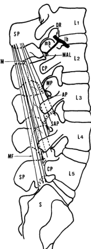
Fig. 1 Schematic illustration of the lumbar multifidus muscle as seen in a lateral view. One segment fascicle attached to the spinous process of the first lumbar verte-bra. 突 起 を起 始 とす る多 裂 筋 の筋 群 は そ の横 突 起 よ り 1分 節 ず つ 高 位 の 異 な る3分 節 の神 経 に 支配 され て い る.一 方,(2)1つ の棘 突 起 に停 止 す る多裂 筋 に注 目す れ ば,す べ て そ の棘 突 起 と同 高 位 の 後 枝 内側 枝 に よっ て1分 節 性 に支 配 さ れ る.(3)こ の 分 節 性 の神 経 支 配様 式 と,筋 束 が共 通 腱 とな り棘 突 起 に付 着 して い る形 態 な どを考 慮 して,多 裂 筋 は 棘 突 起 を 起 始 と した 方 が 合 理 的 で あ る と し て い る.し か し,筋 の 作 用 か らみれ ば,本 筋 は横 突起 を起 始 と し,棘 突 起 を停 止 とす べ きで あ るの で, この 点 に つ い て は い まだ 問 題 点 は残 る. 一方,西15)や佐 藤 ら16)は,筋の 系 統 的 分 類 は 神 経 支 配 を 中 心 に して な され る こ と,筋 と神 経 は 最 も 恒 常 的 な 関 係 に あ る こ と,固 有 背 筋 は きわ め て 原 始 的形 態 を残 して い る こ と,な ど を指 摘 して い る. この考 え方 も,棘 突 起 を起 始 と した ほ うが 横 突 起 を 起 始 と す る よ り 合 理 的 で あ る と す るMa-chintosh7)や,Bogduk8,9)の 説 を支 持 す る.な お, 本 筋 の腰 椎 横 突 起 部 位 で の付 着 領 域 は乳 頭 突 起, 副 突 起,さ ら に は椎 間 関 節 の 関節 包 で 広 範 囲 にお よ ん で い た.本 筋 が 椎 間 関 節 の関 節 包 に付 着 す る 形 態 は,多 裂 筋 に よ っ て 引 き起 こ され る運 動 の 際 に関 節 包 が 関 節 内 に引 き込 まれ る の を防 いで い る と考 え られ,興 味 深 い. 2.成 人 と胎 児 の 違 い に つ い て 立 位 を と らな い胎 児 の腰 部 多 裂 筋 の筋 腹 は,成 人 と比 較 して薄 く,未 発 達 で あ る.後 枝 内側 枝 は, 成 人 で は 多 裂 筋 の下 を く ぐ り腹 側 よ り分 布 す る が,胎 児 で は 多裂 筋 の 棘 突 起 付 着 部 の わ ず か 下 方 の 外 側 か ら直 接 進 入 し分 布 す る.明 らか に 多裂 筋 へ の進 入 部 位 は,胎 児 と成 人 で 異 な っ て い た. 比 較 解 剖 学 で は,固 有 背 筋 を内 側 と外 側 の二 つ の 縦 束 に 区分 して い る.そ して,脊 髄 神 経 後 枝 の
Fig. 2 Morphological classification of the trunk musculature and the their relation to the course of the spinal nerves. (after Nisi) D3: Submultifidus, D2: multifidus, D1: Longissimus, L: Illiocostalis
(443) 31 -内 側 枝 と外 側 枝 は それ ぞ れ の 筋 束 を貫 き,各 々 を さ らに二 分 してL・D1-D3の 四 筋 に分 け て い る15) (Fig.2).図 の よ う に後 枝 内側 枝 はD2-D3の 間 を 貫 く.こ れ は,筋 腹 の外 側 か らの進 入 を意 味 し, 胎 児 の 神 経 分 布 形 態 と一 致 す る. 幼 児 期 の形 態 を検 索 で き な か っ た の で,ど の よ うに して 成 人 の 神 経 分 布 形 態 に変 わ って い くの か 不 明 で あ る が,立 位 を と り腰 椎 の 生 理 的 前 弯 が増 強 し て い く過 程 で の大 き な変 化 で あ る と考 え られ る. 3.変 異 に つ い て 後 枝 内側 枝 が 低 位 の腰 椎 棘 突 起 か ら起 始 す る多 裂 筋 に分 枝 す る変 異 が,成 人 と胎 児 の 検 索 例 を合 わ せ た200束 中5束 にみ られ た.こ の よ うな変 異 に関 す る報 告 は な い. ほ とん どの 腰 部 多裂 筋 は,起 始 す る棘 突 起 と同 高 位 の後 枝 内 側 枝 に 分 節 性 に 支 配 され て い る.し か し,こ の 分 節 性 の神 経 支 配 は,肉 眼 解 剖 学 的 に の み確 認 され て い る が,組 織 学 また は生 理 学 な ど の他 の 方 法 に お け る確 認 は さ れ て い な い.特 に, 後 枝 内側 枝 を電 気 刺 激 し て分 節 状 に多 裂 筋 が収 縮 す る 実験 は大 変 重 要 と思 わ れ るが,動 物 実 験 以 外 は倫 理 上,ま た,技 術 的 に困 難 な た め に ヒ トの生 体 で は行 わ れ て い な い17,18).また,多 裂 筋 は,均 一 な筋 形 態 で あ る た め,組 織 学 的 な レベ ル で の多 重 支 配 の可 能 性 も否 定 で き な い. 結 論 成 人 遺 体10体 と5∼10カ 月 齢 の ヒ ト胎 児10 体 を用 い て,支 配 神 経 の 分 布 形 態 を中 心 に し て腰 部 多 裂 筋 の解 剖 を行 い,以 下 の結 果 を得 た. 1)棘 突 起 か ら起 始 す る多 裂 筋 は,同 高位 の 後 枝 内 側 枝 に 分 節 性 に支 配 され て い る. 2)成 人 多裂 筋 へ の 支 配 神 経 の 進 入 部 位 は棘 突 起 付 着 部 の わ ず か 下 方 の腹 側 で あ っ た. 3)胎 児 多 裂 筋 は 成 人 に比 べ筋 腹 が 薄 く,支配 神 経 の 進 入 部 位 は,棘 突 起 付 着 部 の わ ず か下 方 の 外 側 縁 で あ っ た.こ れ は,胎 児 が 立 位 を と らな い た め に抗 重 力 筋 が 未 発 達 で あ る こ と に よ る と考 え た. 4)後 枝 内 側 枝 が 低 位 の 棘 突 起 か ら起 始 す る 多 裂 筋 に 分 枝 す る 変 異 が 胎 児 と成 人 を 合 わ せ て5束 に み ら れ た. 本 研究 は,日 本 医科 大 学 に お け る倫 理委 員 会 の承 認 を得 た もの で あ る. 稿 を終 わ るに あた り,御 懇 篤 な る御 指導,御 助 言 を賜 っ た 日本 医科 大学 解 剖学 第2教 室 伊藤 博 信教 授 な らびに整 形 外科 学教 室 白井 康 正教 授 に深 く感謝 い た します.ま た,本 研 究 を行 うに あ た り,終 始御 協 力 を頂 い た 日本 医 科大 学 解 剖 学 第2教 室員 各 位,特 に 田沼 久美 子助 教 授 に感 謝 いた し ます. なお,本 論 文 の要 旨 は第98回 日本 解剖 学 会総 会(1993年 7月)に おい て,発 表 した. 文 献
1) Bagnall, K.M., Ford, D.M, McFadden, K.D., Creen-hill, B.J., Raso, U.J., and Eng, P.: The histo-chemical composition of human vertebral muscle. Spine, 9, 470-473, 1984.
2) Fidler, M.W., Jowett, L., and Troup, J.D.G.: Myosin ATPase activity in multifidus muscle from cases of spinal derangement. J. Bone Joint Surg., 57 B, 220-227, 1976.
3) Hides, J.A., Cooper, D.H., and Stokes, M.J.: Diag-nostic ultrasound imaging for measurement of the lumbar multifidus muscle in normal young adults. Physiother Theory Pract., 8, 19-26, 1992. 4) Hides, J.A., Stokes, M.J., Said, M. Jull, G.A., and
Cooper, D.H.: Evidence of lumbar multifidus muscle wasting ipsilateral to symptoms in patients with acute/subacute low back pain. Spine, 19, 165-172, 1994.
5) Donish, E.W., and Basmajian, J.V.: Electromyo-graphy of deep back muscle in man.Am.J. Anat., 133, 25-36, 1972.
6) Valencia, F.P., and Munro, R.R.: An electromyo-graphic study of the lumbar multifidus in man. Electromyogr. Clin. Neurophysiol., 25, 205-221,
1985.
7) Machintosh, J.E., Valencia, F., Bogduk, N., and Munro, R.R.: The morphology of the human lum-bar multifidus. Clin. Biomech., 1, 196-204, 1986. 8) Bogduk, N., Wilson, A.S., and Tynan, W.: The
human lumbar dorsal rami. J. Anat., 134, 383-397, 1982.
9) Machintosh, J.E., Bogduk, N.: The biomechanics of the lumbar multifidus. Clin. Biomech., 1, 205-213, 1986,
ana-- 32 ana-- (444)
tomy.35th.edn.p.470-471, London, Longmans, 1973.
11) Lewin, T., Moffet, B., and Viidik, A.: The mor-phology of the lumbar synovial intervertebral joints. Acta Morphol. Neerlando-Scandinay., 4, 299-319, 1962.
12) Bogduk, N.: The lumbar mamillo-accessory liga-ment. Its anatomical and Neurosurgical significance. Spine, 6, 162-167, 1981.
13) Hollinshead, W.H.: The back and limbs. Anato-my for surgeons. Harper. Row., 3, 158-159, 1969. 14) Warwick, R., and Williams, P.L.: Gray's
anat-omy.35th.edn.p.511-513, London, Longmans, 1969.
15) 西 成 甫:筋 系 統 の 類 型 解 剖 学.日 新 医 学,48, 137(-145, 1963.
16) 佐 藤 達 夫:肋 骨 挙 筋 と横 突 間 筋 の 形 態 学 的 研 究.解 剖 雑 誌,43,305-325,1968.
17) Bogduk, N.: The dorsal lumbar muscles of the cat. Anat. Anz., 148, 55-67, 1980.
18) Carlson, fr.: Morphology and contraction prop-erties of cat lumbar back muscles. Acta Physiol. Scand., 103, 180-197, 1978,
Abbreviations SP: Spinous process
CP: Costal process MP: Mamillary process AP: Accessory process
MAL: Mamillo-accessory ligament SAP: Superior articular process IAP: Inferior articular process ZJ: Zygapophysial joint DR: Dorsal ramus
mb: Medial branch of dorsal ramus lb: Lateral branch of dorsal ramus VR: Ventral rami
MF: Multifidus muscle S: Sacrum
M: Motor point
Legends of Photos
Photo 1 Dorsal view of the multifidus muscle in an adult.
Photo 2 Postero-lateral view of the multifidus muscle attached to the spinous process of the second lumbar vertebra in an adult. The medial branch of the dorsal ramus of the second lumbar nerve
pass-ing below the mamillo-accessory liga-ment runs medio-dorsally to the multifidus muscle atached to the spinous process of the second lumbar vertebra.
Photo 3 The multifidus muscle attached to the spinous process of the second lumbar vertebra is turned laterally in an adult. The medial branch of the dorsal ramus of the second lumbar nerve innervating the multifidus muscle attached to the spinous process of the second lumbar vertebra is indicated by arrows. The nerve enters this muscle from the ventral aspect of the upper portion.
Photo 4 Dorsal view of the right side of the multifidus muscles in a five months old fetus. The multifidus muscles are flat in shape and less developed than those in adults.
Photo 5 A piece of the right multifidus muscle attached to the spinous process of the first lumbar vertebra in a five months old fetus. The nerve innervating this muscle enters from the lateral margin of the upper portion.
Photo 6 A variation case of the medial branch of the dorsal ramus of the second lumbar nerve innervating the multifidus muscles of two segments attached to the spinous processes of the second and third lumbar vertebra in an adult. The medial branch of the dorsal ramus of the second lumbar nerve innervating the multifidus muscle attached to the spinous process of the second lumbar vertebra enters the mus-cle from the ventral aspect of the upper portion, and also enters the multifidus muscle attached to the spinous process of the third lumbar vertebra from the dorso-lateral porsion. The nerve inner-vating the multifidus muscles with two segments is indicated by arrows. Photo 7 A variation case of the medial branch of
the dorsal ramus of the third lumbar nerve innervating the multifidus muscles with two segments attached to the spinous processes of the third and fourth lumbar vertebrae in a ten months old fetus. The nerve innervating these mus-cles is indicated by arows.
Photo 8 Showing the ossified mamillo-accessory ligament and the medial branch of the dorsal ramus of the lumbar nerve pass-ing bellow it with a piece of wire in its place in a skeletal specimen.
(445) 33
-Plate (1)
- 34 - (446)