Mitotic Langerhans Cells in the Normal Human Epidermis: Light and
Electron Microscopic Observations in All Mitotic Phases
Shigeo Oota
First Department of Anatomy, Faculty of Medicine, Tottori University, Yonago 683-0826, Japan In three experiments of the normal human epidermis under different biopsy and fixative buffer conditions, Langerhans cells (LCs) in all mitotic phases were found with the Thin-Section-Reembedding and Ultrathin-Sectioning (TRUS) technique; two in the prophase, five in the metaphase, two in the anaphase and two in the telophase. The serial light microscopic examination showed many characteristic features, namely the shapes of mitotic LCs in each section were quite different in one cell as well as between individual cells, even in the same mitotic phase. No round mitotic LCs were seen. Two LCs in the prophase were detected in the specimens prefixed in a fixative dissolved in dialysate (Kindaly 2) instead of buffer solution. Birbeck granules were witnessed in all phases.
Key words: electron microscopy; epidermis; human; Langerhans cells; mitosis
Abbreviations: CB, cacodylate buffer; LC, Langerhans cell; M, mol/L; PB, phosphate buffer; TRUS, Thin-Section-Reembedding and Ultrathin-Sectioning
It has been considered difficult to observe mito-tic Langerhans cells (LCs) in the normal human epidermis, and so far only one electron micro-scopic observation of the mitotic LC (in the anaphase) has been reported by Konrad and Hönigsmann (1973). In the present study, com-prising three experiments under different biop-sy and fixative buffer conditions, 11 LCs in various mitotic phases were observed with the Thin-Section-Reembedding and Ultrathin-Sectioning (TRUS) technique (Oota, 1999). In all phases, the mitotic LCs showed markedly characteristic features. These features of all phases in the human epidermis were obtained for the first time by light and electron micros-copy.
Materials and Methods
Skin biopsies (5 mm × 15 mm) were taken 3 times, the 2nd one year after the 1st and the 3rd two months after the 2nd, from the forearm of a 63-year-old healthy male volunteer, who had given informed consent. The 1st and the 2nd
biopsy specimens were obtained under lidocaine anesthesia without epinephrine, and the 3rd bi-opsy specimen was excised under anesthesia with epinephrine. The biopsy specimens were treated in the same manner every time: immersed in physiological saline immediately after oper-ating at the dermatological biopsy-operoper-ating room in this Faculty. After 20 min, the speci-men was divided into 3 to 5 equal parts and pre-fixed in the fixative solutions shown in Table 1. Here Kindaly 2 (Fuso Pharmaceutical Industry, Ltd., Osaka, Japan), acetic dialysate solution for hemodialysis, was tried as a physiological vehicle instead of the buffer. The components and osmolarity of the chemical fixatives in Ex-periments 1 and 2 are shown in Table 2. After 1 h, each specimen was dissected into small pieces and the fixation was continued for 24 h at 4˚C. Subsequent procedures were previously described as the TRUS technique (Oota, 1999). Although in the technique the so-called “invert-ed gelatin capsule technique” (Robbins and Gonatas, 1964; Miyauchi and Hashimoto, 1987) was used twice, the polyethylene capsules (TAAB Lab. Equip. Ltd., Berks., United Kingdom)
were employed instead of gelatin capsules. All thin-sections in the 3 experiments were cut parallel to the surface of the epidermis. The oil immersion pictures were taken from the sec-tions on slides; a drop of water was poured be-tween the section and a cover glass for use of the section for electron microscopy. The ultra-thin sections were placed on a copper grid, stain-ed with uranyl acetate and lead citrate, and ob-served with a Hitachi electron microscope, H 500, at 80 kV.
Results
Inthe 1st experiment, 6 mitotic LCs (Fig. 1: Rows 3, 4, 5, 8, 10 and 11) were found in the materials prefixed with the fixatives in 0.1 M and 0.15 M cacodylate buffer (CB) (Table 1). In the 2nd experiment, only 1 LC in the pro-phase (Fig. 1: Row 1) was observed in the ma-terials prefixed in Kindaly 2, which is common-ly used for artificial kidneys, and none in the materials prefixed with the fixatives in phos-phate buffer (PB) (Table 1). Why no mitotic LCs were observed in these materials is
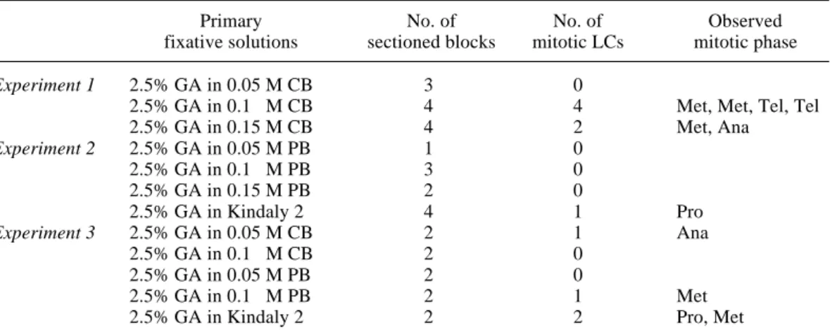
ob-Table 1. Primary fixative solutions and the number of sectioned blocks and of mitotic LCs found in the materials prefixed with the fixatives
Primary No. of No. of Observed
fixative solutions sectioned blocks mitotic LCs mitotic phase
Experiment 1 2.5% GA in 0.05 M CB 3 0
2.5% GA in 0.1 M CB 4 4 Met, Met, Tel, Tel
2.5% GA in 0.15 M CB 4 2 Met, Ana Experiment 2 2.5% GA in 0.05 M PB 1 0 2.5% GA in 0.1 M PB 3 0 2.5% GA in 0.15 M PB 2 0 2.5% GA in Kindaly 2 4 1 Pro Experiment 3 2.5% GA in 0.05 M CB 2 1 Ana 2.5% GA in 0.1 M CB 2 0 2.5% GA in 0.05 M PB 2 0 2.5% GA in 0.1 M PB 2 1 Met
2.5% GA in Kindaly 2 2 2 Pro, Met
Ana, anaphase; CB, cacodylate buffer; GA, glutaraldehyde; LC, Langerhans cell; M, mol/L; Met, metaphase; PB, phosphate buffer; Pro, prophase; Tel, telophase.
Table 2. Components and osmolarity of chemical fixatives Fixative solution Osmolarity of Total osmolarity
buffer (vehicle) (mOsm) (mOsm) Experiment 1 2.5% GA in 0.05 M CB 103 345 2.5% GA in 0.1 M CB 202 430 2.5% GA in 0.15 M CB 285 510 Experiment 2 2.5% GA in 0.05 M PB 94 373 2.5% GA in 0.1 M PB 203 470 2.5% GA in 0.15 M PB 296 566 2.5% GA in Kindaly 2 255 484
CB, cacodylate buffer; GA, glutaraldehyde; LC, Langerhans cell; M, mol/L; PB, phosphate buffer.
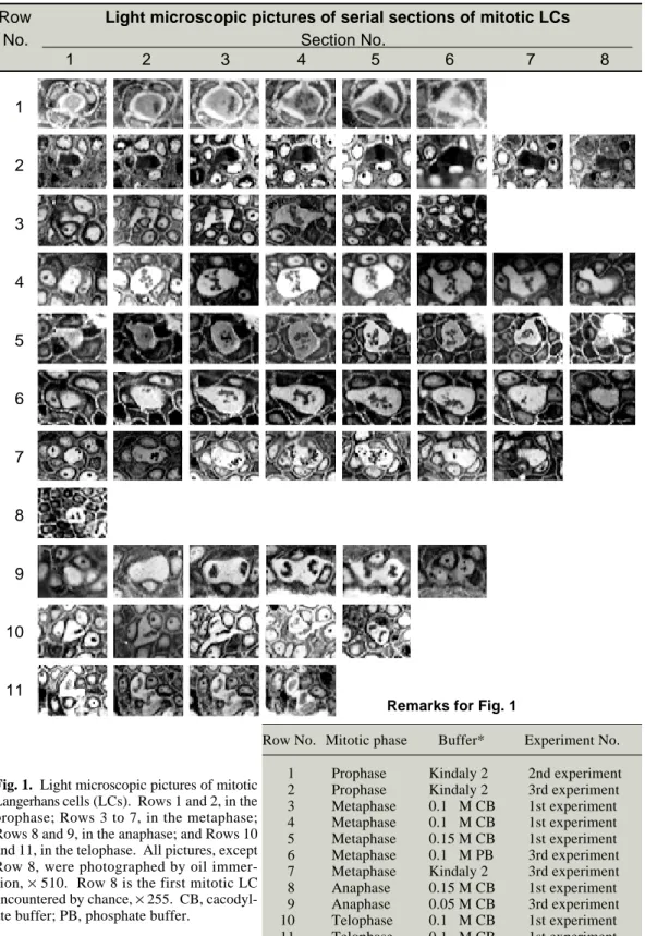
Fig. 1. Light microscopic pictures of mitotic Langerhans cells (LCs). Rows 1 and 2, in the prophase; Rows 3 to 7, in the metaphase; Rows 8 and 9, in the anaphase; and Rows 10 and 11, in the telophase. All pictures, except Row 8, were photographed by oil immer-sion, × 510. Row 8 is the first mitotic LC encountered by chance, × 255. CB, cacodyl-ate buffer; PB, phosphcacodyl-ate buffer.
Row Light microscopic pictures of serial sections of mitotic LCs
No. Section No.
1 2 3 4 5 6 7 8 1 2 3 4 5 6 7 8 9 10 11
Remarks for Fig. 1
Row No. Mitotic phase Buffer* Experiment No. 1 Prophase Kindaly 2 2nd experiment 2 Prophase Kindaly 2 3rd experiment 3 Metaphase 0.1 M CB 1st experiment 4 Metaphase 0.1 M CB 1st experiment 5 Metaphase 0.15 M CB 1st experiment 6 Metaphase 0.1 M PB 3rd experiment 7 Metaphase Kindaly 2 3rd experiment 8 Anaphase 0.15 M CB 1st experiment 9 Anaphase 0.05 M CB 3rd experiment 10 Telophase 0.1 M CB 1st experiment 11 Telophase 0.1 M CB 1st experiment *Buffer or vehicle used in fixative solution.
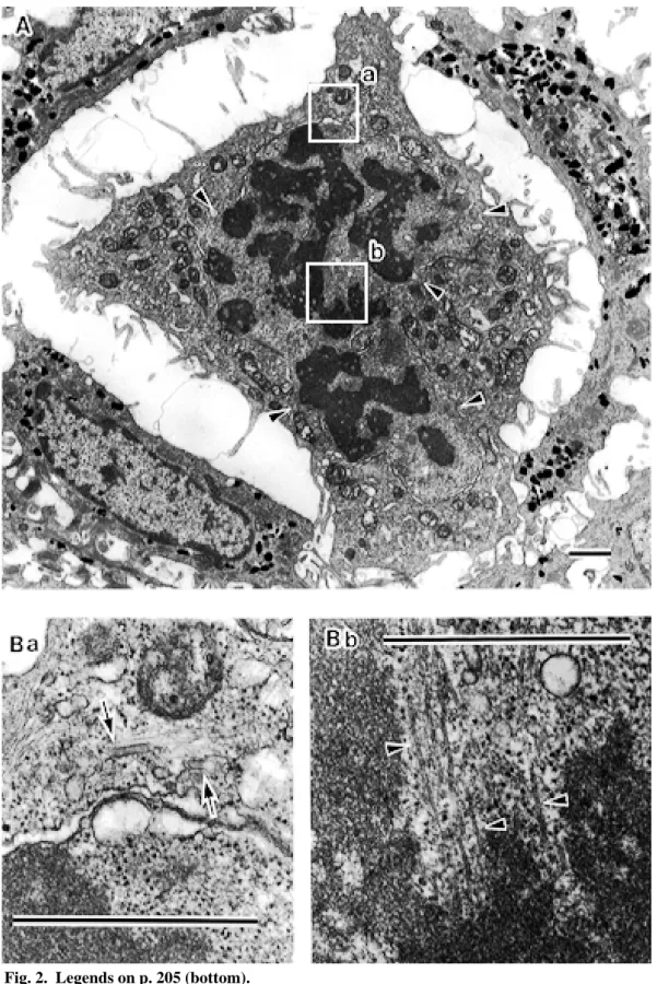
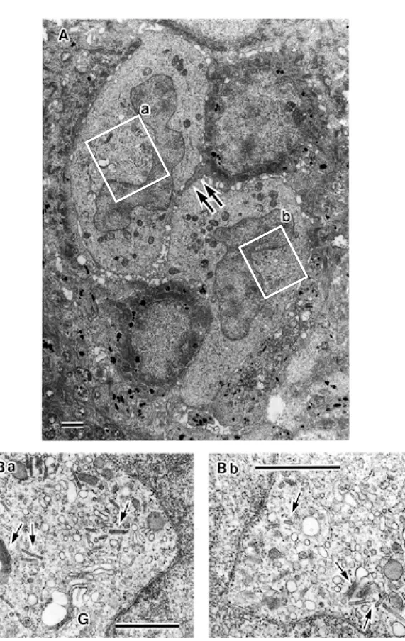
Fig. 2 (p. 204) A: An electron micrograph of a Langerhans cell (LC) in the prophase (Fig. 1: Row 1-No. 4), × 9,000. In places, the nuclear envelope disappears (arrowheads) and, spindle microtubules pass towards chromosomes which have begun to condense (Bb: arrowheads), × 52,500. Birbeck granules are seen (Ba: arrows), × 52,500. Scale bar = 1 µm.
Fig. 3. A: A Langerhans cell (LC) in the metaphase (Fig. 1: Row 3-No. 5), × 5,400. Ba and b: High magni-fication photographs of the rectangular areas in A. Parallel-arranged LC granules (Birbeck granules) and many vesicular structures of various sizes are visible at both polar regions, × 36,000. Scale bar = 1 µm.
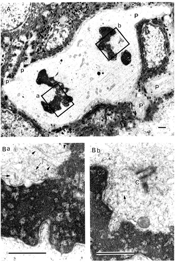
Fig. 4 (p. 207) A: A Langerhans cell (LC) in the anaphase (Fig. 1: Row 9-No. 4), × 6,000. Ba and b: High magnification photographs of the rectangular areas in A, × 30,000. Newly opposed segments of the nuclear envelope have appeared on the partially fused chromatin mass. Spindle microtubules (Ba: arrowheads) and chromatin mass form junctions at points still devoid of nuclear envelope. In this section, orthogonal arranged centrioles are seen at one side of poles (Bb: C). Birbeck granules are sparsely seen in the cytoplasm (Ba and b: arrows). Scale bar = 1 µm.
scure. So, in the 3rd experiment, the addition of epinephrine as a local anesthetic during the skin biopsy was tried, and CB, PB and Kindaly 2 were again used for fixative vehicle. At this time, 4 mitotic LCs (Fig. 1: Rows 2, 6, 7 and 9) were found in the materials prefixed with the fixatives in 0.05 M CB, 0.1 M PB and Kindaly 2 (Table 1).
In these experiments, features observed with a light microscope showed several char-acteristics (Fig. 1): i) Row 8 was the 1st mitotic LC encountered by chance during the test for the effect of buffer osmolarity to the cell struc-tures, and so the serial sections and high mag-nification views were not taken; ii) the cyto-plasm of mitotic LCs, after the metaphase to the telophase, was clearer than that of epidermal mature LCs and highly contrasted with the neighboring keratinocytes and the dark-stained chromosomes, whereas the cytoplasm of LCs in the prophase was stained at the same level as neighboring cells, or more intensely, and so the contrast with the condensing chromatin was low (Rows 1 and 2); iii) the serial sections indi-cated that each mitotic LC possessed several, short or long, slender or podgy, cytoplasmic processes; iv) so in the same way, the shapes of mitotic LCs in each section were quite different in one cell as well as between individual cells, even in the same mitotic phase. From the serial sections of each row, the three-dimensional fea-tures possessing process(es) could be imaged; and v) no round mitotic LCs were seen.
Electron microscopy showed many char-acteristics as follows.
Figure 2 is an electron micrograph of one of the LCs in the prophase observed in the material prefixed with the fixative in Kindaly 2 solution. The intercellular space between this cell and the neighboring keratinocytes has been markedly widened possibly due to a fixative artifact. This might have happened based on peculiar situa-tions involving neighboring keratinocytes. How-ever, the wide bases of the processes of this LC
still remain (Fig. 2A). In places, the nuclear envelope disappeared (Fig. 2A), and spindle microtubules passed towards chromosomes which had begun to condense (Fig. 2Bb). LC granules (Birbeck granules) were seen (Fig. 2Ba).
Figure 3 is one of the LCs in the metaphase observed in the material prefixed with the fixa-tive in 0.1 M CB. In the row of this cell, every section showed considerably different features. The cell of Fig. 3A showed a relatively long and podgy cytoplasmic process, and the cell seemed to be just like a tadpole. Chromosomes were aligned on the metaphase plate. No centriole was caught in the plane of this section, but at the sites regarded as the neighborhood of the poles, parallel-arranged Birbeck granules and many vesicular structures of various sizes were visi-ble (Figs. 3Ba and b).
Figure 4 is one of the LCs in the anaphase. This picture was observed in the material pre-fixed with the fixative in 0.05 M CB. This cell possesses large (P) and small (p) cytoplasmic processes (Fig. 4A). In Figs. 4Ba and b, newly opposed segments of the nuclear envelope appeared on the partially fused chromatin mass. Spindle microtubules and the chromatin mass formed junctions at points still devoid of the nu-clear envelope. Birbeck granules were sparsely seen.
Figure 5 is one of the 2 telophase LCs. This picture was obtained from the material prefixed with the fixative in 0.1 M CB. Reproduced daugh-ter LCs seemed to be separated by the invasion of the neighboring keratinocyte between the daughter cells (Fig. 5A, double arrows). Al-though the nuclei of both daughter LCs showed still young forms (Fig. 5A), their function seemed to have become markedly active (Figs. 5Ba and b).
Through these observations, in all mitotic phases of LCs, Birbeck granules were witnes-sed in the cytoplasm.
Discussion
The features of these mitotic LCs in the normal human epidermis are the first ones observed both by light and electron microscopy. They seem to show actual evidence for the reproduc-tion of LCs in the normal human epidermis. In 1979, 2 groups of investigators (Frelinger et al., 1979; Katz et al., 1979) using bone marrow chimeric animals showed that LCs in the epi-dermis originated from bone marrow, and this concept has since been widely accepted. There-after, however, Czernieleski et al. (1985) using flow cytometry showed that human epidermal LCs were a cycling cell population in the nor-mal (physiological) epidermis, and cited the study by Konrad and Hönigsmann (1973) as morphological evidence. The present observa-tions confirm their view. Czernielewski and Demarchez (1987) further studied the self-reproducing capacity of LCs in the human skin grafted onto the nude mouse. Thereafter, Miyauchi and Hashimoto (1989) using the ATPase staining technique presented the mito-tic activities of epidermal LCs in the normal mouse skin. Compared with these studies, the present study shows the actual features of mito-tic LCs in close proximity to the natural state of the human epidermis. In this study, the relation between the mitosis of LCs and the kind or osmolarity of the fixative buffer (vehicle) could not be clarified, and the relation between the mitosis of LCs and epinephrine in the local anesthetic is also indistinct. However, it is very interesting that the mitotic LCs in the prophase were detected only in the materials which had been prefixed in the fixative dissolved in Kindaly 2 . Why so? This problem remains for further study.
As a whole, it must be one of the most impor-tant observations that Bibeck granules have been witnessed in all mitotic phases of human LCs.
Fig. 5 (p. 208) A: A Langerhans cell (LC) in the telophase (Fig. 1: Row 11-No. 2), × 7,000. Double arrows indicate the process of the neighboring keratinocyte invading between the daughter LCs. Ba and b: High magnification photographs of the rectangular areas in A, × 21,000 and × 28,000, respectively. In the Golgi area, a lot of vesicles and Birbeck granules are observed (G, Golgi apparatus; arrows, Birbeck granules). These show clearly the reopening of the cell function as soon as cell division is completed. Scale bar =1 µm.
By the TRUS technique, it was possible for the first time to carry out a wide survey of the human epidermis, to find a large amount of mitotic LCs at the light microscopic level and to view fine details of the same cells at the elec-tron microscopic level. Thus, anyone hereafter can observe mitotic LCs both in the normal and pathological human epidermis, and consequent-ly, the nature, kinetics and function of LCs can be more clearly understood.
Acknowledgements: The author gratefully acknowl-edges the support of Emeritus Prof. Keiichi Tanaka and Prof. Akihiro Iino, who gave him the opportunity to carry out this study in the Second and First Depts. of Anatomy, Faculty of Medicine, Tottori Univ., respectively. He further expresses his gratitude to Prof. A. Iino for the reading of this manuscript and expresses his appreciation to Prof. Motoyuki Mihara for the kind help in obtaining biopsy specimens. The author also thanks Prof. Takao Inoué and Dr. Sumire Inaga for their heartful discussions, Dr. Tadayuki Aman for the kind gift of dialysate, Kindaly 2, and the measurement of buffer (vehicle) osmolarity.
References
1 Birbeck MS, Breathnach AS, Everall JD. An electron microscope study of basal melanocytes and high-level clear cells (Langerhans cells) in vitiligo. J Invest Dermatol 1961;37:51–64. 2 Czernielewski J, Vaigot P, Prunieras M.
Epider-mal Langerhans cells—A cycling cell population. J Invest Dermatol 1985;84:424–426.
3 Czernielewski JM, Demarchez M. Further evi-dence for the self-reproducing capacity of Langerhans cells in human skin. J Invest Dermatol 1987;88:17–20.
4 Frelinger JG, Hood L, Hill S, Frelinger JA. Mouse epidermal Ia molecules have a bone mar-row origin. Nature 1979;282:321–323.
5 Katz SI, Tamaki K, Sachs DH. Epidermal Langerhans cells are derived from cells originat-ing in the bone marrow. Nature 1979;282:324– 326.
(Received September 1, Accepted September 17, 1999) 6 Konrad K, Hönigsman H.
Electronenmikroskopis-cher Nachweis einer mitotischen Langerhans-Zelle in normaler menschlicher Epidermis. Arch Dermatol Forsch 1973;246:70–76.
7 Miyauchi S, Hashimoto K. Epidermal Langerhans cells undergo mitosis during the early recovery phase after ultraviolet-B irradiation. J Invest Dermatol 1987;88:703–708.
8 Miyauchi S, Hashimoto K. Mitotic activities of normal epidermal Langerhans cells. J Invest
Der-matol 1989;92:120–121.
9 Oota S. Some new aspects of Langerhans cells in the human epidermis: Light and electron micro-scopic observations on the swelling sites seen in the process terminals of the dendritic cells described by Langerhans in 1868. Yonago Acta Med 1999; 42:153–161.
10 Robbins E, Gonatas NK. The ultrastructure of a mammalian cell during the mitotic cycle. J Cell Biol 1964;20:356–359.