Cellular Leiomyoma of the Nasal Cavity: Report of a Case and Review
of Literature
Satoshi Horie, Kensaku Hasegawa* and Tadashi Terada
Second Department of Pathology, Tottori University Faculty of Medicine, Yonago 683-0826 Japan and *Clinic of Otolaryngology, Public Yoka Hospital, Yoka 667-8555 Japan We report a rare case of cellular leiomyoma in the nasal cavity. A 72-year-old Japanese woman was admitted to a hospital. A tumoral lesion was revealed in the left nasal cavity. Angiography showed tumor staining, and the tumor was endoscopically resected after the embolizaion of the feeding artery. At gross inspection, the tumor measured 1.0 × 1.5 × 2.0 cm. Microscopically, the tumor consisted of many spindled cells with blunt ended nuclei. Immunohistochemical examination revealed that the tumor cells were positive for vimentin, alpha smooth muscle actin. We diagnosed this case as cellular leiomyoma. To the best of our knowledge, there have been only 23 reported cases of nasal leiomyoma in English medical literature. We made a brief literature review of the occurrence of this tumor in the nasal cavity.
Key words: cellular leiomyoma; nasal cavity
Leiomyoma is a benign smooth muscle tumor found mainly in the uterus, skin, gastrointes-tinal tract, deep soft tissues, peritoneum and other sites. Enzinger and Weiss (1995) reported that 95% of leiomyoma (7,748 cases) were lo-cated in the female genitalia, 3% in the skin and the remainder in other sites. They are quite un-usual in the nasal cavity and paranasal sinuses, and a search of the literature revealed only 23 reports (Maesaka et al., 1966; Ram, 1971; Kotaka and Furuya, 1973; Schwartzman and Schwartzman, 1973; Timirgaleev, 1973; Wholfowitz and Schmaman, 1973; Fu and Perzin, 1975; Nall et al., 1997; MaCaflley et al., 1978; Papavasiliou and Micheaels, 1981; Lijovetzky et al., 1985; Daisley, 1987; Hanna et al., 1988; Harcourt and Gallimore, 1988; Tang and Tse, 1988; Nam et al., 1989; Barr et al., 1990; Ragbeer and Stone, 1990; Sawada, 1990; Van Ingen et al., 1991; Khan et al., 1994; Ardekian et al., 1996; Llorente et al., 1996).
We report the clinical and histological fea-tures of a rare case of the intranasal leiomyoma including its immunohistochemistry.
Patient report
Clinical Summary
A 72-year-old Japanese woman was admitted to a hospital with a history of temporal epistaxis. She had no other symptoms such as headache, pain, nasal discharge or anosmia. Her past medical history showed only bronchial asthma. On initial physical examination, a mass mea-suring 1.0 × 2.0 cm was located on the nasal septum of the left nasal cavity. A computed to-mography scan of the head showed a solid mass measuring 0.8 × 1.0 × 2.0 cm in the left nasal cavity (Fig. 1). Angiography revealed tumor stainings (Fig. 2). Selective embolization of the feeding artery was performed. Resection of the tumor by endoscopic nasal surgery was per-formed. Postoperative recovery was uneventful with the exception of slight bleeding. She was discharged from the hospital, and there has been no recurrence since.
Fig. 1. Computerized tomography sections of axial (A) and coronal (B) views of the tumor (arrows) seen in the left nasal cavity.
A
B
Pathologic Findings
On gross examination, the resected speci-men consisted of a grayish-pink tissue measuring 1.0 × 1.5 × 2.0 cm. Microscop-ically, the mass consisted of spindle cells which lay in the submucosa and was par-tially covered by non-keratinizing strati-fied squamous epithelium. The cellularity of the tumor was high. The cells had blunt-ended oval nuclei and minimal nuclear pleomorphism (Fig. 3). No mitotic figures were observed.
For confirmation, an immunohisto-chemical study was performed. The tumor cells were negative for epithelial mem-brane antigen, cytokeratin, and S-100, but positive for vimentin and alpha smooth muscle actin (Fig. 4).
Discussion
We diagnosed this case as cellular leio-myoma because of its histology and the strongly positive myogenic marker (alpha smooth muscle actin). Differential diagno-sis included angiofibroma, epithelioid leio-myoma, hemangiopericyioma and schwan-noma. Immunohistochemically, angiofi-broma and schwannoma were not proba-ble. Because most of the tumor cells had a spindle shape, showing a fascicular pat-tern, leiomyoma was probable rather than epithelioid leiomyoma or hemangioperi-cytoma.
To the best our knowledge, there have been previously only 23 reported cases of leiomyoma involving the nasal cavity and paranasal sinuses (Table 1) (Maesaka et al., 1966; Ram, 1971; Kotaka and Furuya, 1973; Schwartzman and Schwartzman, 1973; Timirgaleev, 1973; Wholfowitz and Schmaman, 1973; Fu and Perzin, 1975; Nall et al., 1997; MaCaflley et al., 1978; Papavasiliou and Micheaels, 1981; Lijovetzky et al., 1985; Daisley, 1987; Hanna et al., 1988; Harcourt and Gallimore, 1988; Tang and Tse, 1988; Nam
Fig. 2. Preoperative angiography showing tumor stainings (arrow) from a left internal maxillary artery.
Fig. 3. The tumor cells were composed of spindle cells. Hematoxylin-eosin, original magnification × 200
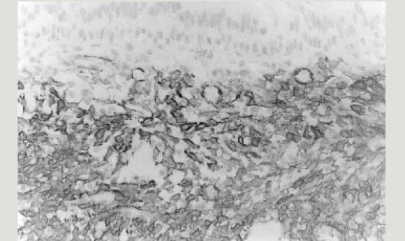
Fig. 4. The tumor cells were diffusely positive for alpha smooth muscle actin. Immunostaining for alpha smooth muscle actin (original magnification × 200).
et al., 1989; Barr et al., 1990; Ragbeer and Stone, 1990; Sawada, 1990; Van Ingen et al., 1991; Khan et al., 1994; Ardekian et al., 1996; Llorente et al., 1996). Therefore, the present case is very rare.
The rarity of smooth muscle tumors in the nasal cavity and paranasal sinuses is probably due to the paucity of smooth muscle fibers in this loca-tion.
Leiomyomas of the skin and subcutis are usually divided into vascular and nonvascular types. In the nasal cavity, three hypotheses have been given for the origin of smooth muscle tumors: from aberrant undifferentiated mesen-chyme; from smooth muscle elements in the wall of blood vessels; or from both sources (Batsakis, 1979). Most of the authors support the idea that the vascular smooth muscle is the origin of the tumor.
In agreement with Barr et al. (1990) and Llorente et al. (1996), we think that the origin of this type of nasal septal leiomyoma is from the smooth muscle component of a blood vessel, because of the absense of the other types of muscle in the septum.
In the present case, the cellularity of the tumor was higher than the usual leiomyoma in other sites. We diagnosed this case as cellular leiomyoma. There have not been any reported cases of cellular leiomyoma of the nasal cavity. Several stains have been used to identify leio-myoma including desmin, vimentin, Masson’s trichrome, actin and myosin (Maeda and Osaki,
1989). In our case, the tumor was strongly positive for alpha smooth muscle actin, support-ing the diagnosis of leiomyoma.
The current treatment is surgical resection and there are only a few reports of recurrence in the literature (Hanna et al., 1988; Khan et al., 1994). Because of the vascularity of the lesion in this case, we elected to embolize the feeding vessels prior to surgical resection.
In summary, leiomyomas of the nasal cavity are extremely rare. This case is the 24th re-ported case in the literature. The exact origin of these tumors is not known, but most agree that the etiology is probably from smooth muscle cells in the walls of blood vessels. Surgical ex-cision of these benign tumors yields high cure rates.
References
1 Ardekian L, Samet N, Talmi YP, Roth Y, Bendet E, Kronenberg J. Vascular leiomyoma of the nasal septum. Otolaryngol Head Neck Surg 1996;114:798–800.
Table 1. Previous cases of leiomyoma of the nasal cabity and paranasal sinuses
Author(s) Reference number Year Location
1 Maesaka et al. 16 1966 Vestibule
2 Ram 21 1971 Inferior turbinate
3 Schwartzman and Schwartzman 23 1973 Sinuses and nasal fossa
4 Wolfwitz and Schmaman 27 1973 Inferior turbinate
5 Kotaka and Furuya 11 1973 Nasal cavity
6 Timirgaleev 25 1973 Nasal septum
7 Fu and Perzin 6 1975 Nasal cavity
8 McCaffrey et al. 14 1978 Inferior turbinate
9 Papavasiliou and Michaels 19 1981 Middle turbinate
10 Lijovetzky et al. 12 1985 Vestibule
11 Daisley 4 1987 Middle turbinate
12 Tang and Tse 24 1988 Inferior turbinate
13 Hanna et al. 8 1988 Inferior turbinate
14 Nam et al. 18 1988 Vestibule
15 Ragbeer and Stone 20 1990 Nasal floor
16 Sawada 22 1990 Vestibule
17 Barr et al. 2 1990 Nasal septum
18 Van Ingen et al. 26 1991 Choana
19 Harcourt and Gallimore 9 1993 Ethnocide sinus
20 Khan et al. 10 1994 Inferior turbinate
21 Llorente et al. 13 1996 Nasal septum
22 Ardekian et al. 1 1996 Nasal septum
23 Nall et al. 17 1997 Superior turbinate
2 Barr GD, More IAR, McCallum HM. Leiomyo-ma of the nasal septum. J Laryngol Otol 1990; 104:891–893.
3 Batsakis JG. Tumors of the head and neck. Clin-ical and pathologClin-ical considerations. 2nd ed. Baltimore: Williams and Wilkins; 1979. p. 354–356. 4 Daisley H. Leiomyoma of the nasal cavity. West
Indian Med J 1987;36:181.
5 Enainger FM, Weiss SW. Soft tissue tumors. 3rd ed. St. Louis: CV Mosby; 1995. p. 467–491. 6 Fu YS, Perzin KH. Non-epithelial tumors of the
nasal cavity, paranasal sinuses, and nasopharynx: a clinicopathologic study. IV. Smooth muscle tumoures (leiomyoma, leiomyosarcoma). Cancer 1975;35:1300–1308.
7 Hachisuga T, Hashimoto H, Enjoji M. Angiolei-omyoma: a clinicopathologic reappraisal of 562 cases. Cancer 1984;84:126– 130.
8 Hanna GS, Akosa AB, Ali MH. Vascular leio-myoma of the inferior turbinate—report of a case and review of the literature. J Laryngol Otol 1988;102:1159–1160.
9 Harcourt JP, Gallimore AP. Leiomyoma of the paranasal sinuses. J Laryngol Otol 1993;107: 740–741.
10 Khan MHZ, Jones AS, Haqqani MT. Angioleio-myoma of the nasal cavity—report of a case and review of the literature. J Laryngol Otol 1994; 108:244–246.
11 Kotaka S, Furuya H. Leiomyoma of the nasal cavity. Otolaryngology (Tokyo) 1973;456:423– 429.
12 Lijovetzky G, Zaarura S, Gai I. Leiomyoma of the nasal cavity. J Laryngol Otol 1985;99:197– 200.
13 Llorente JL, Suarez C, Seco M, Garcia A. Leio-myoma of the nasal septum: report of a case and review of the literature. J Laryngol Otol 1996; 110:65–68.
14 MaCaflley TV, McDonald TJ, Unni KK. Leio-myoma of the nasal cavity. Laryngoscope 1978; 92:817–818.
15 Maeda Y, Osaki T. Angiomyoma of the cheek: a case report. J Oral Maxillofac Surg 1989;47: 1090–1093.
16 Maesaka A, Keyaki Y, Nakahoshi T. Nasal angi-oleiomyoma and leiomyosarcoma: report of 2 cases. Otologia (Fukuoka) 1966;12:42.
17 Nall AV, Stringer SP, Bauglunan RA. Vascular leiomyoma of the superior turbinate: first report-ed case. Head Neck 1997;19:63–67.
18 Nam HK, Kaufinan MW, WoolfAP. Pathology quiz, case 2. Arch Otolaryngol Head Neck Surg 1989;115:244.
19 Papavasiliou A, Micheaels L. Unusual leiomyo-ma of the nose (leiomyoblastoleiomyo-ma): report of a case. J Laryngol Otol 1981;95:1281.
20 Ragbeer SM, Stone J. Vascular leiomyoma of the nasal cavity: report of a case and review of literature. J Oral Maxllofac Surg 1990;48:1113– 1117.
21 Ram M. Fibromyoma of posterior end of inferior turbinal. J Laryngol Otol 1971;85:719–721. 22 Sawada Y. Angioleiomyoma of the nasal cavity.
J Oral Maxillofac Surg 1990;48:1100–1101. 23 Schwartzman J, Schwartzman J. Leiomyoma of
paranasal sinuses;case report. Laryngoscope 1973;83:1856–1858.
24 Tang SO, Tse CH. Leiomyoma of the nasal cavi-ty. J Laryngol Otol 1988;102:831.
25 Timirgaleev MKH. [Angioleiomyoma of the nasal septum.] Vestn Othorinolaringol 1973;35: 106.
26 Van Ingen G, Stel HV, Tiwari RM. Atypical lei-omyoma of the choana. J Laryngol Otol 1991; 105:1065–1067.
27 Wolfowitz BL, Schmaman A. Smooth muscle tumours of the upper respiratory tract. S Afr Med J 1973;47:1189–1191.
Received December 11, 2000; accepted January 5, 2001 Corresponding author: Prof. Tadashi Terada