Japan Advanced Institute of Science and Technology
Title
Site-specific transition of cytosine to uracil
via reversible DNA photoligation
Author(s)
Fujimoto, Kenzo; Matsuda, Shigeo; Yoshimura,
Yoshinaga; Matsumura, Takashi; Hayashi, Masayuki;
Saito, Isao
Citation
Chemical Communications, 2006(30): 3223-3225
Issue Date
2006
Type
Journal Article
Text version
author
URL
http://hdl.handle.net/10119/7905
Rights
Copyright (C) 2006 Royal Society of Chemistry.
Kenzo Fujimoto, Shigeo Matsuda, Yoshinaga
Yoshimura, Takashi Matsumura, Masayuki Hayashi
and Isao Saito, Chemical Communications,
2006(30), 2006, 3223-3225.
http://dx.doi.org/10.1039/b605289d - Reproduced
by permission of The Royal Society of Chemistry
Description
Site-specific Transition of Cytosine to Uracil via Reversible DNA
Photoligation
Kenzo Fujimoto,
*abShigeo Matsuda,
cYoshinaga Yoshimura,
bTakashi Matsumura,
aMasayuki Hayashi
dand Isao Saito
eReceipt/Acceptance Data [DO NOT ALTER/DELETE THIS TEXT] Publication data [DO NOT ALTER/DELETE THIS TEXT] DOI: 10.1039/b000000x [DO NOT ALTER/DELETE THIS TEXT]
We report that deamination coupled with 5-carboxyvinyl-deoxyuridine-mediated photobranching causes the heat-induced transition of cytosine to uracil with high efficiency without any side reaction.
The development of a technique for artificial site-specific mutation will be a useful genetic tool for preparing non-native proteins. The mutation of nucleic acid bases is also caused by intrinsic factors such as sunlight and ultraviolet light.1 In living dells, transition of C
is also caused by the formation of pyrimidine dimer in the 5’-CT-3’, 5’-TC-3’ and 5’-CC-3’ sequence by UV irradiation coupled with enzymatic repair.2,3,4 The deamination of cytosine in cyclobutane
pyrimidine dimers was assigned as a key step in these transitions based on the reduced half-life of deamination to a matter of hours when the 5,6 bond is saturated compared to the 30,000 years’ half-life for the deamination of normal monomeric cytosine. While many kinetic and biological studies on such deamination have already been reported,5–13 a method for C to U transition based on
this deamination process with site-specificity has not been reported. We have recently reported a template-directed DNA photoligation mediated by 5-vinyldeoxyuridine (VU) via [2+2] cycloaddition between the VU base and terminal C.14 In fact, the rapid
deamination of the photoligated dimer in an enzymatic digestion process has been also observed in this study.14 We have also
reported the photochemical synthesis of branched oligodeoxynucleotides (ODNs) via 5-carboxyvinyldeoxyuridine (CVU).14 We therefore report here the artificial site-specific
transition from cytosine to uracil mediated by reversible DNA photoligation. The efficient, selective transition of cytosine to uracil was achieved by photochemical branched ODN formation at the target cytosine and the successive photosplitting of the branched ODN after heat treatment.
CVU-containing ODN was synthesized according to standard
phosphoramidite chemistry on a DNA synthesizer using phosphoroamidite of methyl ester of CVU coupled with the
post-modification procedure after ODN synthesis (Scheme 1).15 Figure 1 shows a schematic chart for the method for site-selective transition from cytosine to uracil and the detection system of uracil as a cleavage band in PAGE analysis. The photoirradiation of cytosine-containing ODN at 366 nm while addressing the CVU-containing
hairpin template ODN introduced the branch structure at the target cytosine via [2+2] cycloaddition between the CVU base and the
target cytosine. The deamination of the target cytosine was promoted site-specifically with heat treatment to produce a uracil analogue in ligated ODN. After heat treatment, uracil-containing ODN was produced by photosplitting at 302-nm irradiation. The uracil base produced in ODN was removed with uracil DNA glycosylase (UDG), and the resulting abasic site was converted into
Scheme 1 ODNs used in this study.
Fig. 1 Outline of the method for site-specific transition and the detection of uracil.
strand breaks with piperidine treatment, which was detected by PAGE analysis (Fig. 1).16 When 32P-5'-end-labeled 11mer 5’-† Electronic Supplementary Information (ESI) available: Experimental
details. See http://www.rsc.org/suppdata/xx/b0/b000000x/ * [email protected]
d(TGTGCAAAAAA)-3’ (ODN 1) and 21mer 5’-d(CVUGCGTGTTTTCACGCAGCACA)-3’ (ODN 2) were
irradiated at 366 nm for 2 h (Fig. 1), the expected ligated 32mer ODN 3 was produced at a 98% yield as determined by the densitometric assay of PAGE (Fig. 2, lane 2). After heating at 90 °C for 2 h (Fig. 2, lane 3), quantitative photosplitting was achieved by 302-nm irradiation for 1 h (Fig. 2, lane 4). A cleavage band was observed by piperidine treatment after enzymatic base excision using UDG after photosplitting (Fig. 2, lane 5). The mobility of this band was equal to the band that was produced after strand cleavage at the abasic sites by the UDG treatment of authentic deoxyuridine-containing ODN 4 (Fig. 2, lane 8). This cleaved band was not observed when the heating of photoligated ODN 3 at 90 °C was not conducted (Fig. 2, lane 6). These results clearly indicate that the cleaved band seen in lane 5 was caused by the transformation from cytosine to uracil at the target site via the deamination of the C<>CVU photoadduct by heating at 90 °C and
photosplitting at 302 nm. This method for the transition of cytosine to uracil proceeded effectively at every step. Uracil formation was also directly confirmed by the HPLC analysis of enzymatic digestion of isolated ligated 17mer after heat treatment.
Fig. 2 Autoradiogram of denaturing polyacrylamide gel electrophoresis of site-selective cytosine to the uracil mutation of
CVU-containing ODN 2 (7 µM) and 32P-5'-end-labeled ODN 1 (1
µM) in a sodium cacodylate buffer (50 mM, pH 7.0) containing NaCl (100 mM). Lane 1, before reaction; lane 2, ODN 1 + ODN 2, irradiation at 366 nm, 3 h on ice; lane 3, incubation at 90 °C for 2 h of lane 2; lane 4, irradiation at 302 nm, 1 h at 40 °C of lane 3; lane 5, UDG treatment at 37 °C for 1 h followed by incubation with piperidine at 90 °C for 20 min of lane 4; lane 6, irradiation at 302 nm, 1 h of lane 2; lane 7, UDG treatment followed by piperidine treatment of lane 6 under the same conditions as lane 5; lane 8, treatment of ODN 4 with UDG and piperidine under the same conditions as lane 5.
The temperature dependency of this deamination process of C<>CVU photoadduct-containing ODN 3 was examined in duplex
in order to understand the deamination kinetics. The deamination reaction was conducted at 37 °C and 90 °C, and the efficiency was estimated by quantifying the intensity of the cleaved band after UDG treatment as described above. The rate constant for deamination was 2.75 × 10⁻⁴ at 90 °C, pH 7.0, which was two
orders of magnitude greater than that of 2.66 × 10⁻⁶ at 37 °C, pH
7.0 for deamination.17 This acceleration in the rate constant for
deamination is likely attributed not only to a gain in its activation energy for deamination but also to the increased accessibility of the cytosine analogue in denatured DNA to protons and hydroxyl ions. The rate constant for deamination at 37 °C was similar to that at 1.8 × 10⁻⁶ observed in the CCCC target sequence containing
M13mp2C141 ds DNA exposed to UV at 37 °C, pH 7.4.7 This suggests the similarity of solvent exposure originating in local structural and conformational changes between C<>VC dimer-containing ODN in duplex and C<>C dimer-dimer-containing ODN in duplex. The effect of pH for deamination was also examined at 37 °C. However no difference was observed in its rate constant for deamination at pH 5.0, pH 7.0 and pH 9.0. This indicates that the protonation of N3 of the cytosine analogue and attack by water or hydroxyl at C4 of the cytosine analogue in this addition elimination pathway of deamination might also play an important role in the kinetics of deamination. These results indicate that the cytosine residue in the C<>CVU photoadduct was easily transformed to uracil residue at higher temperatures.7
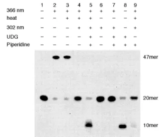
Fig. 3 Autoradiogram of a denaturing polyacrylamide gel electrophoresis of site-specific cytosine to the uracil mutation of
CV
U-containing ODN 6 (7 µM) and 32P-5'-end-labeled ODN 5 (1 µM) in a sodium cacodylate buffer (50 mM, pH 7.0) containing NaCl (100 mM). Lane 1, before reaction; lane 2, ODN 5 + ODN 6, irradiation at 366 nm, 3 h on ice; lane 3, incubation at 90 °C for 2 h of lane 2; lane 4, irradiation at 302 nm, 1 h at 40 °C of lane 3; lane 5, UDG treatment at 37 °C for 1 h followed by incubation with piperidine at 90 °C for 20 min of lane 4; lane 6, irradiation at 302 nm, 1 h of lane 2; lane 7, UDG treatment followed by piperidine treatment of lane 6 under the same conditions as lane 5; lane 8, treatment of ODN 8 with UDG and piperidine under the same conditions as lane 5; lane 9, incubation with piperidine at 90 °C for 20 min of lane 4.
We further demonstrate that deamination coupled with CV U-mediated photobranching can be carried out when ODN containing two consecutive cytosines is used as a target sequence. When 32 P-5'-end-labeled 20mer 5’-d(TGGCTACGAGCCAACAACAA)
(ODN 5) and 27mer
5’-d(CVUGCGTGTTTTCACGCAGCTCGTA GCCA) (ODN 6) were
irradiated at 366 nm for 3 h, the expected ligated 47mer ODN 7 was produced in 80% yield as determined by densitometric assay of PAGE (Fig. 3, lane 2). After heating at 90 °C for 2 h (Fig. 3, lane 3), quantitative photosplitting was achieved by 302-nm irradiation for 1 h (Fig. 3, lane 4). A cleavage band was observed by piperidine treatment after enzymatic base excision using UDG after photosplitting (Fig. 3, lane 5). The mobility of this band was equal to the band that was produced after the strand cleavage at the abasic sites produced by UDG treatment of authentic deoxyuridine-containing ODN 8 (Fig. 3, lane 8). This cleaved band was not observed when heating of photoligated ODN 7 at 90 °C was not carried out (Fig. 3, lane 6) or when piperidine treatment was carried
out without UDG treatment after photosplitting (Fig. 3, lane 9). These results clearly indicated that the cleaved band seen in lane 5 was caused by the transformation from cytosine to uracil at the target site via deamination of C<>CVU photoadduct by heating at
90 °C and photoplitting at 302 nm. It was also found that an efficient transformation from cytosine to uracil occurred at the target cytosine at eleventh position with site-selectivity. These deaminations of Cat eleventh position and C at twelfth position were 88% and 12% yield, respectively.
The heat-induced deamination of the cytosine residue of a cyclobutane dimer followed by splitting of the photolyase7,8,12 or
photoreactivation2,4,11 has thus far been known as a method for converting cytosine to uracil. However, these methods are not site-specific. Our deamination method using reversible DNA photoligation has high sequence selectivity and efficiency at the target cytosine compared with other methods for converting cytosine to uracil using enzymatic or chemical procedures.5,7,11
In summary, we have demonstrated that deamination coupled with CVU-mediated photobranching causes the heat-induced
transition of cytosine to uracil with high efficiency without any side reaction. The present site-specific C to U transition can be widely used for the site-specific mutation of DNA. This method may also find application in mutation not only for DNA but also for messenger RNA.
Kenzo Fujimoto,*ab Shigeo Matsuda,c Yoshinaga Yoshimura,b
Takashi Matsumura,a Masayuki Hayashid and Isao Saitoe
aSchool of Materials Science, Japan Advanced Institute of Science and
Technology, Ishikawa 923-1292, Japan. Fax: +81 761 51 1671; Tel: +81 761 51 1671; E-mail: [email protected]
bPRESTO, Japan Science and Technology Agency, Kawaguchi 332-0012,
Japan
cCurrent address: Department of Chemistry, The Scripps Research
Institute, La Jolla, CA 92037, USA
dDepartment of Synthetic Chemistry and Biological Chemistry, Faculty of
Engineering, Kyoto University, Kyoto 606-8501, Japan
eCurrent address: NEWCAT Institute, School of Engineering, Nihon
University, Koriyama 963-8642, Japan
Notes and references
1 E. C. Friedberg, G. C. Waker and W. Siede, DNA Repair and
Mutagenesis, ASM Press, Washington, D. C., 1995.
2 R. B. Setlow, W. L. Carrier and F. J. Bollum, Proc. Natl. Acad. Sci,
U.S.A., 1965, 53, 1111.
3 B. K. Duncan and J. H. Miller, Nature, 1980, 287, 560.
4 D. F. Fix and R. Bockrath, Mol. Gen. Genet., 1981, 182, 7; D. F. Fix,
Mol. Gen. Genet., 1986, 204, 452; M. Ruiz-Rubio and B. Bockrath, Mutat. Res., 1989, 210, 93; I. Tessman, S. K. Liu and M. A. Kennedy, Proc. Natl. Acad. Sci, U.S.A., 1992, 89, 1159; N. Jiang and J. S.
Taylor, Biochemistry, 1993, 32, 472.
5 T. Lindahl and B. Nyberg, Biochemistry, 1974, 13, 3405.
6 R. Y. H. Wang, K. C. Kuo, C. W. Gehrke, L. H. Huang and M. Ehrlich, Biochim. Biophys Acta, 1982, 697, 371.
7 L. A. Frederico, T. A. Kunkel and B. R. Shaw, Biochemistry, 1990,
29, 2532; L. A. Frederico, T. A. Kunkel and B. R. Shaw,
Biochemistry, 1993, 32, 6523; L. A. Frederico, T. A. Kunkel and B.
R. Shaw, Biochemistry, 1993, 32, 3535; H, Chen and B. R. Shaw,
Biochemistry, 1994, 33, 4121; W. Peng and B. R. Shaw, Biochemistry, 1996, 35, 10172.
8 I. Tessman and M. A. Kennedy, Mol. Gen. Genet., 1991, 227, 144; I. Tessman, Photochem. Photobiol., 1992, 55, 80S; I. Tessman,
Photochem. Photobiol., 1992, 55, 83S; I. Tessman, M. A. Kennedy
and S. K. Liu, J. Mol. Biol., 1994, 235, 807.
9 T. Douki and J. Cadet, J. Photochem. Photobiol. B: Biol., 1992, 15, 199.
10 P. V. Hariharan and H. E. Johns, Photochem. Photobiol., 1968, 8, 11; D. G. Lemaire and B. P. Ruzsicska, Biochemistry, 1993, 32, 2525. 11 Y. Barak, O. Cohen-Fix and Z. Livneh, J. Biol. Chem., 1995, 270,
24174.
12 Y. Tu, R. Dammann and G. P. Pfeifer, J. Mol. Biol., 1998, 284, 297.
13 M. M. Ali, M. R. Alam, T. Kawasaki, S. Nakayama, F. Nagatsugi and S. Sasaki, J. Am. Chem. Soc., 2004, 126. 8864.
14 K. Fujimoto, S. Matsuda, N. Takahashi and I. Saito, J. Am. Chem.
Soc., 2000, 122. 5646; K. Fujimoto, N. Ogawa, M. Hayashi, S.
Matsuda and I. Saito, Tetrahedron Lett., 2000, 41, 9437; I. Saito, Y. Miyauchi, Y. Saito and K. Fujimoto, Tetrahedron Lett., 2005, 46, 97; Y. Yoshimura, Y. Ito and K. Fujimoto, Bioorg. Med. Chem. Lett., 2005, 15, 1299; K. Fujimoto, Y. Yoshimura, T. Ikemoto, A. Nakazawa, M. Hayashi and I. Saito, Chem. Commun., 2005, 3177; M. Ogino, Y. Yoshimura, A. Nakazawa, I. Saito and K. Fujimoto, Org.
Lett., 2005, 7, 2853; S. Ogasawara and K. Fujimoto, ChemBioChem,
2005, 6, 1756; Y. Yoshimura, Y. Noguchi, H. Sato and K. Fujimoto,
ChemBioChem, 2006, 7, 598.
15 Incorporation of CVU into oligonucleotides was confirmed by
enzymatic digestion and MALDI–TOF–MS analysis. MALDI–TOF– MS: calcd. for ODN 2 (C206H259N75O128P20) [(M–H)–] 6452.25; found
6452.60.
16 J. Sambrook, E. F. Fritsch and T. Maniatis, Molecular Cloning: A
Laboratory Manual, 2nd Ed., Cold Spring Harbor Laboratory Press:
New York, 1989.
17 The rate constant (k) for deamination of C<>CVU photoadduct in the
ligation site can be calculated from equation (1) where x is the fraction of cytosine converted into uracil which is determined from the intensities of the cleaved bands and t is time of incubation. k = –ln(1–x)/t (1)