M alignant Pleural Effusion and Osteoblastic M etastases
as the Initial M anifestation of Occult Gastric Cancer:
A Case Report
Yuji Shimizu,
Koji Kurosawa,
Manabu Ueno,
Junichi Nakagawa,
Toshitaka Maeno,
Tatsuo Suga,
Mitsuru Motegi,
Norio Kanesawa
and Masahiko Kurabayashi
A 53-year-old man was admitted to our hospital because of lumbago. Gastrointestinal endoscopic examination performed on admission did not reveal any gross gastric abnormalities. Lumbar radiogra-phy and bone scintigraradiogra-phy revealed multiple bone osteoblastic changes. Chest radiograradiogra-phy showed right pleural effusion. The findings of the chest computed tomography and cytological examination of the pleural effusion were strongly suggestive of lung cancer. The patient was refractory to chemotherapy, and he died of cancer and disseminated intravascular coagulation. The autopsy revealed absence of primary lung cancer and the presence of tumor emboli in the right lung field. Swollen perigastric lymph nodes, tiny signet-ring cell carcinoma at the posterior wall of the stomach corpus, and severe vascular invasion were also observed at autopsy.
Therefore,signet ring cell carcinoma of the stomach should be considered as a possible diagnosis in early-stage gastric cancer patients who develop osteoblastic metastasis and pleural effusion.(Kitakanto Med J 2011;61:515∼518)
Key words: Signet-ring cell carcinoma, Gastric cancer, Osteoblastic bone metastasis, Pleural effusion
Introduction
Metastasis of primary gastric carcinoma to the bone is a rare phenomenon and has been observed in only 1-11% of the gastric carcinoma patients. Bone metastases are classified as osteolytic or osteoblastic. The histological findings for bone metastasis show a predominance of osteolytic changes. Prostate cancer is the most frequent cause of osteoblastic metastasis in men. However, previous studies have reported recur-rent gastric cancer associated with osteoblastic bone metastasis after gastrectomy of poorly differentiated carcinoma. Moreover, several studies have reported cases of osteoblastic bone metastasis that is secondary to signet-ring cell carcinoma of the stomach. Although some studies have reported that malignant pleural effusion occurs as a manifestation of advanced
gastric adenocarcinoma,this effusion is rarely observed even at the advanced stage of gastric cancer. We present a case of occult gastric cancer that initially presented as manifestation of pleural effusion and osteoblastic bone metastases.
Case report
A 53-year-old man with lumbago, appetite loss, and body weight loss (loss of 4 kg in 5 months) was referred to our hospital. His Brinkmann Index was 460 (20 cigarettes/day×23 years). Lumbar radiogra-phy showed osteoblastic metastases in the spine (Fig. 1A), and bone scintigraphy revealed multiple abnor-mal uptakes at the ribs and spines (Fig. 1B). The chest radiograph (Fig.1C)and computed tomography (Fig.1D)image revealed nodular and patchy shadows in the right middle lobe at sites distant from the pleura.
515 Kitakanto Med J
2011;61:515∼518
1 Department of Respiratory Medicine, National Hospital Organization Takasaki General Medical Center, 36 Takamatsu-cho, Takasaki, Gunma 370-0829, Japan 2 Department of Medicine and Biological Science, Gunma University Graduate School of Medicine, 3-39-22 Showa-machi, Maebashi, Gunma 371-8511, Japan
Received : August 22, 2011
Address: YUJI SHIMIZU Department of Respiratory Medicine,National Hospital Organization Takasaki General Medical Center, 36 Takamatsu-cho, Takasaki, Gunma 370-0829, Japan
The findings of the cytological examination of the right pleural effusion were indicative of adenocar-cinoma (class V). Laboratory examination showed markedly elevated levels of serum alkaline phosphatase (4,200IU/l, isozyme III dominant). The level of car-cinoembryonic antigen in the serum was within the normal limits(1.7ng/ml),but was remarkably elevated (1,630ng/ml) in the pleural effusion. The patients prostate was normal, and the serum prostate-specific antigen level was not elevated.
Gastrointestinal endoscopy was performed, but there was no endoscopic appearance of the stomach mucosa. Other than the nodular and patchy shadows
at the right upper lung lobe, no primary lesions capa-ble of causing the malignant right pleural effusion were detected ; therefore we diagnosed primary lung cancer. Subsequently, the patient was administered systemic anti-cancer chemotherapy (cisplatin (60mg/m ) plus docetaxel(60mg/m ),at Day 1). However,the disease followed a progressive course even after 1 course of chemotherapy, and 45 days after the induction of chemotherapy, the patient died of cancer and dis-seminated intravascular coagulation.
An autopsy revealed the absence of primary lung cancer in the lung field. Slightly swollen perigastric lymph nodes were the notable findings at the autopsy.
Osteoblastic metastases in occult gastric cancer
Fig. 1A Fig. 1B
Fig. 1C
Fig. 1D Fig. 1 Spine and pulmonary disorder
Lateral lumbar radiograph (Fig. 1A) and bone scintigram (Fig. 1B) indicate multiple osteoblastic bone metastases. Chest radiograph (Fig. 1C) and chest computed tomography (Fig. 1D) image revealed primary lesions causing malignant pleural effusion in the right middle lobe at sites distant from the pleura.
Fig. 2A
Fig. 2B
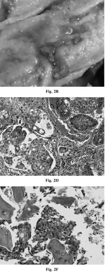
Fig. 2C Fig. 2D
Fig. 2F Fig. 2E
Fig. 2 Pathology of the stomach and spine tissues obtained at autopsy
2A : Macroscopic appearance of the stomach obtained at autopsy,which was suggestive of occult change of cancer. However, hypermagnitude of erosive change (Fig. 2B) was also difficult to diagnose as cancer.
2C : Histology of the gastric mucosa from the stomach corpus at autopsy indicated signet-ring cell carcinoma (original magnification ×200)
2D : Microscopic examination of the lung showed apparent vascular embolism (original magnification ×200). 2E : Macroscopic examination of the spine at autopsy showed osteoblastic metastases in the lumbar spines. 2F : Cancer cells derived from signet ring cell carcinoma were detected in the spine(original magnification ×400)
Macroscopic examination revealed tiny erosive changes at the posterior wall of the stomach corpus (Fig. 2A & 2B). The results of a comprehensive microscopic examination indicated signet-ring cell carcinoma with remarkable vascular invasion (Fig. 2C).
In the lung, the vascular systems showed marked invasion by tumor cells,and were partially filled with tumor emboli. Histological examination revealed that the embolus in the right lung was derived from gastric cancer (Fig. 2D), and was responsible for malignant right pleural effusion. The vertebra showed osteoblastic changes instead of osteolytic changes (Fig. 2E), and signet-ring cell carcinoma was also detected in the spine (Fig. 2F).
In the light of the above mentioned findings, we proposed that occult gastric cancer cells may have directly invaded the gastric vascular system in this patient, thereby inducing osteoblastic metastases and tumor embolism in the right lung and causing pleural effusion.
Discussion
There are 2 interesting aspects in this case: osteoblastic bone metastases and right pleural effusion occurs as the initial manifestation of occult gastric carcinoma, which was histologically diagnosed as signet-ring cell carcinoma. Although a case of occult gastric cancer,in which the histological diagnosis was signet-ring cell carcinoma, has been reported, the obvious bone metastasis and pleural effusion in this case were not observed. In a previous study, 4 patients who developed malignant right pleural effu-sion were diagnosed with gastric adenocarcinoma. However, in our case, the gastrointestinal endoscopic examinations performed on admission showed no gross abnormalities.
Metastasis of gastric cancer to the bone is rarer than that of breast,lung and prostate cancers. Several cases of gastric cancer with osteoblastic bone metas-tasis have been reported, but the endoscopic findings in these cases indicated Borrmann type IV advanced gastric cancer. Kobayashi et al reported a case of recurrent multiple bone metastases after complete resection of signet-ring cell carcinoma of the stomach. They concluded that cancer cells probably invaded the small venule in the mucosa at operation. They also referred to several autopsy reports of mucosal or sub-mucosal signet-ring cell carcinoma of the stomach ; the reports had recorded findings of bone metastasis. Lehnert et al investigated the blood and lymph capil-laries of the human gastric mucosa,and speculated that the blood-borne metastasis in recurrent gastric cancer
may be associated with the rich vascularity of the gastric mucosa. In our case, osteoblastic bone metas-tasis was secondary to occult gastric cancer. We spec-ulate that the cancer cells may have invaded the vascu-lar system at the early stage of gastric cancer,as shown in Fig. 2C, thereby inducing bone matastases.
The mechanism of osteoblastic metastasis and the factors involved in this process are unknown. A continuous circle of events may be involved in osteob-lastic metastasis in which the tumors induce osteoblas-tic activity to release growth factors that increase the tumor progression. Endothelin-1, platelet-derived growth factor, and urokinase may also be involved in this process.
In conclusion, we report a rare case of right pleural effusion and osteoblastic metastases as initial manifestations of occult gastric cancer, which was definitely diagnosed at autopsy. Signet-ring cell car-cinoma of the stomach should be considered as a possible diagnosis in cases of pleural effusion and osteoblastic metastases,even if advanced gastric cancer is not detected.
References
1. Yoshikawa K, Kitaoka H. Bone metastasis of gastric can-cer. Jpn J Surg. 1983; 13: 173-176.
2. Roodman GD. Mechanisms of bone metastasis. N Engl J Med. 2004; 350: 1655-1664.
3. Nakanishi H, Araki N, Kuratsu S, et al. Skeletal metas-tasis in patients with gastric cancer. Clin Orthop. 2004; 423: 208-212.
4. Kobayashi M, Araki K, Matsuura K, et al. Early gastric cancer giving rise to bone and brain metastases-a review of the Japanese literature. Hepatogastroenterology 2002; 49 : 1751-1754.
5. Roussos A,Patsopoulos D,Philippou N. Pleural effusion as the initial manifestation of gastric adenocarcinoma: a report of four cases. Scand J Gastroenterol. 2001; 36: 784.
6. Uchida T,Shikata T,Shimizu SI,et al. Gonadotropin and alkaline phosphatase producing occult gastric carcinoma with widespread metastasis of generalized bone. Cancer. 1981; 48: 140-150.
7. Chung YS, Choi TY, Ha CY, et al. An unusual case of osteoblastic metastasis from gastric carcinoma. Yonsei Med J. 2002; 43: 377-380.
8. Lehnert T, Erlandson RA, Decosse JJ. Lymph and blood capillaries of the human gastric mucosa. A morphologic basis for metastasis in early gastric carcinoma. Gas-troenterology. 1985; 89 : 939-950.
9. Nelson JB,Hedican SP,George DJ,et al. Identification of endothelin-1 in the pathophysiology of metastatic adenocar-cinoma of the prostate. Nat Med. 1995; 1: 944-949. 10. Yi B, Williams PJ, Niewolna M, et al. Tumor-derived
platelet-derived growth factor-BB plays a critical role in osteosclerotic bone metastasis in an animal model of human breast cancer. Cancer Res. 2002; 62: 917-923.