Cyclin-dependent kinase-activating kinases CDKD;1 and CDKD;3 are essential for preserving mitotic activity in Arabidopsis thaliana
49
0
0
全文
(2) Cyclin-dependent kinase-activating kinases CDKD;1 and CDKD;3 are essential for preserving mitotic activity in Arabidopsis thaliana. Hirotomo Takatsuka1, Chikage Umeda-Hara1 and Masaaki Umeda1,2 1. Graduate School of Biological Sciences, Nara Institute of Science and Technology,. 8916-5 Takayama, Ikoma, Nara 630-0192, Japan; 2JST, CREST, 8916-5 Takayama, Ikoma, Nara 630-0192, Japan. For correspondence: Masaaki Umeda (Graduate School of Biological Sciences, Nara Institute of Science and Technology, 8916-5 Takayama, Ikoma, Nara 630-0192, Japan; Tel. +81-743-72-5592; Fax +81-743-72-5599; E-mail: [email protected]) Authors’ e-mail addresses: Hirotomo Takatsuka ([email protected]), [email protected], Masaaki Umeda ([email protected]). Running title: Essential CDK-activating kinases in Arabidopsis Keywords: Cell cycle, cyclin-dependent kinase, CDK-activating kinase, gametogenesis, Arabidopsis thaliana. Word count: 6051 (234 for Summary, 1068 for Introduction, 2103 for Results, 1327 for Discussion, 707 for Experimental Procedures, 20 for Acknowledgements, 688 for Figure Legends), and 1800 for References. 1.
(3) Summary For the full activation of cyclin-dependent kinases (CDKs), not only cyclin binding but also CDK phosphorylation is required. This activating phosphorylation is mediated by CDK-activating kinases (CAKs). Arabidopsis has four genes showing similarity to vertebrate-type CAKs, three CDKDs (CDKD;1–CDKD;3) and one CDKF (CDKF;1). We previously found that the cdkf;1 mutant is defective in post-embryonic development even though the kinase activities of core CDKs remain unchanged relative to the wild-type. This raised a question about the involvement of CDKDs in CDK activation in planta. Here we report that the cdkd;1 cdkd;3 double mutant showed gametophytic lethality. Most cdkd;1-1 cdkd;3-1 pollen grains were defective in pollen mitosis I and II, producing onecell or two-cell pollen grains that lacked fertilization ability. We also found that the double knockout of CDKD;1 and CDKD;3 caused arrest and/or delay in the progression of female gametogenesis at multiple steps. Our genetic analyses revealed that the functions of CDKF;1 and CDKD;1 or CDKD;3 do not overlap, either during gametophyte and embryo development or in post-embryonic development. Consistent with these analyses, CDKF;1 expression in the cdkd;1-1 cdkd;3-1 mutant could not rescue the gametophytic lethality. These results suggest that, in Arabidopsis, CDKD;1 and CDKD;3 function as CAKs controlling mitosis, whereas CDKF;1 plays a distinct role mainly in postembryonic development. We propose that CDKD;1 and CDKD;3 phosphorylate and activate all core CDKs, CDKA, CDKB1 and CDKB2, thereby governing cell cycle progression throughout plant development.. 2.
(4) Introduction Double fertilization is one of the unique features of angiosperms. Two sperm cells are delivered through the pollen tube to the embryo sac; one fuses with the egg cell and generates the zygote, whereas the other fuses with the central cell, giving rise to the endosperm, which is required for embryo development and germination (for a review, see Dumas and Rogowsky, 2008). Precise control of cell division is prerequisite to proper development of both embryo and endosperm. Gametophytes are produced through controlled mitotic divisions. In Arabidopsis, sporogenous initial cells, also called microspore or megaspore mother cells, undergo meiosis to produce a tetrad of microspores in the anther or four megaspores in the ovule (Liu and Qu, 2008). Microspores released from a tetrad by the action of callase further undergo an asymmetric cell division (pollen mitosis I, PM I) to form a bicellular pollen grain carrying two cells with distinct fates: the larger vegetative cell later forms a pollen tube nucleus, while the smaller generative cell undergoes a symmetric division (pollen mitosis II, PM II) to produce two sperm cells (Figure S1) (McCormick, 1993; 2004). The resultant three-celled pollen grain constitutes the male gametophyte. On the other hand, at the onset of female gametophyte development, only one of the four megaspores survives and undergoes three rounds of nuclear division and subsequent cellularization (Yang and Sundaresan, 2000; Drews and Yadegari, 2002). In Arabidopsis, megagametogenesis is divided into seven distinct stages, FG1 to FG7 (Figure S2) (Misra, 1962; Poliakova, 1964; Webb and Gunning, 1990; Mansfield et al., 1991; Murgia et al., 1993; Webb and Gunning, 1994; Schneitz et al., 1995; Christensen et al., 1997). First, the surviving megaspore undergoes mitosis to produce a binucleate cell (FG1 to FG2). Shortly thereafter, the two nuclei separate to the chalazal and micropylar poles, and a 3.
(5) vacuole is formed at the center (FG3). Two subsequent rounds of mitosis produce an eight-nucleate cell with four nuclei at each pole (FG4 to FG5). One nucleus from each pole (polar nuclei) then migrates toward the center, followed by cellularization that localizes three cells at each pole (FG5). The polar nuclei eventually fuse to produce the central cell (FG6), and finally the three antipodal cells undergo cell death, generating the female gametophyte comprising one haploid egg cell, two haploid synergid cells, and one diploid central cell (FG7) (Figure S2). Cell division is regulated by the activity of cyclin-dependent kinases (CDKs). Based on amino acid sequence similarities, plant CDKs have been classified into eight types, CDKA to CDKG and the CDK-like kinase (CKL) (Joubés et al., 2000; Vandepoele et al., 2002; Takahashi and Umeda, 2014). Among them, CDKA and CDKB have a primary function in cell cycle control. CDKA is inferred as a functional ortholog of yeast CDC2/CDC28 and mammalian CDK1, and its activity is required for both G1-to-S and G2-to-M transitions. CDKB is a plant-specific CDK with two subtypes, CDKB1 and CDKB2, and its expression is restricted to the late S-to-M phase. Indeed, previous reports demonstrated that CDKB is involved in G2-to-M progression and suppresses endoreplication, in which cells replicate their chromosomes without mitosis (Boudolf et al., 2009). In Arabidopsis, the cdka;1 knockout mutant undergoes PM I but not PM II during male gametogenesis, producing bicellular pollen grains with a single sperm-like cell and one vegetative cell (Iwakawa et al., 2006; Nowack et al., 2006). This suggests that, in cdka;1, CDKB has some activity in G1-to-S progression. Whereas cdka;1 exhibits no obvious defects in megagametogenesis, the combination with knockout mutations in the two CDKB1 genes caused lethality in both female and male gametogenesis (Nowack et al., 2012), indicating an overlapping function between A- and B-type CDKs during 4.
(6) female gametogenesis. For full activation of CDKs, phosphorylation as well as cyclin binding is required; this phosphorylation is carried out at the conserved threonine residue within the T-loop by a CDK-activating kinase (CAK) (Umeda et al., 2005; Fisher, 2005; Harashima et al., 2007; Dissmeyer et al., 2007). Plants possess two types of CAKs, CDKD and CDKF, both of which exhibit CDK-activating kinase activities in vitro and in yeast cells (Umeda et al., 1998; Yamaguchi et al., 1998; Chao et al., 2007). CDKD is closely related to vertebrate-type CAKs in terms of amino acid sequence similarity and enzyme activity; Arabidopsis has three CDKDs (CDKD;1–CDKD;3), among which CDKD;2 and CDKD;3 display kinase activities toward not only CDKs but also the carboxy-terminal domain (CTD) of the largest subunit of RNA polymerase II, suggesting that, like vertebrate-type CAKs, CDKD controls both CDK activation and basal transcription. The CDK-kinase activity of CDKD;3 is higher than that of CDKD;2, while CDKD;2 has higher CTD-kinase activity than CDKD;3 (Shimotohno et al., 2003). CDKD;1 expressed in insect cells exhibits neither CDK- nor CTD-kinase activity (Shimotohno et al., 2003). On the other hand, Arabidopsis has one CDKF, CDKF;1, which shows a lower similarity to vertebrate-type CAKs than CDKDs and exhibits kinase activity on CDK but not CTD (Shimotohno et al., 2004). A striking feature of CDKF;1 is that it phosphorylates and activates CDKD;2 and CDKD;3, thus functioning as a CAK-activating kinase (CAKAK) (Shimotohno et al., 2004; Umeda et al., 2005). However, in the cdkf;1 knockout mutant, the kinase activities of CDKA, CDKB and CDKDs except CDKD;2 were the same as those in the wild-type (Takatsuka et al., 2009). Moreover, the cdkf;1 mutant has fewer and smaller cells in leaves while the cdka;1 mutant has fewer but larger cells (Dissmeyer et al., 2007), suggesting that CDKF;1 does not play a major role in CDK activation in 5.
(7) planta. Rather, in cdkf;1, the protein level and kinase activity of CDKD;2 were significantly reduced, indicating that CDKF;1 controls basal transcription by maintaining the steady-state level of CDKD;2 (Takatsuka et al., 2009). As described above, CDKA and CDKB are involved in gametophyte development, while the cdkf;1 mutant displays severe growth defects in post-embryonic development, but no obvious defect in seed development or germination (Takatsuka et al., 2009). This suggests that CDKD plays a major role in CDK activation during gametogenesis and embryogenesis. Here we investigated the phenotype of cdkd knockout mutants, and found that the cdkd;1 cdkd;3 double mutant was gametophyte-lethal. This lethality was derived from impaired mitosis during both male and female gametogenesis. Our data showed that CDKF;1, which is dispensable in gametogenesis and embryogenesis, could not substitute for CDKD;3. We propose that CDKD;1 and CDKD;3 are the principal regulators of CDK activation, and are thus essential for cell cycle regulation throughout plant development.. Results Expression analysis of CAKs in gametophytes and embryos To investigate the role of CAKs during gametogenesis and embryogenesis, we first observed their expression patterns using 2-kb promoter regions fused to the glucuronidase (GUS) reporter gene. However, we could see no GUS signal for any of the CAKs, probably because the promoter regions lacked regulatory elements required for transcriptional activation. We therefore generated translational fusion constructs including the 2-kb promoter and full-length coding regions that were fused in-frame to GUS. As we reported previously, strong GUS signals were observed in young flowers of pCDKF;1–CDKF;1::GUS, while in mature flowers the GUS expression was mostly 6.
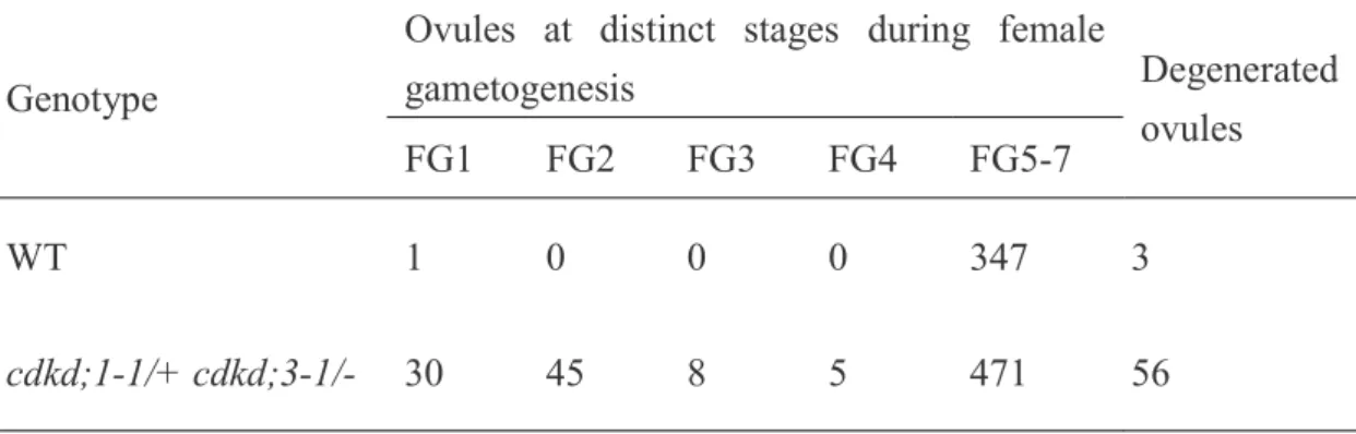
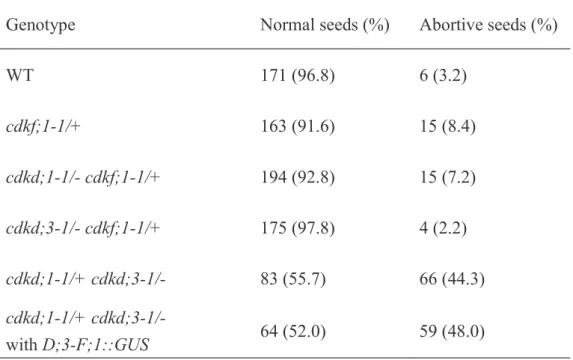
(8) restricted to stamen filaments and pedicels (Figure 1a) (Takatsuka et al., 2009). In pCDKD;1–CDKD;1::GUS and pCDKD;2–CDKD;2::GUS, GUS signals were detected in young flowers and in pistils of mature flowers; pedicels also showed signals for CDKD;2. pCDKD;3–CDKD;3::GUS displayed the same expression pattern as pCDKD;2–CDKD;2::GUS, but with a much higher expression level (Figure 1a). We next observed expression in gametophytes. GUS signals for CDKD;2 and CDKD;3 were localized at the center of the pollen grain (Figure 1b), which probably represents an assemblage, called the male germ unit, consisting of the vegetative cell nucleus and two sperm cells (Lalanne and Twell, 2002). pCDKD;1–CDKD;1::GUS and pCDKF;1–CDKF;1::GUS showed GUS signals in whole pollen grains, with the strongest signal in the former being detected in the male germ unit (Figure 1b). Expression in ovules was observed for the three CDKDs but not for CDKF;1 (Figure 1c), indicating that CDKF:1 is dispensable for female gametogenesis. The expression level of CDKF;1 was very low throughout embryogenesis; at the late embryo stage, GUS signal was detected only in the hypocotyl, in which cells are not actively dividing (Figure 2a) (Gendreau et al., 1997). This suggests that CDKF;1 is not principally involved in cell cycle regulation during embryogenesis, which is consistent with our previous observation that the cdkf;1 mutant exhibits only post-embryonic defects (Takatsuka et al., 2009). Whereas the expression level of CDKD;2 was very low at the globular and torpedo stages, pCDKD;1–CDKD;1::GUS and pCDKD;3–CDKD;3::GUS showed relatively higher GUS signals during early embryogenesis, and in dividing tissues, such as root and shoot meristems and vascular bundles, at the later stage (Figure 2a and b). These results suggest that CDKD;1 and CDKD;3 are engaged in the control of cell division during embryogenesis. During endosperm development, nuclear divisions occur 7.
(9) at the globular stage in the absence of cellularization (Berger, 1999); we also observed GUS signals in the endosperm at the globular stage. As shown in Figure 2c, CDKD;1 and CDKD;3 showed GUS expression in the endosperm, with higher signals for CDKD;3, whereas we could not detect any such signal for CDKD;2 or CDKF;1. These results suggest that CDKD;1 and CDKD;3 participate in endosperm development as well as embryogenesis.. cdkd;1 and cdkd;3 double mutation is gametophyte-lethal We previously showed that CDKD;2 has a lower CDK-kinase activity than CDKD;3 in vitro (Shimotohno et al., 2004). Moreover, in the cdkf;1 mutant, protein levels and kinase activities of CDKA;1 and CDKBs did not decrease relative to the wild-type although the steady-state level of CDKD;2 protein was reduced (Takatsuka et al., 2009). These results suggested that CDKD;2 does not have a major role in CDK activation. On the other hand, CDKD;1 produced in insect cells did not display CDK or CTD kinase activities (Shimotohno et al., 2003), but, as described above, CDKD;1 is highly expressed during gametogenesis and embryogenesis. Hence, in this study, we focused on in vivo functions of CDKD;1 and CDKD;3. We previously reported that single-knockout mutants of CDKD;1 and CDKD;3 (Figure 3a) exhibited no obvious phenotype (Shimotohno et al., 2006). However, no progeny with both mutations was obtained by crossing, and we therefore observed siliques of plants with heterozygous cdkd;1-1 and homozygous cdkd;3-1 mutations (cdkd;1-1/+ cdkd;3-1/-). We found empty spaces in an alignment of mature seeds (Figure 3b); about 50% of the seeds were abortive and did not form embryolike structures (Table 1 and Figure 3c). This phenotype was complemented by expression of CDKD;3 under its own promoter (Figure 3b and Table 1). Since half of the seeds were 8.
(10) defective in cdkd;1-1/+ cdkd;3-1/- plants, we inferred that cdkd;1-1 and cdkd;3-1 double mutation was lethal in the gametophyte, but not in the embryo. After double fertilization, at least three rounds of nuclear divisions normally occur in the wild-type endosperm before the first zygotic division; the zygote then starts cell division to form an embryo, while the endosperm nuclei continue to proliferate (Figure 3d). However, in young siliques of cdkd;1-1/+ cdkd;3-1/- plants, we found unfertilized ovules that were arrested at early stages of female gametogenesis, such as FG1 and FG2, and at FG7 (Figure 3e). This indicates that CDKD;1 and CDKD;3 are required for female gametophyte development. We also observed seeds containing a oneor two-cell-stage embryo with a reduced number of endosperm nuclei, but at very low frequencies (Figure 3f). To further investigate the observed lethality, we conducted reciprocal crosses between the wild-type and cdkd;1-1/+ cdkd;3-1/- (Table 2). As technical controls, pistils of wild-type and cdkd;1-1/+ cdkd;3-1/- plants were pollinated with pollen from wild-type and cdkd;1-1/+ cdkd;3-1/- plants, respectively. When wild-type pistils were pollinated with pollen from cdkd;1-1/+ cdkd;3-1/- plants, 33.8% of seeds were aborted. Similarly, 39.9% of seeds were aborted in siliques of cdkd;1-1/+ cdkd;3-1/- plants pollinated with wild-type pollen. These results indicate that not all but a large percentage of male and female gametophytes fail to survive when they lack both CDKD;1 and CDKD;3.. cdkd;1 and cdkd;3 mutations impair pollen mitosis To test pollen viability, we conducted Alexander staining, which can distinguish viable and nonviable pollen grains. Pollen grains from cdkd;1 or cdkd;3 knockout mutants were stained similarly to those from the wild-type, and most pollen grains from cdkd;1-1/+ 9.
(11) cdkd;3-1/- plants were also normally stained and did not show any morphological defect (Figure S3). We then observed the cells in each pollen. Wild-type pollen grains contain three cells, a vegetative cell and two smaller sperm cells (Figure S1 and Figure 4a); indeed, DAPI staining visualized three nuclei in nearly 90% of wild-type pollen grains collected at floral stage 13, when the bud opens and anthesis occurs (Irish, 2010) (Table 3 and Figure 4b). However, at the same floral stage, only 56.7% of pollen grains from cdkd;11/+ cdkd;3-1/- plants possessed three nuclei; 23.6% contained two, 7.4% contained one nucleus, and 12.3% did not have any nucleus that was clearly stained with DAPI (Table 3, Figure 4a and b). This indicates that most cdkd;1-1 cdkd;3-1 pollen was defective in PM I and PM II, producing one-cell or two-cell pollen grains, while about 13% [(56.7 – 50) x 2 = 13.4%] of pollen grains with both cdkd;1-1 and cdkd;3-1 mutations completed two rounds of cell division. To examine whether cdkd;1-1 cdkd;3-1 pollen is able to fertilize, we observed transmission of the mutant alleles in wild-type pistils pollinated with pollen from cdkd;1-1/+ cdkd;3-1/- plants. The ratio of cdkd;1-1/- to cdkd;1-1/+ in F1 plants was 17.5% (Table S1), which is close to the percentage (13.4%) of three-cell pollen grains with both cdkd;1-1 and cdkd;3-1 mutations as described above. This suggests that cdkd;1-1 cdkd;3-1 pollen with three nuclei retains fertilization ability. As mentioned above, 33.8% of seeds in wild-type siliques pollinated with pollen from cdkd;1-1/+ cdkd;3-1/- plants were abortive (Table 2). Moreover, 38.0% of embryos were abortive at three days after pollination (DAP) in this crossing (n = 400), suggesting that cdkd;1-1 cdkd;3-1 pollen with no more than two cells lacks fertilization ability. To further examine the defect in fertility, we observed abortive seeds produced by crossing. In wild-type pistils pollinated with wild-type pollen, an 8- or 16-cell embryo with many endosperm nuclei was formed at 3 DAP (Figure 4c). By contrast, when wild-type pistils 10.
(12) were pollinated with pollen from cdkd;1-1/+ cdkd;3-1/- plants, 44% of the resultant abortive seeds contained one central cell and one egg cell, but no synergid cell, at 3 DAP (Figure 4c). It is known that one of the two synergid cells is physically disrupted by pollen penetration, and that the persisting one becomes difficult to identify at 2 DAP (Christensen et al., 2002). Therefore, it is probable that some of the cdkd;1-1 cdkd;3-1 pollen tubes were attracted by synergid cells, but that a sperm-like cell could not fuse with the central cell or the egg cell. On the other hand, 56% of the abortive seeds were degenerated and shrunken, and we could not find any cell structure in these seeds by microscopic observation. It has been reported that unfertilized ovules became degenerated (Chaudhury et al., 1997), as observed in wild-type siliques pollinated with pollen from cdkd;1-1/+ cdkd;3-1/- plants, suggesting that the above-mentioned degenerated phenotype of abortive seeds is the outcome of unsuccessful fertilization. These results support the idea that most of the cdkd;1-1 cdkd;3-1 pollen grains harboring no more than two cells lack fertilization ability.. Nuclear division in female gametogenesis is defective in cdkd;1 cdkd;3 ovules We next analyzed female gametophyte development in cdkd;1-1/+ cdkd;3-1/- plants. In wild-type plants, ovules had reached stages FG5 to FG7 when the flowers opened and the stamens nearly reached the pistils (Onodera et al., 2008) (Table 4 and Figure 5a). However, in cdkd;1-1/+ cdkd;3-1/-, some ovules were still at FG1 to FG4, and we found FG7 ovules having central cells with abnormal, shrunken nuclei (Table 4 and Figure 5b). When pistils of cdkd;1-1/+ cdkd;3-1/- plants were pollinated with wild-type pollen, resultant abortive seeds contained ovules at FG5 to FG7 (Table S2). Although we could not determine whether gametogenesis was delayed or arrested, these results indicate that CDKD;1 and 11.
(13) CDKD;3 are essential for nuclear division in ovules. We sometimes found seeds containing a two-cell stage embryo with only two endosperm nuclei in self-pollinated siliques of cdkd;1-1/+ cdkd;3-1/- plants as well as in cdkd;1-1/+ cdkd;3-1/- siliques pollinated with wild-type pollen (Figure 5c), but not in wild-type pistils pollinated with pollen from cdkd;1-1/+ cdkd;3-1/- plants. This indicates that such a phenotype is caused by defects on the female side, and that some cdkd;1-1 cdkd;3-1 ovules can undergo fertilization but do not proceed with nuclear divisions in the endosperm.. CDKF;1 is not genetically related to CDKD;1 or CDKD;3 Although the cdkf:1 knockout mutant exhibits defects only in post-embryonic development (Takatsuka et al., 2009), CDKF;1 is expressed in pollen grains and during embryogenesis, as described above. Therefore, to examine whether CDKF;1 has a redundant function with CDKDs during gametogenesis and/or embryogenesis, we generated double knockout mutants of CDKF;1 and either CDKD;1 or CDKD;3. While cdkd;1-1/- cdkf;1-1/+ and cdkd;3-1/- cdkf;1-1/+ seedlings grew normally, the double mutants of cdkd;1-1 cdkf;1-1 and cdkd;3-1 cdkf;1-1 showed growth inhibition similar to that observed in the cdkf;1-1 mutant (Figure 6a). In the root tip of cdkd;1-1 cdkf;1-1 and cdkd;3-1 cdkf;1-1, the meristem size was reduced and the number of columella cell layers decreased, as observed in cdkf;1-1 (Figure 6b and c). These results suggest that, although CDKF;1 is involved in post-embryonic development, its function is distinct from that of CDKD;1 or CDKD;3. Moreover, almost all seeds developed normally in siliques of cdkd;1-1/- cdkf;1-1/+ and cdkd;3-1/- cdkf;1-1/+ plants (Figure S4 and Table 5), implying that double knockout of CDKF;1 and CDKD;1 or CDKD;3 does not lead to defects in gametogenesis or embryogenesis as seen in mutants lacking both CDKD;1 and CDKD;3. 12.
(14) This suggests that the functions of CDKF;1 and CDKDs do not overlap either during gametophyte and embryo development or in post-embryonic development.. CDKF;1 cannot rescue the gametophyte lethality of the cdkd;1 cdkd;3 double mutant To further examine the functional divergence between CDKF;1 and CDKDs, we conducted a promoter-swapping experiment, in which the CDKF;1 coding region fused to GUS was expressed using the CDKD;3 promoter (D;3-F;1::GUS; Figure 7a). We first confirmed the functionality of the CDKF;1::GUS fusion protein by expressing GUS-fused CDKF;1 using the CDKF;1 promoter (F;1-F;1::GUS; Figure 7a); it fully rescued the growth defect of the cdkf;1-1 mutant (Figure 7b). We then examined the CDKD;3 promoter activity in D;3-F;1::GUS. While the promoter of CDKF;1, but not of CDKD;3, is active in mature columella cells (Takatsuka et al., 2009), the GUS signal of D;3F;1::GUS was absent in the columella (Figure 7c). Moreover, the GUS signal of D;3F;1::GUS was observed in ovules, where the promoter of CDKD;3, but not of CDKF;1, is active (Figure 7d and Figure 1c). These results indicate that D;3-F;1::GUS produces functional CDKF;1::GUS protein in tissues where exogenous CDKD;3 is expressed. Finally, we observed seed development in cdkd;1-1/+ cdkd;3-1/- plants with or without D;3-F;1::GUS. As shown in Table 5 and Figure 7e, almost the same percentage (44–48%) of seeds were abortive regardless of the expression of D;3-F;1::GUS, demonstrating that CDKF;1 cannot substitute for CDKD;1 or CDKD;3 during gametogenesis or embryogenesis.. Discussion GUS-fused CDKD;1 and CDKD;3 expressed under their own promoters accumulated in 13.
(15) pollen grains and ovules. We also found that the combination of cdkd;1-1 and cdkd;3-1 mutations disrupted pollen mitosis. Previous reports showed that pollen grains from the cdka;1 knockout mutant were defective in mitosis: they failed in PM II, but not PM I, producing one vegetative cell and one sperm-like cell (Iwakawa et al., 2006; Nowack et al., 2006). However, in cdkd;1-1/+ cdkd;3-1/- plants, we found pollen grains with only one vegetative cell, as well as others with one vegetative cell and one sperm-like cell, suggesting that pollen with both cdkd;1 and cdkd;3 mutations is defective in both PM I and PM II. Nowack et al. (2012) reported that pollen from the cdka;1/+ cdkb1;1/cdkb1;2/- mutant displayed early germline arrest, and that 3% of pollen grains were unicellular, indicating an impairment in PM I. Therefore, it is likely that CDKD;1 and CDKD;3 control PM I by activating CDKB1 as well as CDKA;1. Since 7.4% of pollen grains from cdkd;1-1/+ cdkd;3-1/- plants were unicellular (Table 3), the defect in PM I is apparently severer in cdkd;1-1 cdkd;3-1 than in cdka;1 cdkb1;1 cdkb1;2. Because plants have another type of CDK, CDKB2, that regulates G2-to-M progression (Menges et al., 2005), CDKD;1 and CDKD;3 may also control CDKB2 during pollen mitosis. Arabidopsis mutants with defects in pollen mitosis vary in their fertilization ability. Chen et al. (2008) reported that pollen grains with defects in MSI1, which encodes a factor associating with the core subunits of chromatin assembly factor 1 (CAF1) complex, are impaired in PM II. However, when wild-type ovules were pollinated with msi1 pollen, the sperm-like cell fused with either the egg cell or the central cell, generating seeds with either an embryo lacking endosperm or endosperm lacking an embryo. Another example is mutants of DUO1 and DUO3, which encode an R2R3-type MYB transcription factor and a protein related to a cell-lineage regulator in gonadogenesis in Caenorhabditis elegans, respectively (Brownfield et al., 2009a; 2009b). Both duo1 and 14.
(16) duo3 mutants produce a single sperm-like cell due to impaired pollen mitosis; however, fertilization never occurs with either the egg cell or the central cell because of defective differentiation of the sperm cell (Brownfield et al., 2009a; 2009b). When we pollinated wild-type pistils with pollen from cdkd;1-1/+ cdkd;3-1/- plants, we could not find any seeds with either embryo or endosperm, indicating that the single sperm-like cell produced in cdkd;1 cdkd;3 pollen grains does not successfully fuse with the egg cell or the central cell. Therefore, as reported for duo1 and duo3, the double knockout of CDKD;1 and CDKD;3 perturbs sperm cell differentiation during gametogenesis, causing failure in fertilization with both the egg cell and the central cell. Liu et al. (2008) demonstrated that dysfunctions of the RING-type E3 ubiquitin ligases RHF1a and RHF2a caused male and female gametophyte lethality due to the accumulation of Kip-related protein 6 (KRP6), an inhibitor of CDK-cyclin complexes. In the rhf1a rhf2a double mutant, development of female gametophytes was arrested at early stages, making a good contrast with the cdka;1 knockout mutant in which no defect in female gametogenesis is evident. This suggests that, in rhf1a rhf2a, accumulated KRP6 inhibits not only CDKA;1 but also other CDKs which have an essential role in female gametophyte development. This is consistent with the previous observation that triple mutations in CDKA;1, CDKB1;1 and CDKB1;2 caused defects in female gametogenesis; namely, ovules contained only one to four free nuclei, suggesting arrest of nuclear division before FG4 (Nowack et al., 2012). In this study, we revealed that the combination of cdkd;1 and cdkd;3 mutations also caused arrest and/or delay in the progression of female gametogenesis at multiple steps. Therefore, it is probable that CDKD;1 and CDKD;3 control both A- and B-type CDKs during female as well as male gametogenesis. During endosperm development, expression was detected for CDKD;1 and 15.
(17) CDKD;3, but not for CDKD;2 or CDKF;1 (Figure 2c) (Day et al., 2008). Siliques of cdkd;1-1/+ cdkd;3-1/- plants pollinated with wild-type pollen contained endosperm with fewer nuclei, and some central cells harbored distorted nuclei (Figure 5b). Proper development of the central cell is known to be essential for endosperm formation; for example, the deficiency of AGL61, which encodes a Type I MADS domain protein, leads to morphological defects of the central cell, causing a failure in endosperm development (Bemer et al., 2008; Steffen et al., 2008). It will therefore be of interest to examine whether the central cell is specified in cdkd;1 cdkd;3, and whether nuclear divisions are also impaired during endosperm development. Hajheidari et al. (2012) reported that cdkd;1 cdkd;2 and cdkd;2 cdkd;3 double mutants showed dwarfism in post-embryonic development, but did not describe any gametophytic defect. By contrast, we could not obtain seedlings with both cdkd;1 and cdkd;3 mutations. We found that mitosis in female and male gametogenesis was severely defective when cdkd;1 and cdkd;3 mutations were combined. Taken together, these data indicate that CDKD;1 and CDKD;3 are essential CDK-activating kinases during gametogenesis, and even though CDKD;2 has some function during gametogenesis, it plays a distinct role from CDKD;1 or CDKD;3. In addition, our promoter-swapping experiment revealed that CDKF;1 cannot substitute for CDKD;1 or CDKD;3. During post-embryonic development, CDKF;1 does not have a pivotal role in activating CDKA;1 or CDKBs, but is instead involved in stabilization of CDKD;2 (Takatsuka et al., 2009). Since CDKD;2 has a higher kinase activity toward CTD than CDK (Shimotohno et al., 2004), CDKF;1 may principally control basal transcription by stabilizing CDKD;2. These results are consistent with the above-mentioned idea that CDKD;2 (and CDKF;1) has a distinct function from CDKD;1 or CDKD;3. We propose that, in Arabidopsis, CDKD;1 16.
(18) and CDKD;3 are the CAKs that phosphorylate and activate core CDKs controlling the cell cycle. We previously reported that CDKD;1 expressed in insect cells exhibits neither CDK- nor CTD-kinase activity (Shimotohno et al., 2003). However, our genetic data indicate that CDKD;1 has a redundant function with CDKD;3 during gametogenesis. This contradiction can be explained by the possibility that CDKD;1 requires some unknown regulator(s) for its enzyme activity which is, conversely, dispensable for CDKD;3. An important subject for future research will be to investigate functional similarity and divergence between these two core CAKs in plants. In mammals, CDK7 functions as a CAK, exhibiting in vitro phosphorylation activity on all CDKs that directly regulate the cell cycle, such as CDK1, CDK2, CDK4 and CDK6 (for a review, see Fisher, 2005). On the other hand, some of the CDKs are phosphorylated and activated in vivo by kinases other than CDK7 (Larochelle et al., 1998; Bockstaele et al., 2009): p42, for example, acts as a CAK for CDK2 (Liu et al., 2004). Plants possess two types of CDKs, CDKD and CDKF, both of which display sequence similarity to yeast and mammalian CAKs and exhibit kinase activities on human CDK2 in vitro (Shimotohno et al., 2004). However, as discussed above, CDKF;1 is not an essential CAK, whereas CDKD;1 and CDKD;3 are crucial for CDK activation. Are CDKD;1 and CDKD;3 redundant in terms of the control of cell division during plant development? Liu et al. (2004) proposed that a multiple-CAK system is beneficial for an elastic response to various conditions: one CAK may participate in maintaining CDK activity, for example, while other(s) may be engaged in the response to internal and/or external stimuli. The co-expression database ATTED-II (http://atted.jp/) shows that CDKD;3 is closely associated with E2F target genes that include crucial regulators of G1to-S progression. Indeed, the promoter region of CDKD;3 has an E2F-binding sequence 17.
(19) (TTTCCCGG) (Vandepoele et al., 2005). E2F activity is inhibited by retinoblastomarelated (RBR) protein, which is inactivated by CDK phosphorylation in response to growth stimuli (Nakagami et al., 2002; Hirano et al., 2008). It is therefore probable that Arabidopsis CDKD;3 mediates between growth signals and the cell cycle, and adjusts cell division activity to suit various environmental conditions. Further studies will reveal how CDKD;3 activity is controlled in response to internal and external signals, and how its function differs from that of CDKD;1.. Experimental Procedures Plant materials and growth conditions Arabidopsis thaliana (ecotype Col-0) was first grown at 23°C on a Murashige and Skoog agar plate (Murashige et al., 1962), and then transferred onto soil and grown under continuous light conditions. The cdkf;1-1 mutant was grown on an MSAR agar plate (Koncz et al., 1990) under short-day conditions (16 h dark; 8 h light) (Takatsuka et al., 2009). The cdkd;1-1, cdkd;3-1 and cdkf;1-1 mutants were described previously (Shimotohno et al., 2006; Takatsuka et al., 2009). When double mutants were generated, cdkf;1-1 was identified using sulfadiazine, and cdkd;1-1 and cdkd;3-1 were tested by genomic PCR using primers described previously (Shimotohno et al., 2006; Takatsuka et al., 2009). Transgenic plants expressing pCDKD;2–CDKD;2::GUS, pCDKD;3– CDKD;3::GUS and pCDKF;1–CDKF;1::GUS were described by Takatsuka et al. (2009).. Plasmid construction for plant transformation To make pCDKD;1–CDKD;1::GUS, a genomic fragment of CDKD;1 from 1697 bp upstream of the start codon to 1 bp before the stop codon was amplified by PCR and 18.
(20) cloned into the GATEWAY entry vector pDONR221 (Invitrogen). A recombination reaction was conducted between the entry clone and the Gateway destination vector pGWB3 (Nakagawa et al., 2007) using LR clonase (Invitrogen), generating a GUS-fusion gene. For the complementation test of cdkd;1-1/+ cdkd;3-1/-, pCDKD;3–CDKD;3 was constructed by cloning a genomic fragment of CDKD;3 from 2011 bp upstream of the start codon to 2018 bp downstream of the stop codon into pGWB3. For the promoterswapping experiment, promoter fragments of CDKF;1, CDKD;1 and CDKD;3 from 2032, 1697 and 2000 bp upstream of the start codon, respectively, to 1 bp before the start codon were amplified and cloned into the entry vector pDONR P4-P1R (Invitrogen). The coding regions of CDKF;1, CDKD;1 and CDKD;3 from the start codon to 1 bp before the stop codon were amplified and cloned into the entry vector pDONR221 (Invitrogen). The resultant entry clones carrying promoter or coding regions were reacted with the destination vector R4pGWB533 (Nakagawa et al., 2008) using LR clonase, generating fusion gene comprising a promoter, a coding region and the GUS gene.. Microscopy To observe seed development in siliques, green mature siliques were harvested and fixed in a solution of 90% ethanol and 10% acetic acid at 4°C overnight, hydrated through a graded series of ethanol and stored in water at 4°C. The samples were mounted on a glass slide and cleared overnight in chloral hydrate solution (71% chloral hydrate and 11% glycerol). Alexander staining was performed to test pollen viability (Alexander, 1969; Ravi et al., 2008). When anthers were about to dehisce, they were collected and dissected in Alexander staining solution (9.5% ethanol, 0.1 mg/ml Malachite Green, 25% glycerol, 19.
(21) 0.05 g/ml phenol, 0.05 g/ml chloral hydrate, 0.5 mg/ml acid fuchsin, 0.05 mg/ml Orange G, and 2% acetic acid, pH 2.3). The samples were then transferred to a drop of Alexander staining solution on a glass slide, and gently pressed with a coverslip so that the staining solution penetrated the anthers. After incubation at 37°C for 6 h, images were acquired under bright-field microscopy. Nuclei in pollen grains were visualized as described by Park et al. (1998) and Iwakawa et al. (2006). Open flowers were collected and immersed in a DAPI staining solution [100 mM sodium phosphate (pH 7.0), 1 mM EDTA, 0.1% Triton X-100, 0.4 µg/ml DAPI (4',6-diamidino-2-phenylindole)]. After brief mixing and centrifugation, pelleted pollen grains were transferred onto glass slides. Microscopic observation was performed using an LSM510 confocal laser scanning microscope system (Zeiss, Thornwood, NY, USA) with UV excitation. To observe female gametophyte development, sepals, petals and stamens were removed from collected flowers, and the pistils were fixed and cleared and subjected to microscopic analyses. At the early phase of floral stage 13, when flowers were opening and the stamens nearly reached the pistils (Bowman, 1994), female gametogenesis progressed into stages FG5 to FG7 as described by Yu et al. (2005).. GUS staining For GUS staining, siliques and pollen were collected from flowers at floral stage 13. Tissues were fixed in 90% acetone for 15 min on ice, washed with GUS buffer [100 mM sodium phosphate (pH 7.0), 5 mM K3Fe(CN)6, 5 mM K4Fe(CN)6], and immersed in the same buffer containing 0.5 mg/ml X-gluc (5-bromo-4-chloro-3-indolyl--D-glucuronide). The samples were then degassed for 45 min, and incubated at 37°C overnight.. 20.
(22) Acknowledgements This work was supported by MEXT KAKENHI (Grant Numbers 21027026 and 22119009), JSPS KAKENHI (Grant Number 26291061) and JST, CREST.. Supporting Information Figure S1. Male gametogenesis in Arabidopsis. Figure S2. Female gametogenesis in Arabidopsis. Figure S3. Examination of pollen viability. Figure S4. Seed formation in cdkd;1 cdkf;1 and cdkd;3 cdkf;1. Table S1. Transmission efficiency of cdkd;1-1 and cdkd;3-1 mutations from pollen. Table S2. Ovule number in cdkd;1 cdkd;3 pistils pollinated with wild-type pollens.. 21.
(23) References Alexander, M.P. (1969) Differential staining of aborted and nonaborted pollen. Stain Technol., 44, 117–122. Bemer, M., Wolters-Arts, M., Grossniklaus, U. and Angenent, G.C. (2008) The MADS domain protein DIANA acts together with AGAMOUS-LIKE80 to specify the central cell in Arabidopsis ovules. Plant Cell, 20, 2088-2101. Berger, F. (1999) Endosperm development. Curr. Opin. Plant. Biol., 2, 28-32. Bockstaele, L., Bisteau, X., Paternot, S. and Roger, P.P. (2009) Differential regulation of cyclin-dependent kinase 4 (CDK4) and CDK6, evidence that CDK4 might not be activated by CDK7, and design of a CDK6 activating mutation. Mol. Cell. Biol., 29, 4188-4200. Boudolf, V., Lammens, T., Boruc, J., Van Leene, J., Van Den Daele, H., Maes, S., Van Isterdael, G., Russinova, E., Kondorosi, E., Witters, E., De Jaeger, G., Inzé, D. and De Veylder, L. (2009) CDKB1;1 forms a functional complex with CYCA2;3 to suppress endocycle onset. Plant Physiol., 150, 1482-1493. Bowman, J. (1994) Arabidopsis: An Atlas of Morphology and Development. Springer. Brownfield, L., Hafidh, S., Borg, M., Sidorova, A., Mori, T. and Twell, D. (2009a) A plant germline-specific integrator of sperm specification and cell cycle progression. PLoS Genet., 5, e10000430. Brownfield, L., Hafidh, S., Durbarry, A., Khatab, H., Sidorova, A., Doerner, P. and Twell, D. (2009b) Arabidopsis DUO POLLEN3 is a key regulator of male germline development and embryogenesis. Plant Cell, 21, 1940-1956. Chao, W.S., Serpe, M.D., Jia, Y., Shelver, W.L., Anderson, J.V. and Umeda, M. (2007) Potential roles for autophosphorylation, kinase activity, and abundance of a CDKactivating kinase (Ee;CDKF;1) during growth in leafy spurge. Plant Mol. Biol., 22.
(24) 63, 365-379. Chaudhury, A.M., Ming, L., Miller, C., Craig, S., Dennis, E.S. and Peacock, W.J. (1997) Fertilization-independent seed development in Arabidopsis thaliana. Proc. Natl. Acad. Sci. U S A., 94, 4223-4228. Chen, Z., Tan, J.L., Ingouff, M., Sundaresan, V. and Berger, F. (2008) Chromatin assembly factor 1 regulates the cell cycle but not cell fate during male gametogenesis in Arabidopsis thaliana. Development, 135, 65-73. Christensen,. C.A.,. King,. E.J.,. Jordan,. J.R.. and. Drews,. G.N.. (1997). Megagametogenesis in Arabidopsis wild type and the Gf mutant. Sex. Plant Reprod., 10, 49–64. Christensen, C.A., Gorsich, S.W., Brown, R.H., Jones, L.G., Brown, J., Shaw, J.M. and Drews, G.N. (2002) Mitochondrial GFA2 is required for synergid cell death in Arabidopsis. Plant Cell, 14, 2215-2232. Day, R.C., Herridge, R.P., Ambrose, B.A. and Macknight, R.C. (2008) Transcriptome Analysis of Proliferating Arabidopsis Endosperm Reveals Biological Implications for the Control of Syncytial Division, Cytokinin Signaling, and Gene Expression Regulation. Plant Physiol., 148, 1964-1984. Dissmeyer, N., Nowack, M.K., Pusch, S., Stals, H., Inzé, D., Grini, P.E. and Schnittger, A. (2007) T-loop phosphorylation of Arabidopsis CDKA;1 is required for its function and can be partially substituted by an aspartate residue. Plant Cell, 19, 972-985. Drews, G.N. and Yadegari, R. (2002) Development and function of the angiosperm female gametophyte. Annu. Rev. Genet., 36, 99–124. Dumas, C. and Rogowsky, P. (2008) Fertilization and early seed formation. C. R. Biol., 23.
(25) 331, 715-25. Fisher, R.P. (2005) Secrets of a double agent: CDK7 in cell-cycle control and transcription. J. Cell Sci., 118, 5171-5180. Gendreau, E., Traas, J., Desnos, T., Grandjean, O., Caboche, M. and Höfte, H. (1997) Cellular basis of hypocotyl growth in Arabidopsis thaliana. Plant Physiol., 114, 295-305. Hajheidari, M., Farrona, S., Huettel, B., Koncz, Z. and Koncz, C. (2012) CDKF;1 and CDKD protein kinases regulate phosphorylation of serine residues in the Cterminal domain of Arabidopsis RNA polymerase II. Plant Cell, 24, 1626-1642. Harashima, H., Shinmyo, A. and Sekine, M. (2007) Phosphorylation of threonine 161 in plant cyclin-dependent kinase A is required for cell division by activation of its associated kinase. Plant J., 52, 435-448. Hirano, H., Harashima, H., Shinmyo, A. and Sekine, M. (2008) Arabidopsis RETINOBLASTOMA-RELATED PROTEIN 1 is involved in G1 phase cell cycle arrest caused by sucrose starvation. Plant Mol. Biol., 66, 259-275. Howden, R., Park, S.K., Moore, J.M., Orme, J., Grossniklaus, U. and Twell, D. (1998) Selection of T-DNA-tagged male and female gametophytic mutants by segregation distortion in Arabidopsis. Genetics, 149, 621-631. Irish, V.F. (2010) The flowering of Arabidopsis flower development. Plant J., 61, 10141028. Iwakawa, H., Shinmyo, A. and Sekine, M. (2006) Arabidopsis CDKA;1, a cdc2 homologue, controls proliferation of generative cells in male gametogenesis. Plant J., 45, 819–831. Joubés, J., Chevalier, C., Dudits, D., Heberle-Bors, E., Inzé, D., Umeda, M. and 24.
(26) Renaudi, J.P. (2000) CDK-related protein kinases in plants. Plant Mol. Biol., 43, 607–620. Koncz, C., Mayerhofer, R., Koncz-Kalman, Z., Nawrath, C., Reiss, B., Redei, G.P. and Schell, J. (1990) Isolation of a gene encoding a novel chloroplast protein by T-DNA tagging in Arabidopsis thaliana. EMBO J., 9, 1337-1346. Lalanne, E. and Twell, D. (2002) Genetic control of male germ unit organization in Arabidopsis. Plant Physiol., 129, 865-875. Larochelle, S., Pandur, J., Fisher, R., Salz, H. and Suter, B. (1998) Cdk7 is essential for mitosis and for in vivo Cdk-activating kinase activity. Genes Dev., 12, 370381. Liu, Y., Wu, C. and Galaktionov, K. (2004) p42, a novel cyclin-dependent kinaseactivating kinase in mammalian cells. J. Biol. Chem., 279, 4507-4514. Liu, J., Zhang, Y., Qin, G., Tsuge, T., Sakaguchi, N., Luo, G., Sun, K., Shi, D., Aki, S., Zheng, N., Aoyama, T., Oka, A., Yang, W., Umeda, M., Xie, Q., Gu, H. and Qu L.J. (2008) Targeted degradation of the cyclin-dependent kinase inhibitor ICK4/KRP6 by RING-type E3 ligases is essential for mitotic cell cycle progression during Arabidopsis gametogenesis. Plant Cell, 20, 1538–1554. Liu, J. and Qu, L.J. (2008) Meiotic and mitotic cell cycle mutants involved in gametophyte development in Arabidopsis. Mol. Plant, 4, 564-574. Mansfield, S.G., Briarty, L.G., and Erni, S. (1991) Early embryogenesis in Arabidopsis thaliana. I. The mature embryo sac. Can. J. Bot., 69, 447–460. McCormick, S. (1993) Male gametophyte development. Plant Cell, 5, 1265–1275. McCormick, S. (2004) Control of male gametophyte development. Plant Cell, 16, Suppl, S142–S153. 25.
(27) Menges, M., de Jager, S.M., Gruissem, W. and Murray, J.A.H. (2005) Global analysis of the core cell cycle regulators of Arabidopsis identifies novel genes, reveals multiple and highly specific profiles of expression and provides a coherent model for plant cell cycle control. Plant J., 41, 546-566. Misra, R.C. (1962) Contribution to the embryology of Arabidopsis thaliana (Gay and Monn.). Agra Univ. J. Res. Sci., 11, 191–199. Murashige, T. and Skoog, F. (1962) A revised medium for rapid growth and bioassays with tobacco tissue culture. Physiol. Plant, 15, 473-497. Murgia, M., Huang, B.Q., Tucker, S.C. and Musgrave, M.E. (1993) Embryo sac lacking antipodal cells in Arabidopsis thaliana (Brassicaceae). Am. J. Bot., 80, 824–838. Nakagami, H., Kawamura, K., Sugisaka, K., Sekine, M. and Shinmyo, A. (2002) Phosphorylation of retinoblastoma-related protein by the cyclin D/cyclindependent kinase complex is activated at the G1/S-phase transition in tobacco. Plant Cell, 14, 1847-1857. Nakagawa, T., Kurose, T., Hino, T., Tanaka, K., Kawamukai, M., Niwa, Y., Toyooka, K., Matsuoka, K., Jinbo, T. and Kimura T. (2007) Development of series of gateway binary vectors, pGWBs, for realizing efficient construction of fusion genes for plant transformation. J. Biosci. Bioeng., 104, 34-41. Nakagawa, T., Nakamura, S., Tanaka, K., Kawamukai, M., Suzuki, T., Nakamura, K., Kimura, T. and Ishiguro, S. (2008) Development of R4 gateway binary vectors (R4pGWB) enabling high-throughput promoter swapping for plant research. Biosci. Biotechnol. Biochem., 72, 624-629. Nowack, M.K., Grini, P.E., Jakoby, M.J., Lafos, M., Koncz, C. and Schnittger, A. 26.
(28) (2006) A positive signal from the fertilization of the egg cell sets off endosperm proliferation in angiosperm embryogenesis. Nat. Genet., 38, 63–67. Nowack, M.K., Harashima, H., Dissmeyer, N., Zhao, X., Bouyer, D., Weimer, A.K., De Winter, F., Yang, F. and Schnittger, A. (2012) Genetic framework of cyclindependent kinase function in Arabidopsis. Dev. Cell, 22, 1030-1040. Onodera, Y., Nakagawa, K., Haag, J.R., Pikaard, D., Mikami, T., Ream, T., Ito, Y. and Pikaard, C.S. (2008) Sex-biased lethality or transmission of defective transcription machinery in Arabidopsis. Genetics, 180, 207-218. Park, S.K., Howden, R. and Twell, D. (1998) The Arabidopsis thaliana gametophytic mutation gemini pollen1 disrupts microspore polarity, division asymmetry and pollen cell fate. Development, 125, 3789–3799. Poliakova, T.F. (1964). Development of the male and female gametophytes of Arabidopsis thaliana (L.) Heynh. Issled. Genet. USSR, 2, 125–133. Ravi, M., Marimuthu, M.P.A. and Siddiqi, I. (2008) Gamete formation without meiosis in Arabidopsis. Nature, 451, 1121–1124. Schneitz, K., Hülskamp, M. and Pruitt, R.E. (1995) Wild-type ovule development in Arabidopsis thaliana: A light microscope study of cleared whole-mount tissue. Plant J., 7, 731–749. Shimotohno, A., Matsubayashi, S., Yamaguchi, M., Uchimiya, H. and Umeda, M. (2003) Differential phosphorylation activities of CDK-activating kinases in Arabidopsis thaliana. FEBS Lett., 534, 69–74. Shimotohno, A., Umeda-Hara, C., Bisova, K., Uchimiya, H. and Umeda, M. (2004) The plant-specific kinase CDKF;1 is involved in activating phosphorylation of cyclin-dependent kinase-activating kinases in Arabidopsis. Plant Cell, 16, 295427.
(29) 2966. Shimotohno, A., Ohno, R., Bisova, K., Sakaguchi, N., Huang, J., Koncz, C., Uchimiya, H. and Umeda, M. (2006) Diverse phosphoregulatory mechanisms controlling cyclin-dependent kinase-activating kinases in Arabidopsis. Plant J., 47, 701–710. Steffen, J.G., Kang, I.H., Portereiko, M.F., Lloyd, A. and Drews, G.N. (2008) AGL61 interacts with AGL80 and is required for central cell development in Arabidopsis. Plant Physiol., 148, 259-268. Takahashi, N. and Umeda, M. (2014) Cell Cycle. Cell Biology, Edited by Assmann, S. and Liu, B. pp.1 - 19. Springer New York, DOI: 10.1007/978-1-4614-7881-2_111. Takatsuka, H., Ohno, R. and Umeda, M. (2009) The Arabidopsis cyclin-dependent kinase-activating kinase CDKF;1 is a major regulator of cell proliferation and cell expansion but is dispensable for CDKA activation. Plant J., 59, 475-487. Umeda, M., Bhalerao, R.P., Schell, J., Uchimiya, H. and Koncz, C. (1998) A distinct cyclin-dependent kinase-activating kinase of Arabidopsis thaliana. Proc. Natl Acad. Sci. USA, 95, 5021–5026. Umeda, M., Shimotohno, A. and Yamaguchi, M. (2005) Control of cell division and transcription by cyclin-dependent kinase-activating kinases in plants. Plant Cell Physiol., 46, 1437-1442. Vandepoele, K., Raes, J., De Veylder, L., Rouzé, P., Rombauts, S. and Inzé, D. (2002) Genome-wide analysis of core cell cycle genes in Arabidopsis. Plant Cell, 14, 903-916. Vandepoele, K., Vlieghe, K., Florquin, K., Hennig, L., Beemster, G.T., Gruissem, W., 28.
(30) Van de Peer, Y., Inzé, D. and De Veylder, L. (2005) Genome-wide identification of potential plant E2F target genes. Plant Physiol., 139, 316-328. Webb, M.C., and Gunning, B.E.S. (1990) Embryo sac development in Arabidopsis thaliana. I. Megasporogenesis, including the microtubular cytoskeleton. Sex. Plant Reprod., 3, 244–256. Webb, M.C. and Gunning, B.E.S. (1994) Embryo sac development in Arabidopsis thaliana. II. The cytoskeleton during megagametogenesis. Sex. Plant Reprod., 7, 153–163. Yamaguchi, M., Umeda, M. and Uchimiya, H. (1998) A rice homolog of Cdk7/MO15 phosphorylates both cyclin-dependent protein kinases and the carboxy-terminal domain of RNA polymerase II. Plant J., 16, 613–619. Yang, W.C. and Sundaresan, V. (2000) Genetics of gametophyte biogenesis in Arabidopsis. Curr. Opin. Plant Biol., 3, 53–57. Yu H, J., Hogan, P. and Sundaresan, V. (2005) Analysis of the female gametophyte transcriptome of Arabidopsis by comparative expression profiling. Plant Physiol., 139, 1853–1869.. 29.
(31) Table 1. Number of seeds produced in cdkd;1-1/+ cdkd;3-1/- plants Genotype. Normal seeds (%). Abortive seeds (%). WT. 222 (97.8). 5 (2.2). cdkd;1-1/+ cdkd;3-1/-. 311 (50.2). 302 (49.8). 215 (91.9). 19 (8.1). cdkd;1-1/+ cdkd;3-1/with pCDKD;3-CDKD;3. Seeds containing no embryo at the stage when normal seeds reach maturity were defined as abortive seeds.. 30.
(32) Table 2. Number of seeds produced by reciprocal crosses between wild-type and cdkd;1-1/+ cdkd;3-1/- plants Parental genotype (Female x Male). Normal seeds (%). Abortive seeds (%). WT selfed. 765 (92.2). 65 (7.8). WT x WT. 131 (89.1). 16 (10.9). cdkd;1-1/+ cdkd;3-1/- selfed. 353 (53.4). 308 (46.7). cdkd;1-1/+ cdkd;3-1/- x cdkd;1-1/+ cdkd;3-1/-. 102 (50.5). 100 (49.5). WT x cdkd;1-1/+ cdkd;3-1/-. 738 (66.2). 376 (33.8). cdkd;1-1/+ cdkd;3-1/- x WT. 776 (60.1). 515 (39.9). 31.
(33) Table 3. Percentage of tri-, bi- and mononucleate and degenerated pollen grains produced in wild-type and cdkd;1-1/+ cdkd;3-1/- plants Genotype. Three nuclei. Two nuclei. One nucleus Degenerated. WT. 88.7%. 6.6%. 1.9%. 2.8%. cdkd;1-1/+ cdkd;3-1/-. 56.7%. 23.6%. 7.4%. 12.3%. These data are also shown in Figure 4b. n =106 (WT) and 203 (cdkd;1-1/+ cdkd;3-1/-).. 32.
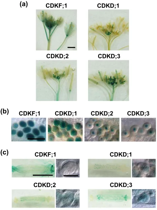
(34) Table 4. Number of ovules produced in wild-type and cdkd;1-1/+ cdkd;3-1/- plants. Genotype. Ovules at distinct stages during female Degenerated gametogenesis ovules FG1 FG2 FG3 FG4 FG5-7. WT. 1. 0. 0. 0. 347. 3. cdkd;1-1/+ cdkd;3-1/-. 30. 45. 8. 5. 471. 56. 33.
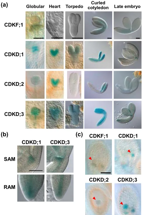
(35) Table 5. Number of seeds produced in cdkd;1-1/- cdkf;1-1/+, cdkd;3-1/- cdkf;1-1/+ and cdkd;1-1/+ cdkd;3-1/- plants expressing D;3-F;1::GUS Genotype. Normal seeds (%). Abortive seeds (%). WT. 171 (96.8). 6 (3.2). cdkf;1-1/+. 163 (91.6). 15 (8.4). cdkd;1-1/- cdkf;1-1/+. 194 (92.8). 15 (7.2). cdkd;3-1/- cdkf;1-1/+. 175 (97.8). 4 (2.2). cdkd;1-1/+ cdkd;3-1/-. 83 (55.7). 66 (44.3). cdkd;1-1/+ cdkd;3-1/with D;3-F;1::GUS. 64 (52.0). 59 (48.0). 34.
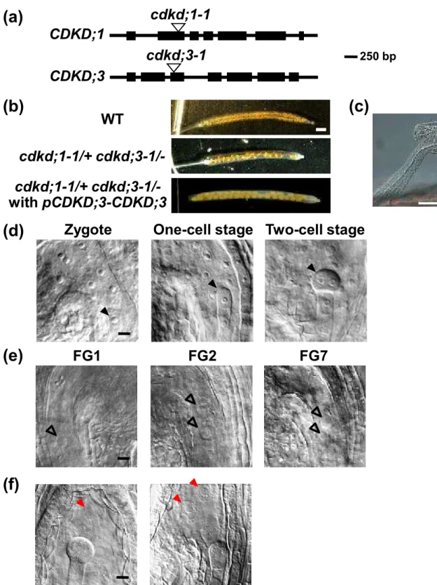
(36) Figure legends Figure 1. Expression patterns of CAKs in flowers and gametophytes. GUS staining of transgenic plants harboring pCDKF;1–CDKF;1::GUS, pCDKD;1– CDKD;1::GUS, pCDKD;2–CDKD;2::GUS or pCDKD;3–CDKD;3::GUS. (a) Flowers. (b) Mature pollen. (c) Siliques (left) and ovules (right). Bars = 1 mm (a), 50 µm (b), 1 mm (c, left) and 50 µm (c, right).. Figure 2. Expression patterns of CAKs in embryo and endosperm. GUS staining of transgenic plants harboring pCDKF;1–CDKF;1::GUS, pCDKD;1– CDKD;1::GUS, pCDKD;2–CDKD;2::GUS or. pCDKD;3–CDKD;3::GUS. (a). Embryos at different stages. (b) Enlargement of the regions around the shoot apical meristem (SAM) and the root apical meristem (RAM) in late embryos. (c) Ovules with embryo at the globular stage. Red arrowheads indicate embryos. Bars = 50 µm (a, globular stage), 100 µm (a, heart stage to late embryo) and 100 µm (b, c).. Figure 3. Gametophyte-lethal phenotype of the cdkd;1 cdkd;3 double mutant. (a) T-DNA insertion sites in cdkd;1-1 and cdkd;3-1. Exons are indicated by black boxes. (b) Seeds in siliques of cdkd;1-1/+ cdkd;3-1/- plants with or without complementation by the genomic fragment of CDKD;3. (c) Seed abortion observed in siliques of cdkd;1-1/+ cdkd;3-1/- plants. (d) Early embryo and endosperm development in wild-type ovules. Before the first zygotic division, at least three rounds of nuclear divisions occur in the endosperm (left). After mitosis starts in the embryo, endosperm nuclei continue to divide (middle and right). Black arrowheads indicate the zygote and embryos. (e) Unfertilized ovules at stages FG1, FG2 and FG7 observed in siliques of cdkd;1-1/+ cdkd;3-1/- plants. 35.
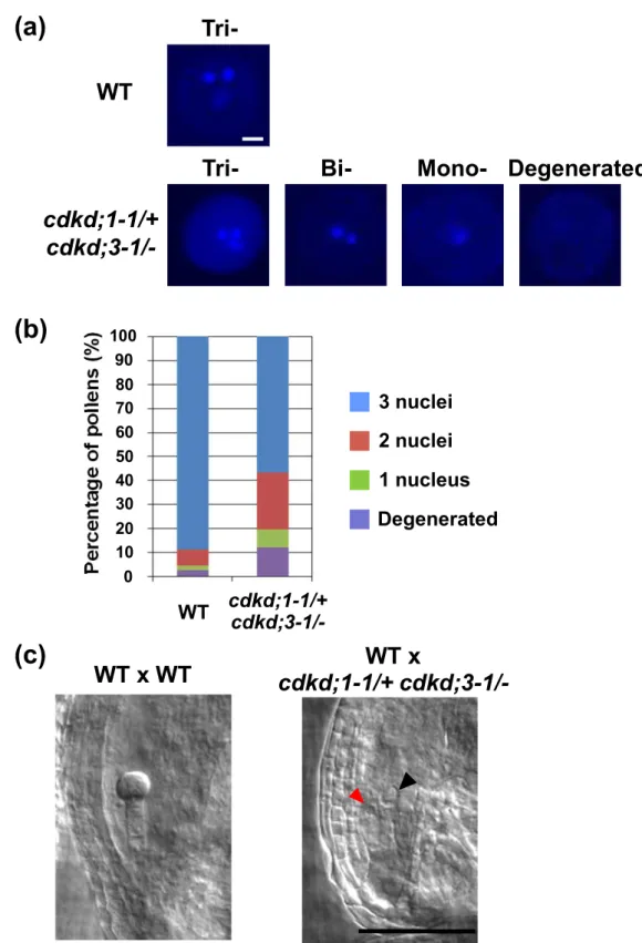
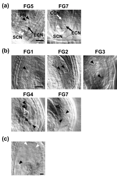
(37) Open arrowheads indicate nuclei. (f) Ovules with decreased number of endosperm nuclei observed in siliques of cdkd;1-1/+ cdkd;3-1/- plants. Red arrowheads indicate endosperm nuclei. Bars = 1 mm (b), 100 µm (c) and 10 µm (d-f).. Figure 4. Disturbed pollen mitosis in cdkd;1 cdkd;3. (a) DAPI staining of mature pollen from anthers of wild-type and cdkd;1/+ cdkd;3/plants. Tri-, bi- and mononucleate and degenerated pollen grains are shown. (b) Percentage of tri-, bi- and mononucleate and degenerated pollen grains. n = 106 (wildtype) and 203 (cdkd;1/+ cdkd;3/-). (c) Three-DAP ovules of wild-type plants pollinated with pollen from wild-type (left) or cdkd;1-1/+ cdkd;3-1/- (right) plants. Black and red arrowheads indicate nuclei of the central cell and the egg cell, respectively. Bars = 5 µm (a) and 50 µm (c).. Figure 5. Defects in female gametogenesis in cdkd;1 cdkd;3. (a) Wild-type ovules when flowers have opened and stamens nearly reached pistils. Two ovules at stages FG5 and FG7 are shown. PN, polar nucleus (black arrowheads); ECN, egg cell nucleus (black arrows); SCN, synergid cell nucleus (white arrowheads); CCN, central cell nucleus (white arrows). (b) Ovules of cdkd;1/+ cdkd;3/- plants. Ovules at FG1 to FG7 were observed when flowers had opened and stamens nearly reached pistils. Black and open arrowheads indicate nuclei and a central cell with a distorted nucleus, respectively. (c) Aberrant endosperm development in a 3-DAP ovule of a cdkd;1/+ cdkd;3/- plant pollinated with wild-type pollen. Black and white arrowheads indicate an embryo and endosperm nuclei, respectively. Bars = 20 µm.. 36.
(38) Figure 6. Phenotypes of cdkd;1 cdkf;1 and cdkd;3 cdkf;1. (a) Four-day-old seedlings of cdkf;1-1/+, cdkf;1-1/-, cdkd;1-1/- cdkf;1-1/+, cdkd;1-1/cdkf;1-1/-, cdkd;3-1/- cdkf;1-1/+, and cdkd;3-1/- cdkf;1-1/-. (b) Root tips of 4-day-old seedlings. Arrowheads indicate the quiescent center (lower) and the first elongated cell in the cortex cell file (upper). (c) Columella cell layers of 4-day-old seedlings. Arrowheads indicate the quiescent center (white) and columella cell layers including the initial cell layer (black). Bars = 5 mm (a) and 50 µm (b, c).. Figure 7. CDKF;1 cannot complement cdkd;1 cdkd;3. (a) Schematic diagrams of GUS-fusion genes. Yellow and orange boxes indicate the promoter regions and exons of, respectively, CDKD;3 and CDKF;1, and blue boxes indicate the GUS gene. Lines represent introns. (b) Complementation of cdkf;1-1 by expression of pCDKF;1-CDKF;1::GUS (F;1-F;1::GUS). Ten-day-old seedlings are shown. (c, d) GUS staining of transgenic plants expressing F;1-F;1::GUS, pCDKD;3CDKD;3::GUS (D;3-D;3::GUS) and pCDKD;3-CDKF;1::GUS (D;3-F;1::GUS). Root tips of 4-day-old seedlings (c) and pistils just before pollination (d) are shown. (e) Cleared mature siliques of wild-type and cdkd;1-1/+ cdkd;3-1/- plants, and of cdkd;1-1/+ cdkd;31/- harboring D;3-F;1::GUS. Bars = 5 mm (b), 50 µm (c) and 1 mm (d, e).. 37.
(39) (a). (b). (c). CDKF;1. CDKF;1. CDKD;1. CDKD;2. CDKD;3. CDKD;1. CDKD;2. CDKD;3. CDKF;1. CDKD;1. CDKD;2. CDKD;3. Figure 1. Expression patterns of CAKs in flowers and gametophytes. GUS staining of transgenic plants harboring pCDKF;1–CDKF;1::GUS, pCDKD;1– CDKD;1::GUS, pCDKD;2–CDKD;2::GUS or pCDKD;3–CDKD;3::GUS. (a) Flowers. (b) Mature pollen. (c) Siliques (left) and ovules (right). Bars = 1 mm (a), 50 µm (b), 1 mm (c, left) and 50 µm (c, right)..
(40) (a). Globular Heart. Curled Torpedo cotyledon. Late embryo. CDKF;1. CDKD;1. CDKD;2. CDKD;3. (b). (c) CDKD;1. CDKF;1. CDKD;1. CDKD;2. CDKD;3. CDKD;3. SAM. RAM. Figure 2. Expression patterns of CAKs in embryo and endosperm. GUS staining of transgenic plants harboring pCDKF;1–CDKF;1::GUS, pCDKD;1– CDKD;1::GUS, pCDKD;2–CDKD;2::GUS or pCDKD;3–CDKD;3::GUS. (a) Embryos at different stages. (b) Enlargement of the regions around the shoot apical meristem (SAM) and the root apical meristem (RAM) in late embryos. (c) Ovules with embryo at the globular stage. Red arrowheads indicate embryo. Bars = 50 µm (a, globular stage), 100 µm (a, heart stage to late embryo) and 100 µm (b, c)..
(41) (a). cdkd;1-1 CDKD;1 cdkd;3-1. 250 bp. CDKD;3. (b). (c). WT. cdkd;1-1/+ cdkd;3-1/cdkd;1-1/+ cdkd;3-1/with pCDKD;3-CDKD;3. (d). Zygote. (e). FG1. One-cell stage Two-cell stage. FG2. FG7. (f). Figure 3. Gametophyte-lethal phenotype of the cdkd;1 cdkd;3 double mutant. (a) T-DNA insertion sites in cdkd;1-1 and cdkd;3-1. Exons are indicated by black boxes. (b) Seeds in siliques of cdkd;1-1/+ cdkd;3-1/- plants with or without complementation by the genomic fragment of CDKD;3. (c) Seed abortion observed in siliques of cdkd;1-1/+ cdkd;3-1/- plants. (d) Early embryo and endosperm development in wild-type ovules. Before the first zygotic division, at least three rounds of nuclear divisions occur in the endosperm (left). After mitosis starts in the embryo, endosperm nuclei continue to divide (middle and right). Black arrowheads indicate the zygote and embryos. (e) Unfertilized ovules at stages FG1, FG2 and FG7 observed in siliques of cdkd;1-1/+ cdkd;3-1/- plants. Open arrowheads indicate nuclei. (f) Ovules with decreased number of endosperm nuclei observed in siliques of cdkd;1-1/+ cdkd;3-1/- plants. Red arrowheads indicate endosperm nuclei. Bars = 1 mm (b), 100 µm (c) and 10 µm (d-f)..
(42) (a). TriWT Tri-. Bi-. Mono- Degenerated. (b). Percentage of pollens (%). cdkd;1-1/+ cdkd;3-1/-. 100 90 80 70 60 50 40 30 20 10 0. 3 nuclei 2 nuclei 1 nucleus Degenerated. WT. (c). WT x WT. cdkd;1-1/+ cdkd;3-1/-. WT x cdkd;1-1/+ cdkd;3-1/-. Figure 4. Disturbed pollen mitosis in cdkd;1 cdkd;3. (a) DAPI staining of mature pollen from anthers of wild-type and cdkd;1/+ cdkd;3/- plants. Tri-, bi- and mononucleate and degenerated pollen grains are shown. (b) Percentage of tri-, bi- and mononucleate and degenerated pollen grains. n = 106 (wild-type) and 203 (cdkd;1/+ cdkd;3/-). (c) Three-DAP ovules of wild-type plants pollinated with pollen from wild-type (left) or cdkd;11/+ cdkd;3-1/- (right) plants. Black and red arrowheads indicate nuclei of the central cell and the egg cell, respectively. Bars = 5 µm (a) and 50 µm (c)..
(43) (a). FG5. FG7 CCN. PN. ECN. SCN. (b). SCN. FG1. FG2. FG4. FG7. ECN. FG3. (c). Figure 5. Defects in female gametogenesis in cdkd;1 cdkd;3. (a) Wild-type ovules when flowers have opened and stamens nearly reached pistils. Two ovules at stages FG5 and FG7 are shown. PN, polar nucleus (black arrowheads); ECN, egg cell nucleus (black arrows); SCN, synergid cell nucleus (white arrowheads); CCN, central cell nucleus (white arrows). (b) Ovules of cdkd;1/+ cdkd;3/- plants. Ovules at FG1 to FG7 were observed when flowers had opened and stamens nearly reached pistils. Black and open arrowheads indicate nuclei and a central cell with a distorted nucleus, respectively. (c) Aberrant endosperm development in a 3-DAP ovule of a cdkd;1/+ cdkd;3/- plant pollinated with wild-type pollen. Black and white arrowheads indicate an embryo and endosperm nuclei, respectively. Bars = 20 µm..
(44) (a). (b). (c). cdkf;1-1/+. cdkd;1-1/- cdkf;1-1/+ cdkd;3-1/- cdkf;1-1/+. cdkf;1-1/-. cdkd;1-1/- cdkf;1-1/- cdkd;3-1/- cdkf;1-1/-. cdkd;1-1/- cdkd;1-1/- cdkd;3-1/- cdkd;3-1/cdkf;1-1/+ cdkf;1-1/- cdkf;1-1/+ cdkf;1-1/- cdkf;1-1/+ cdkf;1-1/-. cdkf;1-1/+. cdkd;1-1/- cdkf;1-1/+ cdkd;3-1/- cdkf;1-1/+. cdkf;1-1/-. cdkd;1-1/- cdkf;1-1/- cdkd;3-1/- cdkf;1-1/-. Figure 6. Phenotypes of cdkd;1 cdkf;1 and cdkd;3 cdkf;1. (a) Four-day-old seedlings of cdkf;1-1/+, cdkf;1-1/-, cdkd;1-1/- cdkf;1-1/+, cdkd;1-1/- cdkf;1-1/-, cdkd;3-1/- cdkf;1-1/+, and cdkd;3-1/- cdkf;1-1/-. (b) Root tips of 4-day-old seedlings. Arrowheads indicate the quiescent center (lower) and the first elongated cell in the cortex cell file (upper). (c) Columella cell layers of 4-day-old seedlings. Arrowheads indicate the quiescent center (white) and columella cell layers including the initial cell layer (black). Bars = 5 mm (a) and 50 µm (b, c)..
(45) (a). pCDKD;3-CDKD;3::GUS (D;3-D;3::GUS) GUS. Promoter. pCDKF;1-CDKF;1::GUS (F;1-F;1::GUS) Promoter. GUS. pCDKD;3-CDKF;1::GUS (D;3-F;1::GUS) Promoter. (b). (c). cdkf;1-1/+. GUS. 250 bp. cdkf;1-1 with F;1-F;1::GUS. cdkf;1-1/-. F;1-F;1::GUS D;3-D;3::GUS D;3-F;1::GUS. (d) F;1-F;1::GUS. D;3-D;3::GUS D;3-F;1::GUS. (e) WT cdkd;1-1/+ cdkd;3-1/cdkd;1-1/+ cdkd;3-1/with D;3-F;1::GUS Figure 7. CDKF;1 cannot complement cdkd;1 cdkd;3. (a) Schematic diagrams of GUS-fusion genes. Yellow and orange boxes indicate the promoter regions and exons of, respectively, CDKD;3 and CDKF;1, and blue boxes indicate the GUS gene. Lines represent introns. (b) Complementation of cdkf;1-1 by expression of pCDKF;1CDKF;1::GUS (F;1-F;1::GUS). Ten-day-old seedlings are shown. (c, d) GUS staining of transgenic plants expressing F;1-F;1::GUS, pCDKD;3-CDKD;3::GUS (D;3-D;3::GUS) and pCDKD;3-CDKF;1::GUS (D;3-F;1::GUS). Root tips of 4-day-old seedlings (c) and pistils just before pollination (d) are shown. (e) Cleared mature siliques of wild-type and cdkd;1-1/+ cdkd;31/- plants, and of cdkd;1-1/+ cdkd;3-1/- harboring D;3-F;1::GUS. Bars = 5 mm (b), 50 µm (c) and 1 mm (d, e)..
(46) PM I. Vegetative cell. PM II. Generative cell. Sperm cells. Figure S1. Male gametogenesis in Arabidopsis. A vegetative cell divides asymmetrically to produce a daughter vegetative cell and a generative cell in pollen mitosis I (PM I). The generative cell then divides symmetrically to produce two sperm cells in pollen mitosis II (PM II)..
(47) Nuclear division. FG1. Vacuolation. FG2. Nuclear division. Nuclear division. FG4. FG3. FG5. Antipodal cells. Cellularization. Nuclear migration and fusion. Loss of antipodal cells. Polar nuclei. Central cell. FG5. FG6. Egg cell. FG7. Synergid cell. Figure S2. Female gametogenesis in Arabidopsis. At FG6, the central cell is produced by fusion of two polar nuclei, followed by loss of three antipodal cells. Black and gray circles indicate nuclei and the central vacuole in the embryo sac, respectively..
(48) (a). WT. cdkd;3-1/-. (b). WT. cdkd;1-1/-. cdkd;1-1/+ cdkd;3-1/-. cdkd;1-1/+ cdkd;3-1/-. Figure S3. Viability of pollen from wild-type, cdkd;1-1/-, cdkd;3-1/-, and cdkd;1-1/+ cdkd;3-1/- plants. (a) Alexander staining of pollen grains in anthers just before dehiscence. Violet- and bluestained pollen grains are viable and nonviable, respectively. (b) Pollen grains from mature flowers of wild-type and cdkd;1-1/+ cdkd;3-1/- plants. Bars = 100 µm..
(49) WT. cdkf;1-1/+. cdkd;1-1/- cdkf;1-1/+. cdkd;3-1/- cdkf;1-1/+. Figure S4. Seed formation in siliques of wild-type, cdkf;1-1/+, cdkd;1-1/- cdkf;1-1/+, and cdkd;3-1/- cdkf;1-1/+ plants. Bar = 1 mm..
(50)
図


+7
関連したドキュメント
Here we are interested in studying the weakly coupled system ( 1. 1 ) in the critical case. In particular we want to find solutions which concentrate in some points of in the sense
Using general ideas from Theorem 4 of [3] and the Schwarz symmetrization, we obtain the following theorem on radial symmetry in the case of p > 1..
In a previous paper [1] we have shown that the Steiner tree problem for 3 points with one point being constrained on a straight line, referred to as two-point-and-one-line Steiner
As the study in the single-valued case has dealt with projected DS, we recall in Section 2 the notion of projected differential inclusion (as in [1]), together with the ba- sic
Abstract. In Section 1 we introduce Frobenius coordinates in the general setting that includes Hopf subalgebras. In Sections 2 and 3 we review briefly the theories of Frobenius
In our previous papers (Nishimura [2001 and 2003]) we dealt with jet bundles from a synthetic perch by regarding a 1-jet as something like a pin- pointed (nonlinear) connection
“Breuil-M´ezard conjecture and modularity lifting for potentially semistable deformations after
In particular, we are able to prove that for Volterra scalar systems with a creep kernel a(t) such that a(0 + ) > 0; the finite-time and the infinite-time L 1 -admissibility
