INTRODUCTION
It is well known that cruciferous vegetables con-tain anti-cancer agents (1). In fact, one epidemi-ological study has indicated that the consumption
of cruciferous vegetables is linked to the reductions in the risks of developing breast, prostate and colon cancer (2). These vegetables, especially cabbage, broccoli, Brussels sprout and cauliflower, are rich in glucosinolate, one of the anti-cancer agents in cruciferous vegetables (3).
Glucosinolate is hydrolyzed by myrosinase (also known as thioglucoside glycohydrolase). Since my-rosinase localizes inside plant cells, the cutting and chewing of vegetables release the enzyme and en-hance the hydrolysis of glucosinolate. Inversely,
ORIGINAL
Effects of indole-3-carbinol and phenethyl isothiocyanate
on bile and pancreatic juice excretion in rats
Hiroki Ishibashi
1), Tomomi Kuwahara
2), Haruyuki Nakayama-Imaohji
3),
Yoshinari Ohnishi
3), Hiroki Mori
1), and Mitsuo Shimada
1) 1)Department of Surgery, Institute of Health Biosciences, The University of Tokushima Graduate School, Tokushima, Japan, 2)
Department of Microbiology, Faculty of Medicine, Kagawa University, Kagawa, Japan, 3)
Department of Molecular Bacteriology, Institute of Health Biosciences, The University of Tokushima Graduate School, Tokushima, Japan
Abstract : Bile and pancreatic juice contain a number of parameters for cancer chemo-prevention. Indole-3-carbinol (I3C) and phenethyl isothiocyanate (PEITC), which are hydrolytic products of brassica plants, have been established to be anti-cancer agents. Here, we developed a method for the continuous and selective sampling of bile and pan-creatic juice, and the effects of I3C and PEITC on bile and panpan-creatic excretion and γ-glutamyl transpeptidase (γ-GTP) activity in the samples were investigated. Male Fisher 344 rats (eight weeks of age) were challenged intragastrically with I3C (150 mg/kg) or PEITC (160 mg/kg) for five days. Twenty-four hours after the final administration, can-nulation was undertaken into the rats’ bile and pancreatic ducts, and the bile and pan-creatic juice were separately collected for 48 h. In this rat model, bile was stably ex-creted, and the bile and pancreatic excretion of the control rats was 21.9 1.4 ml/48 h and 12.8 1.7 ml/48 h, respectively. Bile excretion for the first 24 h significantly in-creased in the I3C- or PEITC-treated rats compared with the control rats. In the case of pancreatic juice, excretion during the first 24 h significantly increased in the PEITC-treated rats. In bile,γ-GTP activity was significantly increased for the first 24 h in the I3C- and PEITC-treated rats, but no difference was observed in the pancreatic juice. Increases of bile excretion andγ-GTP activity in bile might be a factor involved in the anti-cancer effect of I3C and PEITC. Our rat model described here is a useful tool for the study of cancer chemoprevention. J. Med. Invest. 59 : 246-252, August, 2012
Keywords : indole-3-carbinol, phenethyl isothiocyanate, bile, pancreatic juice, cancer chemoprevention
Received for publication February 10, 2012 ; accepted February 28, 2012.
Address correspondence and reprint requests to Hiroki Ishibashi, M.D., Department of Surgery, Institute of Health Biosciences, The University of Tokushima Graduate School, 3 18 15, Kuramoto -cho, Tokushima 770 - 8503, Japan and Fax : + 81 - 88 - 631 - 9698.
cooking by heat inactivates myrosinase, resulting in a 30-60% loss of glucosinolate bioavailability. As a result of the glucosinolate hydrolysis by my-rosinase, isothiocyanate and indole derivatives (such as indole-3-carbinol and indole-3-acetonitril) are produced (4). These metabolites from glucosinolate are considered to play a major role in cancer che-moprevention through the induction of detoxifica-tion enzymes (5). Many studies have reported that administration of glucosinolate metabolites (isothio-cyanate and indol derivatives) and brassica plant extract elevates the phase-I and phase-II enzymes in the rat livers (6, 7).
Bile and pancreatic juice contain a number of pa-rameters for drug metabolism associated with can-cer chemoprevention, which include various types of conjugates and detoxifying enzymes. Glutathione-conjugates generate in the liver by glutathione S-transferase and excrete into the gastrointestinal tract through bile. Glutathione-conjugates undergoes to the stepwise cleavage in the small intestine by γ-glutamyl transferase (γ-GTP) that is derived mainly from the pancreas (8, 9). To evaluate the drug me-tabolism in liver, bile and pancreatic juice should be collected separately because the latter contains high γ-GTP activity (10) that degrades the glu-tathione-conjugates in bile. Gluglu-tathione-conjugates begin to degrade in the bile duct in rats with panc-teaticobiliary maljunction. However, there are no reports that analyzes the bile and pancreatic juice
separately in rodent models such as mice and rats. The aim of this study is to develop a method for the selective collection of bile and pancreatic juice in rats. Using this rat model, we evaluated the ef-fects of indole-3-carbinol (I3C) and phenethyl iso-thiocyanate (PEITC) on excretion rates of bile and pancreatic juice andγ-GTP activity in these samples.
MATERIALS AND METHODS
Chemicals and analytical procedureThe I3C, PEITC, bovine serum albumin, γ-gluta-myl-p-nitroanilide, glycylglycine and p -dimethyl-aminocinnaldehyde used in this study were pur-chased from Sigma Chemicals (St Louis, MO, USA). Protein concentration was determined in duplicate by the method of Lowry et al. (11) using bovine serum albumin as a standard.γ-GTP activity was measured according to the method described by Igarashi et al. (12) employing γ-glutamyl-p-ni-troanilide as a donor substrate and glycylglycine as a acceptor substrate. The production of p-nitroani-line was quantified by measuring the absorbance at 565 nm after chromogenization by the addition of p-dimethylaminocinnaldehyde.
Experimental design
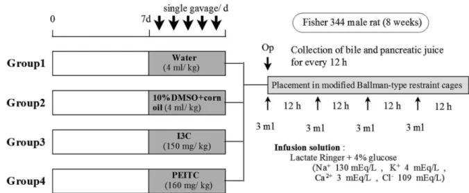
The protocol for the animal experiment per-formed in this study is shown in Figure 1. Male
Figure 1. Experimental protocol and design for the bile and pancreatic juice collections. After acclimation for one week, male Fisher344 rats (eight weeks of age) were divided into four groups and intragastrically administered with tap water, 10% DMSO plus corn oil (vehicle), I3C (150 mg/kg), or PEITC (160 mg/kg) for five days. The rats were operated on at 24 h after the final admini-stration as shown in Figure 2, and the bile and pancreatic juice were separately collected for 48 h. The bile and pancreatic juice ex-cretions were measured every 12 h.
Fisher344 rats (eight weeks of age) were purchased from Japan SLC and housed in polycarbonate cages (four rats/cage) containing hard-wood chip bed-ding. The rats were allowed to access to standard chows and tap water ad libitum. After acclimation for one week, the rats were divided into four groups and intragastrically administered with tap water, 10% DMSO plus corn oil (vehicle), I3C (150 mg/kg), or PEITC (160 mg/kg) for five days. The rats were operated on at 24 h after the final administration as described below, and the bile and pancreatic juice were separately collected for 48 h. The bile and pan-creatic juice excretions were measured every 12 h. The samples were frozen at -80C until use.
The present study was conducted in compli-ance with the Division for Animal Research Re-sources, Institute of Health Biosciences, University of Tokushima. The experiments and procedures were approved by the Animal Care and Use Com-mittee of the University of Tokushima.
Operative procedure
Adult male F344 rats with body weights around 200 g underwent laparotomies under anesthesia,
and a 24G infusion needle was inserted into the caudal vein for drop infusion. As shown in Figure 2A, after the duodenum was turned to the left, the common bile duct and pancreatic duct were exposed from the back side of the pancreas. A polyethylene tube with a 0.8-mm diameter was inserted into the tip of a silicon tube with 1-mm diameter to make the 3-mm overhang (Figure 2B). Under observation with a magnifying glass (
!
3.3), the end of the com-mon bile duct was ligated proximally to the ampulla of Vater, and the cannula shown in Figure 2B was inserted above the ligature to collect pure pancre-atic juice. The cannula was fixed by 5-0 silk. An-other cannula was inserted into the common bile duct at upstream of the pancreatic duct joint to collect pure bile (Figure 2C). The other end of the inserted tube was passed subcutaneously behind the neck and taken outside the body. Both pure bile and pancreatic juice were collected in syringes sur-rounded by ice.After the operation, the rats were placed in modi-fied Bollman-type restraint cages and allowed free access to standard chows and tap water. The room temperature was controlled at 22 to 25!!. The rats
Figure 2. Anatomy of common bile duct and pancreatic duct in rats (A), cannulated tube used in this study (B), and procedure for cannulation (C). Adult male F344 rats with body weights around 200 g underwent laparotomies under anesthesia and after the duo-denum was turned to the left, the common bile duct and pancreatic duct were exposed from the back side of the pancreas (panel A). A polyethylene tube with a 0.8 - mm diameter was inserted into the tip of a silicon tube with 1 - mm diameter to make the 3 - mm over-hang (panel B). As shown in panel C, the end of the common bile duct was ligated proximally to the ampulla of Vater, and the can-nula was inserted above the ligature to collect pure pancreatic juice. The cancan-nula was fixed by 5 - 0 silk. Another cancan-nula was inserted into the common bile duct at upstream of the pancreatic duct joint to collect pure bile.
A A C C B B
received intermittent infusion (Lactate Ringer plus 4% glucose) every 12 h. The injection volume was 3 ml/infusion (for 30 min), and the total infusion volume was 12 ml /48 h.
Statistical analysis
Statistical analysis was performed by one-way ANOVA. All statistical analyses were performed us-ing statistical software (JMP 8.0.1., SAS Campus Drive, Cary, 27513 NC, USA). A p-value of less than 0.05 was considered to be statistically significant.
RESULTS
Rat model for collection of bile and pancreatic juice Cannulation and collection of bile and pancreatic juice were successful in all cases when rats of over 200 g in body weight were used. The excretion vol-umes in the control rats administered with dis-tilled water were 13.2!1.2 ml/24 h and 21.9!1.4 ml/48 h (34.3!1.7μl/kg/min) for bile and 4.8! 1.7 ml/24 h and 12.8!1.7 ml/48 h (20.4!2.9μl/ kg/min) for pancreatic juice, respectively.
Effects of I3C and PEITC on bile and pancreatic juice excretion
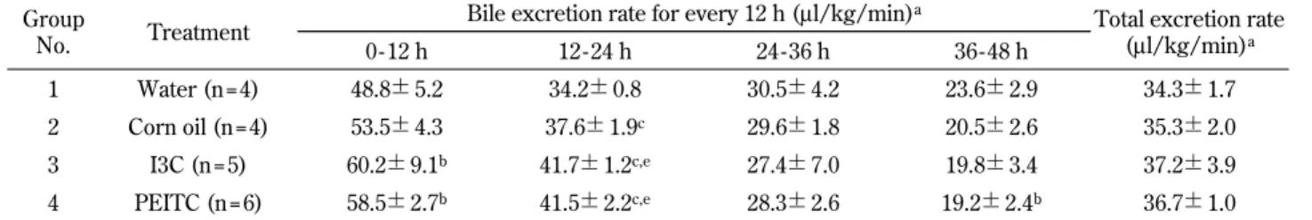
As shown in Table 1, I3C and PEITC administra-tion significantly increased the bile excreadministra-tion rate for
the first 24 h compared with the control rats (corn oil administration). However, bile excretion rate in the rats of these experimental groups tended to de-crease for the next 24 h, and no significant differ-ence was observed in the total bile excretion vol-umes. The total bile excretion for 48 h reached 22.6!2.4 ml (37.2!3.9 μl/kg/min) in the I3C-adnimistered rats and 21.9!1.6 ml (36.7!1.0μl/ kg/min) in the PEITC-administered rats. The pro-tein concentration in bile significantly increased for the first 12 h in the I3C- and PEITC-treated groups but no significant change was observed after 36 h.
The pancreatic juice excretion rate is summarized in Table 2. There was no significant difference in the pancreatic juice excretion rate for the first 36 h between the I3C-treated group and the control group. The total excretion rate significantly de-creased when compared with the corn oil-treated group. In the case of PEITC administration, the pan-creatic juice excretion rate significantly increased when compared with the corn oil-treated group al-though no significant difference was found in the to-tal excretion volume. The toto-tal pancreatic juice ex-cretion for 48 h reached 14.5!3.2 ml (24.0!5.6μl/ kg/min) in the I3C - administered rats and 20.9! 2.6 ml (34.9!2.5μl/kg/min) in the PEITC-admin-istered rats. The protein concentration in the pan-creatic juice significantly decreased for the first 24 h in the PEITC-treated group but no significant
Table 1. Changes of the bile excretion rate Group
No. Treatment
Bile excretion rate for every 12 h (μl/kg/min)a
Total excretion rate (μl/kg/min)a 0 - 12 h 12 - 24 h 24 - 36 h 36 - 48 h 1 Water (n = 4) 48.8!5.2 34.2!0.8 30.5!4.2 23.6!2.9 34.3!1.7 2 Corn oil (n = 4) 53.5!4.3 37.6!1.9c 29.6!1.8 20.5!2.6 35.3!2.0 3 I3C (n = 5) 60.2!9.1b 41.7!1.2c,e 27.4!7.0 19.8!3.4 37.2!3.9 4 PEITC (n = 6) 58.5!2.7b 41.5!2.2c,e 28.3!2.6 19.2!2.4b 36.7!1.0 aMean!S.D.
b, cSignificantly different from group 1 (b : p!0.05, c : p!0.01) d, eSignificantly different from group 2 (d : p!0.05, e : p!0.01)
Table 2. Changes of the pancreatic juice excretion rate Group
No. Treatment
Pancreatic excretion rate for every 12 h (μl/kg/min)a
Total excretion rate (μl/kg/min)a 0 - 12 h 12 - 24 h 24 - 36 h 36 - 48 h 1 Water (n = 4) 12.3!6.2 18.2!4.0 21.8!6.8 29.4!8.7 20.4!2.9 2 Corn oil (n = 4) 15.7!5.0 31.0!2.9b 39.6!3.9b 34.1!7.4 30.1!0.8b 3 I3C (n = 5) 15.7!7.3 29.4!9.2b 29.6!8.2 21.6!6.1e 24.0!5.6e 4 PEITC (n = 6) 28.8!1.6b,d 42.4!2.8b,d 42.5!7.7b 27.5!5.5 34.9!2.5b aMean!S.D.
b, cSignificantly different from group 1 (b : p!0.01, c : p!0.05) d, eSignificantly different from group 2 (d : p!0.01, e : p!0.05)
change was observed after 24 h.
γ-GTP activities in bile and pancreatic juice γ-GTP activity in pancreatic juice was about 100 times higher than that in bile when the activities were compared in the control group (water admini-stration). As shown in Figure 3,γ-GTP activities in the bile collector from the I3C- and PEITC-treated groups were significantly higher than those in the control group for the first 24 h, but no significant difference was observed after 24 h. On the other hand, as shown in Figure 4,γ-GTP activities in the pancreatic juice of PEITC-treated groups were sig-nificantly lower than those in the control group during the 12-24 h period. However, both I3C- and PEITC-treatment provided little difference toγ-GTP activity in pancreatic juice.
DISCCUSION
Methods for the collection of both bile (13) and pancreatic juice in a pancreatitis model (14) have been reported, respectively. However, no reports are available for the selective and simultaneous collec-tion of bile and pancreatic juice. In the method de-scribed by Toriumi et al., rats with tethered pancre-atic fistula were partially restrained by tube (14). We have tried this model but the rats sometimes bit and removed the catheter. To solve this problem, we employed Bollman cages for their short-term restraint. Since restraint using Bollman cage results in dehydration due to insufficient water intake, which leads to the reduction of bile and pancreatic juice excretion, infusions with Lactate Ringer were performed to keep the bile and pancreatic juice ex-cretion stable. For the cannulation into the bile or pancreatic duct, we constructed a modified lation tube that comprised of a hard tip for cannu-lation into the bile or pancreatic duct and a soft tube for drainage to an extra body (Figure 2B). This cannula made it easy to stably collect the bile and pancreatic juice, and we encountered no cannulation-associated complications. Because the bile and pan-creatic ducts of rats are thin (around 1 mm in diame-ter), training is necessary for cannulation. However, once established, the short-term collection of bile and pancreatic juice can be stably performed.
Prevention of cancer initiation can be achieved by limiting the exposure of cells to carcinogenic substances by either by inhibiting their activation or increasing their detoxification and subsequent removal (15). Moreover, cancer chemopreventive compounds can suppress promotion and progres-sion of carcinogenesis by interfering with various signaling pathways involving oxidative stress (16), inflammation (17), and cellular proliferation. Addi-tionally, some of these compounds can show their cancer chemopriventive effects at all of the stages of carcinogenesis (initiation, promotion, and progres-sion) by inducing the cell cycle arrest and apoptosis (18, 19).
Epidemiological studies have shown that con-sumption of brassica plants during adolescence was associated with a 72% reduction of the risk of breast cancer (20). Cabbage consumption has also been reported to be associated with a reduction of the risks of developing prostate and pancreatic cancers (21, 22). The anti-cancer effect of brassica plants derives partly from the glucosinolate and its hydro-lytic metabolites such as I3C and PEITC (ref 4). Figure 3. γ-GTP activities in pure bile. γ-GTP activities in the
bile collected from I3C - and PEITC - treated groups were signifi-cantly higher than those in control group for the first 24 h, but no significant difference was observed after 24 h.
Figure 4. γ-GTP activities in pure pancreatic juice. γ-GTP ac-tivities in pancreatic juice of PEITC - treated groups were signifi-cantly lower than those in control group during the 12 - 24 h period. However, both I3C - and PEITC - treatment provided little difference toγ-GTP activity in pancreatic juice.
These anti-cancer agents are considered to induce detoxification enzymes (5-7), of which the Phase II enzymes such as glutathione-S-transferase es-pecially play an eses-pecially important role (23, 24). As a result of Phase II reaction, the glucuronate-, sulfate- and glutathione-conjugates are produced and excreted into bile or urine. It has been reported that highγ-GTP activity is present in the pancreas and in pancreatic juice (8-10), and glutathione-conjugates are converted into cysteinylglycine-conjugates by this enzyme activity in pancreatic juice. Cysteinylglycine-conjugates are further me-tabolized to cysteine-conjugates by aminopeptidase in pancreatic juice (phase III reaction). Thus, γ-GTP is one of the key enzymes for detoxification, which degradesγ-glutamyl compounds and cata-lyzes the first reaction of the metabolism of glu-tathione conjugates to mercapturic acid in Phase II reaction.
In this study, we investigated the effect of the anti-cancer agents, I3C and PEITC, on the excre-tion of bile and pancreatic juice. Bile excreexcre-tion was significantly increased for the first 24 h in both the I3C- and PEITC-treated groups. In addition,γ-GTP activities were also elevated in these groups. These results indicate that I3C and PEITC enhance bile excretion and billiaryγ-GTP activity. Furthermore, the excretion of pancreatic juice was also increased for the first 24 h in PEITC-treated rats although the γ-GTP activity in pancreatic juice was decreased by PEITC-treatment, possibly leading to the suppres-sion of Phase II reaction. I3C and PEITC might en-hance the detoxification and excretion of the me-tabolites of mutagenic compounds by increasing bile and pancreatic juice excretion and changing the γ-GTP activity in bile and pancreatic juice.
In summary, our rat model for the selective and simultaneous collection of bile and pancreatic juice is expected to work as a useful tool for the analyses of drug metabolism and anti-cancer agents like I3C and PEITC.
CONFLICT OF INTEREST
The authors have no conflict of interest associ-ated with the present study.
REFERENCES
1. McNaughton SA, Marks GC : Development of
a food composition database for the estimation of dietary intakes of glucosinolates, the biologi-cally active constituents of cruciferous vegeta-bles. Br J Nutr 90 : 687-697, 2003
2. Higdon JV, Delage B, Williams DE, Dashwood RH : Cruciferous vegetables and human cancer risk : epidemiologic evidence and mechanistic basis. Pharmacol Res 55 : 224-236, 2007 3. Verhoeven DT, Verhagen H, Goldbohm RA,
van den Brandt PA, van Poppel G : A review of mechanisms underlying anticarcinogenecity by brassica vegetables. Chem Biol Interact 103 : 79-129, 1997
4. Holst B, Williamson G : A critical review of the bioavailability of glucosinolates and related compounds. Nat Prod Rep 21 : 425-447, 2004 5. Staack R, Kingston S, Wallig MA, Jeffery EH :
A comparison of the individual and collective effects of four glucosinolate breakdown prod-ucts from brussels sprouts on induction of de-toxification enzymes. Toxicol Appl Pharmacol. 149 : 17-23, 1998
´
6. Szaefer H, Krajka-Kuzniak V, Bartoszek A, Baer-Dubowska W : Modulation of Carcinogen Metabolizing Cytochromes P450 in Rat Liver and Kidney by Cabbage and Sauerkraut Juices : Comparison with the Effects of Indole-3-carbi-nol and Phenethyl Isothiocyanate. Phytother Res. 2011 Dec 15. [Epub ahead of print]
´
7. Krajka-Kuzniak V, Szaefer H, Bartoszek A, Baer-Dubowska W : Modulation of rat hepatic and kidney phase II enzymes by cabbage juices : comparison with the effects of indole-3-carbinol and phenethyl isothiocyanate. Br J Nutr 105 : 816-826, 2011
8. Kinouchi T, Kataoka K, Miyanishi K, Akimoto S, Ohnishi Y : Biological activities of the intes-tinal microflora in mice treated with antibiotics or untreated and the effect of the microflora on absorption and metabolic activation of orally ad-ministered glutathione conjugates of K-region epoxides of 1-nitropyrene. Carcinogenesis 14 : 869-874, 1993
9. Kinouchi T, Nishifuji K, Ohnishi Y : Biliary ex-cretion of glutathione conjugates of 4,5-epoxy-4,5-dihydro-1-nitropyrene and 9,10-epoxy-9, 10-dihydro-1-nitropyrene in rats administered 1-nitropyrene orally and their further metabo-lism in the intestinal tract. Carcinogenesis 11 : 1381-1387, 1990
10. Battistini B, Chailler P, Brière N, Beaudoin AR. Secretion of gamma-glutamyltranspeptidase by
the pancreas : evidence for a membrane shed-ding process during exocytosis. Life Sci. 47 : 2435-2441, 1990
11. Lowry OH, Rosebrough NJ, Farr AL, Randall RJ : Protein measurement with the Folin phe-nol reagent. J Biol Chem 193 : 265-275, 1951 12. Igarashi T, Satoh T, Ueno K, Kitagawa H :
Spe-cies difference in glutathione level and glu-tathione related enzyme activities in rats, mice, guinea pigs and hamsters. J Pharmacobiodyn 6 : 941-949, 1983
13. Tomlinson PW, Jeffery DJ, Filer CW : A novel technique for assessment of biliary secretion and enterohepatic circulation in the unre-strained conscious rat. Xenobiotica 11 : 863-870, 1981
14. Toriumi Y, Samuel I, Wilcockson DP, Joehl RJ : A new model for study of pancreatic exocrine secretion : the tethered pancreatic fistula rat. Lab Anim Sci 44 : 270-273, 1994
15. Hanausek M, Walaszek Z, Slaga TJ : Detoxify-ing cancer causDetoxify-ing agents to prevent cancer. Integr Cancer Ther 2 : 139-144, 2003
16. Klaunig JE, Kamendulis LM : The role of oxi-dative stress in carcinogenesis. Annu Rev Phar-macol Toxicol 44 : 239-267, 2004
17. Karin M : Nuclear factor-kappaB in cancer de-velopment and progression. Nature 441 : 431-436, 2006
18. Cheung KL, Kong AN : Molecular targets of die-tary phenethyl isothiocyanate and sulforaphane
for cancer chemoprevention. AAPS J 12 : 87-97, 2010
19. Aggarwal BB, Ichikawa H : Molecular targets and anticancer potential of indole-3-carbinol and its derivatives. Cell Cycle 4 : 1201-1215, 2005
20. Nelson NJ : Migrant studies aid the search for factors linked to breast cancer risk. J Natl Cancer Inst 98 : 436-438, 2006
21. Kristal AR, Lampe JW : Brassica vegetables and prostate cancer risk : a review of the epi-demiological evidence. Nutr Cancer 42 : 1-9, 2002
!
22. Larsson SC, Hakansson N, Näslund I, Bergkvist L, Wolk A : Fruit and vegetable consumption in relation to pancreatic cancer : a prospective study. Cancer Epidemiol Biomarkers Prev 15 : 301-305, 2006
23. Nijhoff WA, Grubben MJ, Nagengast FM, Jansen JB, Verhagen H, van Poppel G, Peters WH : Effects of consumption of Brussels sprouts on intestinal and lymphocytic glutathione S-transferases in humans. Carcinogenesis 16 : 2125-2128, 1995
24. Staack R, Kingston S, Wallig MA, Jeffery EH : A comparison of the individual and collective effects of four glucosinolate breakdown prod-ucts from brussels sprouts on induction of de-toxification enzymes. Toxicol Appl Pharmacol 149 : 17-23, 1998