Clinics in Surgery
Step-by-Step Procedure to Test Photoelectric Dye-Coupled
Polyethylene Film as Retinal Prosthesis to Induce
Light-Evoked Spikes in Isolated Retinal Dystrophic Tissue of rd1
Mice
OPEN ACCESS
*Correspondence: Toshihiko Matsuo, Regenerativeand Reconstructive Medicine (Ophthalmology), Okayama University Medical School and Graduate School of Interdisciplinary Science and Engineering in Health Systems, Shikata-cho 2-5-1, Okayama City
700-8558, Japan, Tel: +81-86-251-8106; E-mail: matsuot@cc.okayama-u.ac.jp Received Date: 21 Jun 2020 Accepted Date: 03 Aug 2020 Published Date: 10 Aug 2020 Citation: Matsuo T, Terada K, Sakurai M, Liu S, Yamashita K, Uchida T. Step-by-Step Procedure to Test Photoelectric Dye-Coupled Polyethylene Film as Retinal
Prosthesis to Induce Light-Evoked Spikes in Isolated Retinal Dystrophic Tissue of rd1 Mice. Clin Surg. 2020; 5: 2903. Copyright © 2020 Matsuo T. This is an
open access article distributed under the Creative Commons Attribution License, which permits unrestricted use, distribution, and reproduction in any medium, provided the original work is properly cited.
Research Article Published: 10 Aug, 2020
Abstract
Purpose: Multielectrode array recording for electric activity in cardiac and neuronal cells has
been developed as preclinical tests for drug screening. This study aims to establish an in vitro assay system, using the multielectrode array, to record light-evoked spikes in isolated degenerative retinal tissues of retinal dystrophic rd1 mouse, as a preclinical test to examine the efficacy of photoelectric dye-coupled thin film retinal prosthesis.
Methods: Light-evoked spike response was tested for 1 min at first step in the isolated degenerative
retinal tissue of retinal dystrophic rd1 mouse only on the multielectrode array, tested in the same retinal tissue overlain with a plain control film for light-off and light-on 10 min each at second step, and tested in the same tissue overlain with a dye-coupled film at third step. The retinal tissues which showed light-evoked response at first or second step were not used for evaluation at third step.
Results: Residual light-evoked spikes were recorded at first or second step in 18 of 35 retinal tissues
(51%) at 6 weeks of the age in rd1 mice, 16 of 44 tissues (36%) at 7 weeks, and 10 of 39 tissues (25%) at 8 weeks. At third step, light-evoked spikes were recorded with dye-coupled films in 8 of 17 retinal tissues (47%) at 6 weeks, 10 of 28 tissues (35%) at 7 weeks, and 8 of 29 tissues (27%) at 8 weeks.
Conclusion: A step-by-step procedure with internal control was established to measure light-evoked
spikes by the multielectrode array in the isolated degenerative retinal tissue to evaluate photoelectric dye-coupled thin films. This preclinical study would present one line of evidence for the efficacy of photoelectric dye-coupled thin film retinal prosthesis towards a first-in-human clinical trial.
Keywords: Photoelectric dye; Polyethylene thin film; Spike; Degenerative retina; Retinal dystrophic rd1 mouse
Toshihiko Matsuo1,2*, Keiko Terada3, Mikako Sakurai3, Shihui Liu1, Koichiro Yamashita4 and Tetsuya Uchida4
1Regenerative and Reconstructive Medicine (Ophthalmology), Okayama University Medical School and Graduate
School of Interdisciplinary Science and Engineering in Health Systems, Japan
2Department of Ophthalmology, Okayama University Hospital, Japan
3Research Center, Techno Pro, Inc., Tokyo, Japan
4Polymer Materials Science, Okayama University Faculty of Engineering and Graduate School of Natural Science
and Technology, Japan
Introduction
A major limiting step at developing new drugs and medical devices to restore the vision is how to assess the vision in animal models. Frequently used animals are a rat strain (Royal College of Surgeons RCS rat) and a mouse strain (rd1) which show hereditary retinal dystrophy [1-3]. The vision assessment by behavior tests in rats and mice has been established and used for many decades [4,5]. Under the circumstances, individual difference and ambiguous response in each rat or mouse pose a problem in interpreting the outcome in behavior tests.
To overcome the behavioral limitation, electric response to light as an objective sign in the retina and in the brain was recorded in rats and mice as electroretinogram and visual evoked potential, respectively [6,7]. However, there is still a limitation of sensitivity in detecting the difference which would be generated as the result of intervention. One way to solve the limitation in in vivo animal studies is to design an in vitro test using degenerative retinal tissues which are isolated from retinal dystrophic rats or mice. Recently, multielectrode array dish system has been used as an in vitro assay
to screen drug toxicity, for instance, in cultured cardiomyocytes [8]. Patients with retinitis pigmentosa have lost the vision due to retinal photoreceptor cell loss designated as outer retinal degeneration [9]. Retinal prosthesis replaces the function of dead photoreceptor cells and stimulates the remaining retinal neurons which connect with the brain [10]. We have been developing photoelectric dye-coupled thin film retinal prosthesis and have tested the safety and efficacy in animal studies using monkeys, dogs, rabbits, and rats [4,6,7,11-20]. We proved the recovery of visual evoked potential amplitudes in monkeys with macular degeneration in the eye which had the dye-coupled thin film retinal prosthesis implanted in the subretinal space [18]. We also showed vision recovery by behavior test, electroretinogram, and visual evoked potential in retinal dystrophic RCS rats with subretinal dye-coupled thin film implantation [4,6,7]. In this study, we designed an in vitro assay system to detect light-evoked spikes in degenerative retinal tissues which were isolated from retinal dystrophic rd1 mice and placed in the multielectrode array dish. Light-evoked responses induced by the photoelectric dye-coupled thin film retinal prosthesis were assessed in this in vitro system.
Methods
Preparation of dye-coupled polyethylene film
Thin films were made from polyethylene powder and exposed to fuming nitric acid for 17 min to introduce carboxyl moieties on the film surface. Photoelectric dye molecules 2-[2-[4-(dibutylamino) phenyl]ethenyl]-3-carboxymethylbenzothiazolium bromide (NK-5962, Hayashibara, Inc., Okayama, Japan), were coupled to carboxyl moieties of the polyethylene film surface via ethylenediamine, as described previously [4,13,21,22]. The fuming nitric acid-treated only polyethylene film and the photoelectric dye-coupled polyethylene film were designated as the plain film and the dye-coupled film, respectively. Films were manufactured in quality management system at a clean-room facility in Okayama University Incubator. No toxicity of the dye-coupled film was proven in all tests for biological evaluation of medical devices, based on International Standard ISO 10993 (data not shown). To prepare decay-simulated models of dye-coupled polyethylene films with reduced amounts of dye molecules on the surface, the time for fuming nitric acid treatment was reduced to 14, 10, and 5 min to introduce the smaller number of carboxyl moieties on the film surface (Figure 1). In this study, spike response, as described below, was evaluated in two types of decay-simulated dye-coupled films with surface absorption reduced to 30% and 7%, compared with the standard dye-coupled film.
Multielectrode array measurement
Wild male mice (C57BL/6JJcl) and retinal dystrophic rd1 mice (C3H/HeJJcl) at the age of 6 to 8 weeks were used in this study. This study conformed to the ARVO (Association for Research in Vision and Ophthalmology) statement for the use of animals in ophthalmic and vision research, and was approved by the Animal Care and Use Committee in Okayama University and Techno Pro, Inc. To evaluate biological response to the photoelectric dye-coupled polyethylene film, normal retinal tissues of wild mice or degenerative retinal tissues of retinal dystrophic mice were used for in vitro spike recording by 64 channel multielectrode array dish system (MED64 Head Amplifier MED-A64HE1S and Main Amplifier MED-A64MD1A, Alpha MED Scientific, Osaka, Japan). The MED array (MED-R515A) had 64 indium tin oxide electrodes with 50 µm diameter spaced by 150 µm distance between the centers of each electrode (Figure 2). In the general setting of recording, low cut frequency was at 1 Hz, and high
cut frequency was at 10,000 Hz. To record spikes, high pass at100 Hz was used [23-25].
After sacrifice by cervical dislodgement, mice eyes were enucleated and placed in artificial cerebrospinal fluid with bubbling 95% O2 and 5% CO2: 124 mM NaCl, 3 mM KCl, 26 mM NaHCO3, 2 mM CaCl2, 1 mM MgSO4, 1.25 mM KH2PO4, and 10 mM D-glucose in dark condition. The eye ball was cut circumferentially to remove the anterior segment. The sensory retina was isolated as a whole by cutting at the optic nerve disc, and then cut into the superior (dorsal) half and inferior (ventral) half.
The retinal tissues were put in the orientation of photoreceptor layer at top in artificial cerebrospinal fluid in the multielectrode
Figure 1: Surface absorption spectra of standard dye-coupled film and
decay-simulated dye-coupled films with reduced amount of surface dyes which are produced by fuming nitric acid treatment for 17 minutes (standard), 14 minutes, 10 minutes (reduced to 30%), and 5 minutes (reduced to 7%).
Figure 2: Recording system for light-evoked spike response in degenerative
retinal tissue in multielectrode array dish. A. Degenerative retinal tissue with nylon mesh anchor on multielectrode array.
B. Multielectrode array on a dish.
C. Degenerative retinal tissue placed on multielectrode array.
D. Dye-coupled film placed on degenerative retinal tissue with platinum
weight at top on multielectrode array.
E. Continuous perfusion with oxygenated fluid in multielectrode array dish
placed on the stage of dissecting microscope.
F. Light-emitting diode (LED) placed beneath the multielectrode array dish
on the stage at the distance far enough to avoid the noise caused by light.
G. Recording system and dissecting microscope covered with black bag to
array dish placed on the stage of a dissecting microscope (Olympus, Tokyo). Blue Light-emitting Diode (LED) with the wavelength of 440 nm to 460 nm was placed beneath the multielectrode array dish on the stage at the distance far enough not to cause noise in the multielectrode array recording (Figure 2). The light was put on for 1 second and turned off for 10 sec, and this on-and-off cycle was repeated and monitored by output signals of electric current to LED in the equipment. The intensity of light at the level of the multielectrode array was measured as about 5 Lux by a portable illuminometer (LM-230, AS ONE, Osaka, Japan).
In the step-by-step procedure of testing (Figure 3), at first step, the presence or absence of light-evoked spike response was tested for 1 min in the isolated degenerative retinal tissue only which was pressed by a hexagonal nut anchor with nylon mesh (Figure 2). When the light-evoked spikes were observed, the retinal tissue was not used at next step. The nylon mesh anchor was removed and a plain film was overlain on the retinal tissue, and a platinum weight was placed on the film. The electric activity was recorded for 10 min without light stimulation at second step to confirm stable background spontaneous spikes, and was then recorded for 10 min with light flashing at third step. When the light-evoked spikes were observed, the retinal tissue was not used at next step. After the platinum weight and the plain film were removed, a dye-coupled film was overlain on the retinal tissue, with the platinum weight placed on the film. The electric activity was recorded for 10 min without light stimulation at fourth step to confirm stable background spontaneous spikes, and was then recorded for 10 min with light flashing at fifth step (Figure 3). Positive light-evoked response was defined simply as visibly recognizable spikes in the background of stable spontaneous spikes which arose in response to repeat cycles of on-and-off light in the recording window at each electrode. In addition, the positive response had to be recognized at two or more neighboring electrodes of the multielectrode array [25].
Results
Light-evoked response in wild and retinal dystrophic rd1 mice
Light-evoked electroretinogram-like response and spike response after 5-min dark adaptation were recorded by the multielectrode array in isolated normal retinal tissues derived from wild mice at the age of
6 weeks and 8 weeks (Figure 4). The response to the timing of light put on and off was recorded. As the next step, degenerative retinal tissues of retinal dystrophic rd1 mice at the age of 6 weeks and 8 weeks were evaluated for residual light-evoked electroretinogram-like response and spike response (Figure 5). A third of degenerative retinal tissues
Figure 3: Flow chart to show step-by-step measurement of light-evoked
spike response firstly in degenerative retinal tissue only, secondly with plain film placed on degenerative retinal tissue, and thirdly with dye-coupled film placed on degenerative retinal tissue.
Figure 4: Light-evoked electroretinogram-like response (upper recording)
and spike response (lower recording with high pass at 100 Hz) in the same time span (3-8 second) recorded by multielectrode array dish in normal retinal tissue isolated from wild mice (C57BL/6J) at the age of 6 weeks (top panel) and 8 weeks (bottom panel). Light is put on at 5 second (red arrow) and off at 6 second (blue arrow).
Figure 5: Left panel: Residual light-evoked electroretinogram-like response
(upper recording) and spike response (lower recording with high pass at 100 Hz) in the same time span (3 sec to 8 sec) recorded by multielectrode array dish in degenerative retinal tissue isolated from rd1 mice (C3H/HeJ) at the age of 6 weeks. Right top panel: No light-evoked electroretinogram-like response (upper recording) and spike response (lower recording) in a different sample of degenerative retinal tissue at the age of 6 weeks. Right
bottom panel: No light-evoked electroretinogram-like response (upper
recording) and spike response (lower recording) in degenerative retinal tissue at the age of 8 weeks. Light is put on at 5 second (red arrow) and off at 6 second (blue arrow).
at the age of 6 weeks showed light-evoked spike response while no light-evoked spike response were induced in degenerative retinal tissues at the age of 8 weeks (Table 1).
Light-evoked spikes induced by standard dye-coupled film
The experimental procedure which consisted of five steps (Figure 3) was repeated to check residual light-evoked response in degenerative retinal tissues which might have residual photoreceptor cells. The degenerative retinal tissues which showed light-evoked spikes were excluded at first step, and the degenerative tissues overlain
The age of rd1 mice 6 weeks 8 weeks
The number of rd1 mice used 4 4
The number of isolated degenerative retinal tissues used 15 6
Positive light-evoked electroretinogram-like response in retinal tissue 8 0
Positive light-evoked spike response in retinal tissue 5 0
Table 1: Assessment of residual light-evoked response in degenerative retinal tissues isolated from retinal dystrophic rd1 mice at the age of 6 weeks and 8 weeks.
The age of rd1 mice 6 weeks 7 weeks 8 weeks
The number of rd1 mice used 10 4 4
The number of isolated degenerative retinal tissues tested 40 16 16
The number of retinal tissues excluded at steps 1-4 19 7 7
1. Positive light-evoked spike response for 1 min 11 3 6
in degenerative retinal tissue
2. Unstable background spontaneous spikes with no light for 10 min 0 0 0
in degenerative retinal tissue overlain with plain film
3. Positive light-evoked spike response for 10 min 8 4 1
in degenerative retinal tissue overlain with plain film
4. Unstable background spontaneous spikes with no light for 10 min 0 0 0
in degenerative retinal tissue overlain with dye-coupled film
The number of retinal tissues used after exclusion at steps 1-4 21 9 9
5. Positive light-evoked spike response for 10 min 6 3 3
in degenerative retinal tissue overlain with dye-coupled film
The rate (%) of positive light-evoked spike response 28% 33% 33%
in degenerative retinal tissues overlain with dye-coupled film (6/21) (3/9) (3/9)
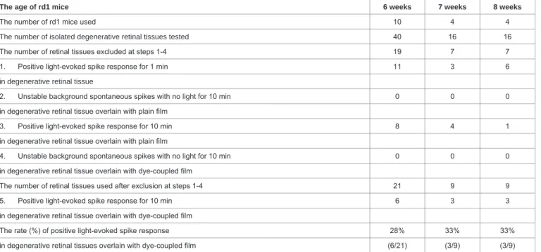
Table 3: The step-by-step procedure for evaluating light-evoked spike response induced by decay-simulated (30% surface absorption) photoelectric dye-coupled
polyethylene film in degenerative retinal tissues isolated from retinal dystrophic rd1 mice at the age of 6, 7, and 8 weeks.
The age of rd1 mice 6 weeks 7 weeks 8 weeks
The number of rd1 mice used 9 11 13
The number of isolated degenerative retinal tissues tested 35 44 39
The number of retinal tissues excluded at steps 1-4 18 16 10
1. Positive light-evoked spike response for 1 min 6 6 6
in degenerative retinal tissue
2. Unstable background spontaneous spikes with no light for 10 min 0 0 0
in degenerative retinal tissue overlain with plain film
3. Positive light-evoked spike response for 10 min 12 10 4
in degenerative retinal tissue overlain with plain film
4. Unstable background spontaneous spikes with no light for 10 min 0 0 0
in degenerative retinal tissue overlain with dye-coupled film
The number of retinal tissues used after exclusion at steps 1-4 17 28 29
5. Positive light-evoked spike response for 10 min 8 10 8
in degenerative retinal tissue overlain with dye-coupled film
The rate (%) of positive light-evoked spike response 47% 35% 27%
in degenerative retinal tissues overlain with dye-coupled film (8/17) (10/28) (8/29)
Table 2: Step-by-step procedure for evaluating light-evoked spike response induced by photoelectric dye-coupled polyethylene film in degenerative retinal tissues
with a plain film which showed light-evoked spikes were excluded at third step. At fifth step, light-evoked spikes were recorded in the degenerative retinal tissues overlain with a dye-coupled film (Figure 6 and 7). Residual light-evoked spikes were recorded at the first and third step in 18 of 35 retinal tissues (51%) at the age of 6 weeks, 16 of 44 retinal tissues (36%) at the age of 7 weeks, and 10 of 39 retinal tissues (25%) at the age of 8 weeks. At fifth step, light-evoked spikes were recorded with dye-coupled films in 8 of 17 retinal tissues (47%) at the age of 6 weeks, 10 of 28 retinal tissues (35%) at the age of 7 weeks, and 8 of 29 retinal tissues (27%) at the age of 8 weeks (Table 2).
Light-evoked spikes induced by decay-simulated dye-coupled film
Dye-coupled films which had the reduced amount of dye molecules coupled on the film surface were tested whether to induce light-evoked spikes in degenerative retinal tissues (Figure 7 and 8). After the exclusion of retinal tissues which showed residual light-evoked spikes at first and third step (Figure 3), dye-coupled films with 30% surface absorption compared with the standard dye-coupled film induced light-evoked spikes in 6 of 21 degenerative retinal tissues (28%) at the age of 6 weeks, 3 of 9 retinal tissues (33%) at the age of 7 weeks, and 3 of 9 retinal tissues (33%) at the age of 8 weeks (Table 3). In contrast, dye-coupled films with 7% surface absorption compared with the standard dye-coupled film induced light-evoked spikes in 0 of 3 degenerative retinal tissues (0%) at the age of 6 weeks, 1 of 8 retinal tissues (12%) at the age of 7 weeks, and 2 of 9 retinal tissues (22%) at the age of 8 weeks (Table 4).
Discussion
The goal of this study is to establish an in vitro assay system to test the efficacy of the photoelectric dye-coupled thin film retinal prosthesis as a preclinical test for a medical device. In our previous study, degenerative retinal tissues isolated from retinal dystrophic RCS rats were placed in the multielectrode array dish to record spikes in response to light [25]. The dye-coupled film was proven to induce light-evoked spikes in degenerative retinal tissues which were isolated from retinal dystrophic RCS rats at the old age of 12 weeks.
The reasons why the RCS rats were used in our preceding study were mainly two fold. Firstly, the RCS rats were used as in vivo animal studies to test the efficacy of the dye-coupled film implantation by behavior tests as well as electroretinographic and visual evoked potential recording [4,6,7,25,26]. Secondly, rats’ eyes are larger than mice eyes and technically easy to isolate retinal tissues in large size. However, it took a longer time to obtain RCS rats at the old age suitable for the testing, and RCS rats were expensive at cost. Therefore, retinal dystrophic rd1 mice which were available easily at low cost were used in this study.
Next, experimental procedures were designed especially from the viewpoint of setting the control. The plain film was used as a control for the dye-coupled film [4,6]. A major question in the experiment was whether residual photoreceptors might be present in the degenerative retinal tissues. Based on this assumption, we designed a step-by-step procedure to check light-evoked response derived from residual photoreceptors firstly in the degenerative tissue only and secondly in the degenerative tissue overlain with the plain film. The degenerative retinal tissues which showed light-evoked response derived from residual photoreceptors were thus excluded, and then, light-evoked response induced by the dye-coupled film was tested in the degenerative retinal tissues which were supposed to have no residual photoreceptors. The reason why the degenerative retinal tissue layered with the plain film showed light-evoked spike response even after the retinal tissues which showed light-evoked response had been excluded at the pretest would be as follows. The plain film with a platinum weight at top would flatten evenly the retinal tissue in close contact with the multielectrode array, leading to better capture of spikes in weaker amplitudes.
In the step-by-step procedure, about a half of the degenerative retinal tissues of rd1 mice at the age of 6 weeks and about a third of the retinal tissues of rd1 mice at the age of 7 weeks and 8 weeks were excluded prior to testing of the dye-coupled films. These facts indicate that residual photoreceptors were present at a higher rate in the degenerative retinal tissues at the younger age of 6 weeks,
The age of rd1 mice 6 weeks 7 weeks 8 weeks
The number of rd1 mice used 4 4 4
The number of isolated degenerative retinal tissues tested 14 16 16
The number of retinal tissues excluded at steps 1-4 11 8 7
1. Positive light-evoked spike response for 1 min 9 7 7
in degenerative retinal tissue
2. Unstable background spontaneous spikes with no light for 10 min 0 0 0
in degenerative retinal tissue overlain with plain film
3. Positive light-evoked spike response for 10 min 2 1 0
in degenerative retinal tissue overlain with plain film
4. Unstable background spontaneous spikes with no light for 10 min 0 0 0
in degenerative retinal tissue overlain with dye-coupled film
The number of retinal tissues used after exclusion at steps 1-4 3 8 9
5. Positive light-evoked spike response for 10 min 0 1 2
in degenerative retinal tissue overlain with dye-coupled film
The rate (%) of positive light-evoked spike response 0% 12% 22%
in degenerative retinal tissues overlain with dye-coupled film (0/3) (1/8) (2/9)
Table 4: Step-by-step procedure for evaluating light-evoked spike response induced by decay-simulated (7% surface absorption) photoelectric dye-coupled
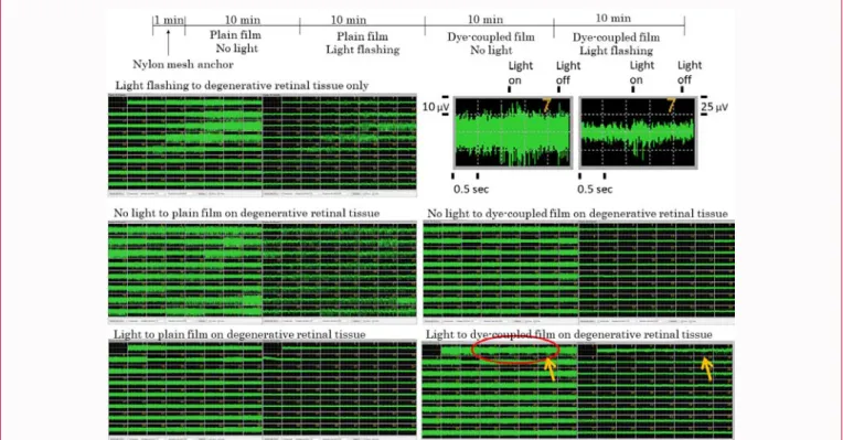
Figure 6: Step-by-step sequential recordings (top left to bottom right) of light-evoked response in degenerative retinal tissue of rd1 mouse at the age of 8 weeks.
Standard dye-coupled film was tested. Spike response with high pass at 100 Hz on left side panels with 64 electrode recording windows and corresponding electroretinogram-like response on right side panels with 64 electrode recording windows in the same time span. The timing of light on and off as well as the scale of vertical line (voltage) and horizontal line (time) are shown in inset figures which are the enlargement of electrode No. 59 recording window (orange arrows). Recording windows which show light-evoked response are marked with red circle.
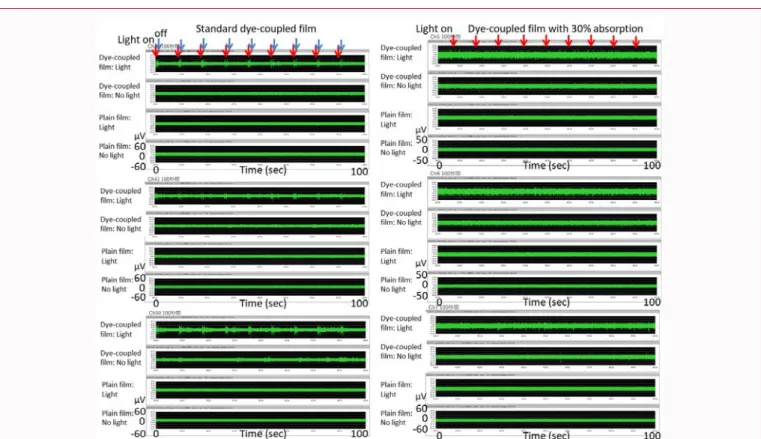
Figure 7: Left panels: Recordings in 100 seconds of light-evoked spike response in 3 channels of electrodes (Channels 26, 41, and 50) for standard dye-coupled
film (corresponding to recording in Fig. 6). Note large amplitudes in response to light on (red arrows) and light off (blue arrows). Right panels: Recordings in 100 seconds of light-evoked spike response in 3 channels of electrodes (Channels 5, 6, and 7) for decay-simulated dye-coupled film with 30% surface absorption (corresponding to recording in Fig. 8). Note smaller and vague amplitudes in response only to light on (red arrows), compared with the standard dye-coupled film (left panels).
Figure 8: Step-by-step sequential recordings (top left to bottom right) of light-evoked response in degenerative retinal tissue of rd1 mouse at the age of 8 weeks.
Decay-simulated dye-coupled film with surface absorption reduced to 30% of the standard was tested. Spike response with high pass at 100 Hz on left side panels with 64 electrode recording windows and corresponding electroretinogram-like response on right side panels with 64 electrode recording windows in the same time span. The timing of light on and off as well as the scale of vertical line (voltage) and horizontal line (time) are shown in inset figures which are the enlargement of electrode No. 7 recording window (orange arrows). Recording windows which show light-evoked response are marked with red circle.
compared with the age of 7 weeks and 8 weeks. After the exclusion of the degenerative tissues with residual photoreceptors, positive light-evoked spikes were induced by the dye-coupled film in about a half of the retinal tissues of rd1 mice at the age of 6 weeks and in about a third of the retinal tissues of rd1 mice at the age of 7 weeks and 8 weeks.
The reasons why only the part of the degenerative retinal tissues showed light-evoked spikes induced by the dye-coupled film are as follows. Firstly, surgical manipulation to extirpate the retinal tissue from the mice eyes would cause serious damage to neurons. Secondly, the absence of light-evoked input to retinal neurons would reduce the neuronal responsiveness. A higher rate of light-evoked response in the degenerative retinal tissues of rd1 mice at the age of 6 weeks, compared with the age of 7 and 8 weeks, suggests the long-term absence of input as a cause for no response in the degenerative retinal tissues. Furthermore, synaptic transmission between bipolar cells and ganglion cells might be down-regulated after the long-term absence of input as the consequence of photoreceptor loss. The dye-coupled film would first induce membrane potential changes in bipolar cells and then, synaptic transmission would induce action potential in retinal ganglion cells in the degenerative retinal tissues. Of course, the dye-coupled film would also stimulate directly the retinal ganglion cells to induce action potential.
As a major limitation in the present study, positive light-evoked response was defined as recording of spikes at two or more neighboring electrodes of the multielectrode array. The reasons why light-evoked spikes were recorded only at the limited number of electrodes are as follows. Firstly, the degenerative retinal tissues would have fatal damage in some parts which would be caused by surgical manipulation. Secondly, the distance from each electrode to
the degenerative retinal tissue would be varying because the retinal tissue beneath the dye-coupled film overlain with a platinum weight was floatable in the fluid. Too much compression of the degenerative retinal tissue by the dye-coupled film would damage the retinal tissue mechanically and would prevent the retinal tissue from oxygenation in the fluid. The varying distance from each electrode would lead to varying amplitudes of spikes recorded by the multielectrode array, leading to ambiguous spikes in the background of spontaneous spike firing. How to put a weight on the degenerative retinal tissue to be brought into close proximity to the multielectrode array remains a technical limiting step in recording light-evoked spikes [27].
Another limitation will arise from the fact that the degenerative retinal tissue in rats and mice has been known to generate spontaneous spikes, irrespective of light stimulation [24]. We had to wait for a while to start the measurement until the spontaneous spikes would become stable. Under the circumstances, light-evoked spikes were defined ideally as increased amplitudes of spikes in response to light in the background of spontaneous spike firing. In fact, the increased amplitudes were judged from visibly recognizable changes corresponding to the timing of light-on and -off in the recording window at each electrode in the passage of 10 min. Therefore, in the present study, screenshots of all electrode recordings and long time-range recordings in time-shrunken style were shown to visualize light-evoked spike response. It should be noted that the dye-coupled film induced spike response in the degenerative retinal tissue to the timing of light-on as well as the timing of light-off. The spike response to light-on and -off could be explained by the fact that surface electric potential on the dye-coupled film changes robustly at the timing of light-on and -off in the measurement by Kelvin probe [11,25]. In future perspectives, positive light-evoked spikes would be defined
more specifically in a quantitative manner, based on the accumulation of recording data which would be analyzed by a new method.
In this study, decay-simulated thin films with reduced surface amount of photoelectric dye were also tested whether to induce light-evoked spikes in the degenerative retinal tissues of rd1 mice. Dye-coupled polyethylene film with 30% reduced absorption, compared with the standard, was set on the basis of similar reduction of surface absorption of the standard dye-coupled films which had been implanted in the subretinal space of monkey eyes for 6 months and then removed surgically for spectroscopic analysis [18]. Positive light-evoked spikes were induced by the 30% reduced absorption dye-coupled film in about a third of the degenerative retinal tissues of rd1 mice at the age of 6, 7, and 8 weeks. In contrast, dye-coupled polyethylene film with further reduction of absorption to 7% could induce positive light-evoked spikes in a limited number of the degenerative retinal tissues of rd1 mice at the age of 6, 7, and 8 weeks. The 7% reduced absorption dye-coupled film would be comparable to decay which would be caused by subretinal implantation for many years. It should be noted that amplitudes of light-evoked spikes induced by the dye-coupled film with reduced surface absorption were smaller, compared with amplitudes induced by the standard dye-coupled film. Overall, functional property of the dye-coupled film with reduced surface absorption suggests that the dye-coupled film in the subretinal space of the eye would be durable at least for half a year to generate spikes in the degenerative retina.
In the present study, the intensity of light to stimulate the dye-coupled film was not changed and was set at about 5 Lux. This luminosity was simply a measured value at the level of multielectrode array dish on the stage when the light source was placed at the bottom of the dissecting microscope. At this distance of the light source, no noise in response to on-and-off timing of light was elicited, as shown on the recordings in retinal tissues with the plain film. Hexagonal nut anchor and platinum weight would not catch electric noise from the light source, based on the part of recordings in retinal tissues with hexagonal nut anchor and in retinal tissues layered by the plain film with platinum weight, respectively. The intensity of light might be weak from the standpoint of electric potential properties of the dye-coupled film, measured by Kelvin probe [11,25]. The weak intensity of light might be one reason for no light-evoked response in some degenerative retinal tissues. We did not attempt to increase the light intensity in the present study since the light-evoked response was certainly induced by the photoelectric dye-coupled film in retinal degenerative tissues.
Ideally, the intensity of light would be changed, and hence, amplitudes and frequency of light-evoked spikes would be evaluated in a quantitative manner. The present study aimed to detect the light-evoked response of the degenerative retinal tissues toward the dye-coupled film in a qualitative manner. Future advance in methodology would lead to more quantitative evaluation of the light-evoked response. The assay system would be further refined to use, for instance, a reagent (2S-amino-4-phosphono-buranoic acid: L-AP4) to block specifically the synaptic transmission between photoreceptor cells and on-type bipolar cells, leading to no influence by residual photoreceptor cells. Furthermore, cultured neuronal cells would be a better alternative sample for the multielectrode array recording since neuronal cells were used to detect the light-evoked response induced by the same photoelectric dye in patch clamp technique [28].
Conclusion
A preclinical in vitro test to assess the efficacy of the photoelectric dye-coupled thin film retinal prosthesis was established in the present study to measure light-evoked spikes by the multielectrode array in the degenerative retinal tissue isolated from retinal dystrophic rd1 mice eyes. As a major limitation, the definition of positive light-evoked response remains to be simply as visibly recognizable spikes in response to repeat cycles of on-and-off light, and thus, would be vulnerable to the distance between the retinal tissue and each recording electrode in the array. The present in vitro biological assay with isolated degenerative retinal tissues could be also used to test samples of the dye-coupled film from the viewpoint of quality management in the manufacture. Together with the results in animal studies, the efficacy of the dye-coupled film which has been proven in this study would solidify the base for a first-in-human clinical trial of the dye-coupled film as retinal prosthesis in patients with retinitis pigmentosa who have lost the vision [4,6,7,18,29,30].
Funding
This study was supported in part by a grant for the Practical Research Program for Rare/Intractable Diseases (18950217, 2018) from the Japan Agency for Medical Research and Development (AMED).
Data Availability
The datasets generated and analyzed in this study are available from the corresponding author upon reasonable request.
Authors Contribution
TM, MS, and TU were involved in conception or design of the work, did data collection, data analysis and interpretation. KT, SL, and KY did data collection, data analysis and interpretation. TM drafted the article. All authors were involved in critical revision of the article and final approval of the version.
References
1. Hetherington L, Benn M, Coffey PJ, Lund RD. Sensory capacity of the Royal College of Surgeons rat. Invest Ophthalmol Vis Sci. 2000;41(12):3979-83. 2. D’Cruz PM, Ysumura D, Weir J, Matthes MT, Abderrahim H, Lavail MM,
et al. Mutation of the receptor tyrosine kinase gene mertk in the retinal dystrophic RCS rat. Hum Mol Genet. 2000;9(4):645-51.
3. Chang B, Hawes NL, Hurd RE, Davisson MT, Nusinowitz S, Heckenlively JR. Retinal degeneration mutants in the mouse. Vision Res. 2002;42(4):517-25.
4. Alamusi, Matsuo T, Hosoya O, Tsutsui KM, Uchida T. Behavior tests and immunohistochemical retinal response analyses in RCS rats with subretinal implantation of Okayama-University-type retinal prosthesis. J Artif Organs. 2013;16(3):343-51.
5. Michalakis S, Koch S, Sothilingam V, Garcia Garrido M, Tanimoto N, Schulze E, et al. Gene therapy restores vision and delays degeneration in the CNGB1(-/-) mouse model of retinitis pigmentosa. Adv Exp Med Biol. 2014;801:733-9.
6. Alamusi, Matsuo T, Hosoya O, Tsutsui KM, Uchida T. Vision maintenance and retinal apoptosis reduction in RCS rats with Okayama University-type retinal prosthesis (OURePTM) implantation. J Artif Organs. 2015;18(3):264-71.
7. Alamusi, Matsuo T, Hosoya O, Uchida T. Visual evoked potential in RCS rats with Okayama University-type retinal prosthesis (OURePTM) implantation. J Artif Organs. 2017;20(2):158-65.
8. Nakamura Y, Matsuo J, Miyamoto N, Ojima A, Ando K, Kanda Y, et al. Assessment of testing methods for drug-induced repolarization delay and arrhythmias in an iPS cell-derived cardiomyocyte sheet: Multi-site validation study. J Pharmacol Sci. 2014;124(4):494-501.
9. Loewenstein JI, Montezuma SR, Rizzo JF III. Outer retinal degeneration: An electronic retinal prosthesis as a treatment strategy. Arch Ophthalmol. 2004;122(4):587-96.
10. Humayun MS, Juan E Jr, Dagnelie G. The bionic eye: A quarter century of retinal prosthesis research and development. Ophthalmology. 2016;123(10S):S89-97.
11. Matsuo T, Uchida T, Takarabe K. Safety, efficacy, and quality control of a photoelectric dye-based retinal prosthesis (Okayama University-type retinal prosthesis) as a medical device. J Artif Organs. 2009;12(4):213-25. 12. Matsuo T. A simple method for screening photoelectric dyes towards their
use for retinal prostheses. Acta Med Okayama. 2003;57(5):257-60. 13. Uchida T, Ishimaru S, Shimamura K, Uji A, Matsuo T, Ohtsuki H.
Immobilization of photoelectric dye on the polyethylene film surface. Mem Fac Eng Okayama Univ. 2005;39:16-20.
14. Matsuo T, Dan-oh Y, Suga S (Inventors). Agent for inducing receptor potential. Assignee: Okayama University. United States Patent. Patent No.: US 7,101,533 B2. Date of Patent. 2006.
15. Uji A, Matsuo T, Ishimaru S, Kajiura A, Shimamura K, Ohtsuki H, et al. Photoelectric dye-coupled polyethylene film as a prototype of retinal prostheses. Aritif Organs. 2005;29(1):53-7.
16. Uji A, Matsuo T, Uchida T, Shimamura K, Ohtsuki H. Intracellular calcium response and adhesiveness of chick embryonic retinal neurons to photoelectric dye-coupled polyethylene films as prototypes of retinal prostheses. Artif Organs. 2006;30(9):695-703.
17. Tamaki T, Matsuo T, Hosoya O, Tsutsui KM, Uchida T, Okamoto K, et al. Glial reaction to photoelectric dye-based retinal prostheses implanted in the subretinal space of rats. J Artif Organs. 2008;11:38-44.
18. Matsuo T, Uchida T, Sakurai J, Yamashita K, Matsuo C, Araki T, et al. Visual evoked potential recovery by subretinal implantation of photoelectric dye-coupled thin film retinal prosthesis in monkey eyes with macular degeneration. Artif Organs. 2018;42(8):E186-203.
19. Matsuo T, Uchida T, Nitta M, Yamashita K, Takei S, Ido D, et al. Subretinal implantation of Okayama University-type retinal prosthesis (OURePTM) in canine eyes by vitrectomy. J Vet Med Sci. 2017;79(12):1939-46.
20. Matsuo T, Uchida T, Yamashita K, Takei S, Ido D, Tanaka M, et al. Visual evoked potential in rabbits’ eyes with subretinal implantation by vitrectomy of Okayama University-type retinal prosthesis (OURePTM). J Vet Med Sci. 2018;80(2):247-59.
21. Okamoto K, Matsuo T, Tamaki T, Uji A, Ohtsuki H. Short-term biological safety of a photoelectric dye used as a component of retinal prostheses. J Artif Organs. 2008;11(1):45-51.
22. Liu S, Matsuo T, Hosoya O, Uchida T. Photoelectric dye used for Okayama University-type retinal prosthesis reduces the apoptosis of photoreceptor cells. J Ocul Pharmacol Ther. 2017;33(3):149-60.
23. Reinhard K, Tikidji-Hamburyan A, Seitter H, Idrees S, Mutter M, Benkner B, et al. Step-by-step instructions for retina recordings with perforated multi electrode arrays. PLoS ONE. 2014;9:e106148.
24. Fujii M, Sunagawa GA, Kondo M, Takahashi M, Mandai M. Evaluation of micro Electroretinograms Recorded with Multiple Electrode Array to Assess Focal Retinal Function. Sci Rep. 2016;6:30719.
25. Matsuo T, Sakurai M, Terada K, Uchida T, Yamashita K, Tanaka T, et al. Photoelectric dye-coupled polyethylene film: Photoresponsive properties evaluated by Kelvin probe and in vitro biological response detected in dystrophic retinal tissue of rats. Adv Biomed Eng. 2019;8:137-44. 26. Matsuo T, Uchida T, Yamashita K, Takei S, Ido D, Fujiwara A, et al. Vision
evaluation by functional observational battery, operant behavior test, and light/dark box test in retinal dystrophic RCS rats versus normal rats. Heliyon. 2019;5(6):e01936.
27. Eleftheriou CG, Zimmermann JB, Kjeldsen HD, David-Pur M, Hanein Y, Sernagor E. Carbon nanotube electrodes for retinal implants: A study of structural and functional integration over time. Biomaterials. 2017;112:108-21.
28. Huang F, Bladon J, Lagoy RC, Shorrock PN Jr, Hronik-Tupaj M, Zoto CA, et al. A photosensitive surface capable of inducing electrophysiological changes in NG108-15 neurons. Acta Biomater. 2015;12:42-50.
29. Matsuo T, Morimoto N. Visual acuity and perimacular retinal layers detected by optical coherence tomography in patients with retinitis pigmentosa. Br J Ophthalmol. 2007;91(7):888-90.
30. Tamaki M, Matsuo T. Optical coherence tomographic parameters as objective signs for visual acuity in patients with retinitis pigmentosa, future candidates for retinal prostheses. J Artif Organs. 2011;14:140-50.