Variable G Protein Determinants of GPCR
Coupling Selectivity
著者
Najeah Okashah, Qingwen Wan, Soumadwip Ghosh,
Manbir Sandhu, Asuka Inoue, Nagarajan Vaidehi,
Nevin A Lambert
journal or
publication title
Proceedings of the National Academy of
Sciences
volume
116
number
24
page range
12054-12059
year
2019-05-29
URL
http://hdl.handle.net/10097/00128250
doi: 10.1073/pnas.1905993116
Variable G protein determinants of GPCR
coupling selectivity
Najeah Okashaha, Qingwen Wana, Soumadwip Ghoshb, Manbir Sandhub, Asuka Inouec, Nagarajan Vaidehib, and Nevin A. Lamberta,1
aDepartment of Pharmacology and Toxicology, Medical College of Georgia, Augusta University, Augusta, GA 30912;bDepartment of Computational and
Quantitative Medicine, Beckman Research Institute of the City of Hope, Duarte, CA 91010; andcGraduate School of Pharmaceutical Sciences, Tohoku
University, Sendai 980-8578, Japan
Edited by Brian K. Kobilka, Stanford University School of Medicine, Stanford, CA, and approved May 8, 2019 (received for review April 9, 2019)
G protein-coupled receptors (GPCRs) activate four families of heterotrimeric G proteins, and individual receptors must select a subset of G proteins to produce appropriate cellular responses. Although the precise mechanisms of coupling selectivity are uncertain, the Gα subunit C terminus is widely believed to be the primary determinant recognized by cognate receptors. Here, we directly assess coupling between 14 representative GPCRs and 16 Gα subunits, including one wild-type Gα subunit from each of the four families and 12 chimeras with exchanged C termini. We use a sensitive bioluminescence resonance energy transfer (BRET) assay that provides control over both ligand and nucleotide bind-ing, and allows direct comparison across G protein families. We
find that the Gs- and Gq-coupled receptors we studied are
rela-tively promiscuous and always couple to some extent to Gi1
het-erotrimers. In contrast, Gi-coupled receptors are more selective.
Our results with Gα subunit chimeras show that the Gα C terminus
is important for coupling selectivity, but no more so than the Gα
subunit core. The relative importance of the Gα subunit core and C
terminus is highly variable and, for some receptors, the Gα core is
more important for selective coupling than the C terminus. Our results suggest general rules for GPCR-G protein coupling and demonstrate that the critical G protein determinants of selectivity vary widely, even for different receptors that couple to the same G protein.
GPCR
|
G protein-coupled receptor|
G protein selectivity|
ternary complexG
protein coupled receptors (GPCRs) exert many of their physiological effects by coupling to and activating hetero-trimeric G proteins. The 16 human Gα subunit genes are classified into four families (Gs, Gi/o, Gq/11, and G12/13), and members of each family interact with different effector molecules to produce distinct cellular responses (1, 2). Individual cells generally express many different G proteins from multiple families, and, therefore, GPCRs must be able to selectively activate subsets of G proteins. Many receptors show a preference for G proteins from just one of the four families, although promiscuous coupling to G proteins from multiple families is also not uncommon (3, 4). The mechanisms of selective GPCR-G protein coupling are not completely understood, and the structural determinants of selectivity have not been de-termined with precision. Analysis of GPCR primary sequences has not revealed simple conserved motifs for coupling to different G protein families (5), and analysis of GPCR–G protein complex structures has only begun to uncover features that may be impor-tant for coupling selectivity (6–12). From the standpoint of the G protein, it has long been appreciated that the Gα carboxy (C) terminus is critical for activation by receptors and also a key de-terminant of selectivity (13–17). The distal part of the C-terminal alpha helix (helix 5; H5) is enveloped by GPCR transmembrane (TM) domains during coupling (6), and several studies have shown that mutations within this region can promote coupling of receptors to noncognate heterotrimers (17–21). Although other Gα regions have been shown to be important for recognition by individual GPCRs (22–25), the Gα C terminus is widely considered to be the most important structural determinant of coupling selectivity.Here, we test this idea by measuring coupling of GPCRs to four representative Gα subunits (Gαs, Gαi1, Gαq, and Gα12) and 12 Gα chimeras with the C terminus of one representative and the Gα core region of another. We monitor coupling using a sensitive energy transfer assay that allows comparison of all GPCR–G protein combinations directly at the coupling step. Our results demonstrate unexpectedly promiscuous coupling of many GPCRs to wild-type G proteins and support the notion that the Gα C terminus is one important determinant of coupling selectivity for many receptors. However, our results also show that the Gα C terminus is a minor contributor to selective cou-pling of several other receptors. Taken together our findings suggest that GPCRs recognize widely distributed structural fea-tures of G proteins and are consistent with the suggestion that different GPCRs recognize different conserved selectivity de-terminants of a given G protein (5).
Results
Promiscuous Coupling to Wild-Type G Proteins.Coupling of a GPCR
to a heterotrimeric G protein can be defined as the allosteric interaction between the ligand binding site of the receptor and the nucleotide binding site of the associated Gα subunit, such that the agonist-bound state of the receptor promotes the nucleotide-free (empty) state of the G protein and vice versa (26, 27) (Fig. 1A). To quantify coupling we monitored agonist-dependent and nucleotide-sensitive association of GPCRs with
Significance
G protein-coupled receptors (GPCRs) regulate a wide variety of important cellular processes and are targeted by a large fraction of approved drugs. GPCRs signal by activating heterotrimeric G proteins and must couple to a select subset of G proteins to produce appropriate intracellular responses. It is not known how GPCRs select G proteins, but it is generally accepted that the Gα subunit C terminus is the primary G protein determinant of coupling selectivity. We systematically studied coupling of GPCRs to four families of G proteins and chimeras with C ter-minal regions that were exchanged between families. We un-covered rules for coupling selectivity and found that different GPCRs can recognize different features of the same G protein for selective coupling.
Author contributions: N.O., N.V., and N.A.L. designed research; N.O., Q.W., S.G., and M.S. performed research; A.I. contributed new reagents/analytic tools; N.O., M.S., A.I., N.V., and N.A.L. analyzed data; and N.O., N.V., and N.A.L. wrote the paper.
The authors declare no conflict of interest. This article is a PNAS Direct Submission.
This open access article is distributed underCreative Commons Attribution-NonCommercial-NoDerivatives License 4.0 (CC BY-NC-ND).
1To whom correspondence may be addressed. Email: [email protected].
This article contains supporting information online atwww.pnas.org/lookup/suppl/doi:10. 1073/pnas.1905993116/-/DCSupplemental.
different heterotrimers using bioluminescence resonance energy transfer (BRET) (28). To interfere as little as possible with receptor–G protein interactions, we positioned BRET donor and acceptor labels far from regions identified in functional and structural studies as important for coupling. A luciferase was fused to the GPCR C terminus, and complementary fragments of the fluorescent protein Venus were fused to Gβ1and Gγ2 sub-units. These components were transfected together with un-labeled Gα subunits into CRISPR/Cas9-edited HEK293 cells deficient in either Gαs, Gαq, and Gα12subunit families (three-family knockouts; 3GKO) or all four Gα families (four-(three-family knockouts; 4GKO) (29). When 3GKO cells were used, the S1 subunit of pertussis toxin (PTX) was coexpressed to prevent coupling to endogenous Gαi/o subunits, except when the over-expressed Gα subunit was itself sensitive to PTX. Cells were permeabilized with digitonin and either supplemented with GDP or treated with apyrase to remove residual nucleotides.
Complexes between agonist-bound GPCRs and G proteins are short-lived when guanine nucleotides are present at concentra-tions similar to those found in the cytosol of intact cells and are greatly stabilized when guanine nucleotides are absent (30). Accordingly, nucleotide depletion significantly enhanced the magnitude of agonist-induced BRET between receptors and G
proteins (Fig. 1B), and maximal BRET was observed when ag-onist was present and GDP was absent (Fig. 1C). As a simple index of coupling, we definedΔBRETmaxas the difference be-tween the BRET observed when receptor–G protein complexes are least stable (agonist absent, GDP present) and that observed when receptor–G protein complexes are most stable (agonist present, GDP absent). This experimental system allows direct comparison of receptor coupling to heterotrimers with Gα sub-units from different families and is sufficiently sensitive to detect secondary coupling to nonpreferred G proteins. Using this assay, we found that receptors generally coupled to the same G pro-teins as indicated by other methods and as annotated in the IUPHAR/BPS Guide to Pharmacology (3, 31). However, many receptors coupled to wild-type G proteins more promiscuously than expected. For example, we found that H1 histamine re-ceptors (H1R), which are generally classified as Gq-coupled (3), couple strongly to both Gqand Giheterotrimers, couple weakly to Gsheterotrimers, and do not couple significantly to G12 het-erotrimers (Fig. 1D). The EC50values for H1R coupling to Gq and Giwere comparable (Fig. 1E), suggesting that activation of Giheterotrimers by this receptor may have physiological signif-icance, as some studies have indicated (32). We carried out similar experiments on a panel of 19 receptors (SI Appendix,
Table S1), 17 of which are classified in the IUPHAR/BPS Guide
to Pharmacology (3) as coupling primarily to Gs, Gi, or Gq proteins, and two of which are unclassified orphan receptors that transduce signals through G12/13proteins (33, 34). We found that promiscuous coupling to more than one wild-type G protein was a universal feature of the Gs-, Gq- and G12-coupled receptors that we studied. In contrast, the Gi-coupled receptors that we studied were much more selective for Giheterotrimers (Fig. 2 andSI Appendix, Fig. S1). From the perspective of the G protein, we found that Giheterotrimers coupled to all of the receptors that we studied. It should be noted that the assay we used for these studies reflects allosteric coupling between agonist and nucleotide binding sites, but this is only a minimum requirement for agonist-dependent G protein signaling. Additional factors may be required for efficient G protein activation and signaling under physiological conditions in cells (see below).
Coupling to G Protein C-Terminal Chimeras.To assess the importance
of the Gα C terminus for coupling selectivity, we constructed 12 Gα chimeras consisting of the main Gα subunit core [HN.1-H5.16 using
C
E
D
- -(PTX) s i q 12 0.00 0.05 0.10 0.15 Gα subunit Δ BRET max n.s. -9 -7 -5 -3 0.0 0.1 0.2 log [HA] M net BRET control Gαq Gαi Gαβγ-Venus GPCR-Rluc8 agonist GDPB
A
20 sec apyrase GDP ΔBRET 0.1 histamine H1R-Rluc8, Gαi net BRET Gαs, PTX 0.0 0.1 0.2 - HA - HA GDP apy Gαi 0.0 0.1 0.2 - HA - HA GDP apy Gαq, PTX 0.0 0.1 0.2 - HA - HA GDP apy Gα12, PTX 0.0 0.1 0.2 - HA - HA GDP apyFig. 1. Coupling to wild-type heterotrimers in permeabilized, nucleotide-depleted cells. (A) Receptors were fused to Renilla luciferase (Rluc8), and Gβγ dimers were fused to Venus. (B) Time course of BRET between H1R-Rluc8 and
Gαi1βγ-Venus in response to 100 μM histamine (HA) in permeabilized cells in
the presence (GDP) and absence (apyrase) of nucleotides. (C and D) H1R
couples to Gs, Gi1, and Gqheterotrimers; **P= 0.0011, ****P < 0.0001, n.s.
not significant; one-way ANOVA, Sidak’s multiple comparisons. Pertussis toxin S1 subunit (PTX) was coexpressed in experiments with Gαs, Gαq, and
Gα12. (E) Similar potency of histamine-induced coupling to Gi1 (EC50 =
258 nM; n= 4) and Gq(EC50= 245 nM; n = 7) heterotrimers.
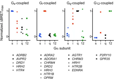
Normalized Δ BRET max Gα subunit s i q 12 0 1 Gs-coupled ADRB2 AVPR2 DRD1 HRH2 HTR4 s i q 12 0 1 Gi-coupled ADRA2 ADORA1 CHRM4 DRD2 HRH3 HTR1B OPRM s i q 12 0 1 Gq-coupled AGTR1 CHRM3 HRH1 HTR2B EDNRA s i q 12 0 1 G12-coupled P2RY10 GPR35
Fig. 2. Gi-coupled receptors are relatively selective. NormalizedΔBRETmax
for a panel of Gs-, Gi-, Gq-, and G12-coupled receptors. Each value represents
the meanΔBRETmax(normalized to the value obtained using the presumed
cognate G protein) from n= 3–6 independent experiments. Individual data points for each receptor are shown inSI Appendix, Fig. S1.
Okashah et al. PNAS | June 11, 2019 | vol. 116 | no. 24 | 12055
P
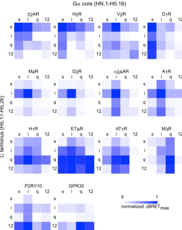
the common G protein numbering (CGN) system] of Gαs(long isoform), Gαi1, Gαq, or Gα12and the final 10 amino acids (H5.17– 26) of each of the others. This portion of the Gα C terminus is deeply embedded in the TM domains of GPCRs in all receptor–G protein complex structures and has been called the interface module of the C-terminal alpha helix (helix 5 or H5) (35). Previous mutagenesis studies have primarily targeted residues within this region to alter coupling selectivity (36). This region is also un-structured in G protein crystal structures (35), suggesting that it is not involved in folding or stability of the Gα subunit core and, therefore, can be exchanged without compromising the structural integrity of heterotrimers. We confirmed the ability of Gα chimeras to form heterotrimers by monitoring sequestration of overexpressed Gβγ dimers (37), and all expressed at comparable levels as in-dicated by immunoprecipitation (SI Appendix, Fig. S2). Conditions were optimized to provide roughly equivalent stoichiometry for each receptor–G protein pair, with the latter in excess. For sim-plicity, we refer to heterotrimers that incorporate these chimeras as Gsi, Gsq, Gs12. We generated full coupling profiles (4 wild-type Gα subunits and 12 chimeras) for 14 GPCRs and plotted the results as ΔBRETmaxheat maps (Fig. 3). Similar heat maps were constructed for GDP-sensitive BRET in the absence of agonist (ΔBRETGDP;SI
Appendix, Fig. S3) and agonist-induced BRET in the presence of
GDP (ΔBRETag;SI Appendix, Fig. S4).
In agreement with our results using wild-type Gα subunits, we found that Gs-, Gq- and G12-coupled receptors coupled more promiscuously to G protein chimeras than Gi-coupled receptors.
The Gs-, Gq- and G12-coupled receptors that we studied always coupled well to several chimeras in addition to wild-type G proteins, sometimes withΔBRETmaxvalues that approached or even exceeded that of the cognate wild-type G protein (Fig. 3). The Gi-coupled receptors that we studied were more selective. For example, the D2dopamine receptor (D2R) failed to couple to any Gα chimera with a ΔBRETmaxvalue that reached half of that observed with wild-type Gαi1. We also observed that the Gα12core was often nonpermissive for coupling, particularly for Gi- and Gq-coupled receptors. This was sometimes partially overcome when a cognate C terminus was present, e.g., for A1R and H1R. By comparison, the Gα12 C terminus was more ac-commodating to most receptors (Fig. 3). This suggests that many receptors reject Gα12core regions rather than the C terminus to select against G12heterotrimers. The most promiscuous receptor that we studied was the endothelin A (ETAR) receptor, which coupled well to Gαi1, Gαq, Gα12, and eight of the nine chimeras bearing the C termini of these subunits. This receptor coupled poorly to Gαsand chimeras bearing the GαsC terminus, sug-gesting that selection against Gs coupling for this receptor is determined primarily by the C terminus.
We anticipated that GPCRs would couple poorly to chimeras bearing noncognate C termini, i.e., adding a noncognate C ter-minus to a cognate Gα core would impair coupling. Indeed, this was observed for all 42 receptor-chimera pairs that we studied. However, the extent of impairment was highly variable, ranging from a near complete loss of coupling to almost no loss (SI Appendix, Fig. S5A). Gi-coupled receptors, in particular the M4 acetylcholine receptor (M4R) and D2R, were the most sensitive to noncognate C termini. Conversely, we expected that GPCRs would generally couple well to chimeras bearing cognate C ter-mini, i.e., noncognate Gα core regions would only modestly impair coupling. However, we found that a noncognate Gα core was often as detrimental to coupling as a noncognate C terminus
(SI Appendix, Fig. S5B). Although adding a cognate C terminus
to a noncognate Gα core usually enhanced coupling, the gain of coupling was again highly variable and rarely a complete “switch.” For most of the receptors that we studied, coupling selectivity was determined both by the Gα subunit core and the C terminus. A clear C terminus-dominant coupling pattern was observed for only a few receptors, the most obvious examples being H1R, P2RY10, and GPR35 (Fig. 3). Other receptors, such as H2R and A1R, showed a more balanced influence of the cognate C terminus and G protein core. These receptors coupled equally well to chimeras bearing either the cognate C terminus or the cognate G protein core, although coupling to these chi-meras was never as efficient as coupling to cognate wild-type G proteins. Unexpectedly, D1R and M3R showed coupling that was determined almost entirely by the G protein core and was vir-tually insensitive to the Gα C terminus (Fig. 3). Full concentra-tion–response curves for these receptors confirmed the patterns observed with single saturating concentrations of agonists. Spe-cifically, for the D1R and M3R agonist potency was greater for chimeras with cognate G protein cores, whereas for the H1R agonist potency was greater for chimeras with cognate C termini (SI Appendix, Fig. S6 and Table S2).
Efficient coupling to G proteins with noncognate Gα C ter-mini was unexpected, and suggested either that the C terminus contributes little to the stability of some receptor–G protein complexes or, alternatively, that some receptors can interact strongly with both cognate and noncognate C termini. To better understand the basis of efficient coupling to Gα subunits with noncognate C termini, we performed atomistic molecular dy-namics (MD) simulations of D1R complexes with Gs, Gsi, Gsq, and Gs12. These simulations revealed that in all cases, the C terminus contributed a substantial portion (at least one-third) of the total Gsprotein interaction energy with D1R (SI Appendix, Fig. S7B) and, therefore, was critical for the overall stability of
s i q 12 s i q 12 s i q 12 s i q 12 s i q 12 s i q 12 s i q 12 s i q 12 M4R D2R α2AAR A1R s i q 12 s i q 12 s i q 12 s i q 12 s i q 12 s i q 12 s i q 12 s i q 12 β2AR H2R V2R D1R Gα core (HN.1-H5.16) C terminus (H5.17-H5.26) normalized ΔBRETmax 0 1 s i q 12 s i q 12 s i q 12 s i q 12 P2RY10 GPR35 s i q 12 s i q 12 s i q 12 s i q 12 s i q 12 s i q 12 H1R AT1R M3R s i q 12 s i q 12 ETAR
Fig. 3. Coupling to heterotrimers with C-terminal Gα subunit chimeras. Heat maps ofΔBRETmax(normalized to the largest value) for Gs-coupled
(Top), Gi-coupled (second row), Gq-coupled (third row), and G12-coupled
(Bottom) receptors. Maps are arranged from left to right depending on coupling that is determined more (e.g., H1R) or less (e.g., D1R) by the Gα C
terminus (H5.17–H5.26). Each value represents the mean normalized ΔBRETmaxfrom n= 3–6 independent experiments.
the complex. However, this receptor accommodated noncognate C termini with small energetic penalties (SI Appendix, Fig. S7), which corresponds to the observation that the C terminus was not critical for D1R selectivity. These simulations also identified several regions of the Gαscore that contribute to the interaction and may also contribute to coupling selectivity for this receptor, including HN, the hns1, s2s3, and h4s6 linkers and N-terminal residues of H5 (SI Appendix, Fig. S7A).
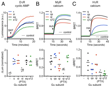
We then asked if receptors that interacted well with nucleotide-free G proteins bearing noncognate Gα C termini in our direct BRET coupling assay and MD simulations could also efficiently activate these G proteins and produce downstream signals. We found that wild-type Gs, Gsi, Gsq, and Gs12all ef-fectively restored potent activation of adenylate cyclase in re-sponse to D1R activation in cells lacking endogenous Gαs subunits (Fig. 4A andSI Appendix, Fig. S8), whereas our assays did not detect signals mediated by wild-type Gi1, Gq, or G12in these cells (SI Appendix, Fig. S9). Likewise, we found that wild-type Gq, Gqi, Gqs, and Gq12 all fully restored calcium release from intracellular stores in response to M3R activation in cells lacking endogenous Gαqsubunits (Fig. 4B andSI Appendix, Fig. S8), whereas our assays did not detect signals mediated by wild-type Gs, Gi1, or G12in these cells (SI Appendix, Fig. S9). Notably, both D1R and M3R coupled significantly to Gi1heterotrimers in our direct BRET assay, with ΔBRETmaxvalues that were ap-proximately half that of the respective cognate heterotrimers (Fig. 2) but failed to inhibit cAMP accumulation through Gi1(SI
Appendix, Fig. S9). This suggests that receptors may couple to
heterotrimers, as defined by formation of an agonist- and nucleotide-sensitive complex, and yet fail to activate a given signaling pathway even when overexpressed. In contrast to what we observed with D1R and M3R, G protein chimeras with non-cognate C termini only partially restored calcium release from intracellular stores in response to H1R activation. The rank or-der of restoration by chimeras was Gqi> Gqs> Gq12(Fig. 4C), which was consistent with the rank order of H1R coupling to
wild-type Gα subunits (Fig. 1). These results confirm that some GPCRs can efficiently couple to and activate heterotrimers with a variety of noncognate Gα C termini. Taken together with our direct coupling BRET results, these findings indicate a highly variable influence of the Gα subunit C terminus on coupling selectivity for different GPCRs.
Discussion
In this study, we used energy transfer between GPCRs and heterotrimeric G proteins to monitor allosteric coupling between agonist and nucleotide binding. This approach allowed us to directly compare coupling of several receptors to multiple G protein families without relying on downstream signals such as second messenger production. The use of G protein-deficient cell lines restricted coupling to defined wild-type and chimeric Gα subunits, and permeabilization allowed us to control occu-pancy of both agonist and nucleotide binding sites.
Our results revealed that many receptors couple more pro-miscuously than expected. For example, every GPCR that we tested coupled to Gi1to some extent, and every Gs-coupled re-ceptor that we tested also coupled somewhat to Gq. As a general rule, it appears that receptors that couple primarily to Gi het-erotrimers are more selective than receptors that couple pri-marily to Gsand Gqheterotrimers. These trends are not readily apparent in annotated databases of coupling selectivity (3, 4). We suspect that the sensitivity of our assay, which detects stable complexes between agonist-occupied receptors and nucleotide-free G proteins, allowed us to detect secondary coupling (par-ticularly to Gi1) that might easily be overlooked in functional studies. Indeed, our results with D1R and M3R coupling to Gi1 suggest that this method can detect interactions that are too inefficient to lead to physiologically significant G protein acti-vation in cells. However, it is also possible that weak secondary coupling interactions have physiological significance under cer-tain circumstances, as previously demonstrated for β2AR ac-tivation of Giheterotrimers (38, 39). We also suspect that a single assay that reports coupling to G proteins directly from all four families is likely to produce a more accurate profile of subtype selectivity than comparison across families with mul-tiple assays based on second messenger accumulation and/or gene expression.
Our results with wild-type Gα subunits are consistent with recent computational and structural studies, which have sug-gested that the outward displacement of transmembrane domain 6 (TM6) is restricted in active Gi-coupled receptors (8, 10–12, 40, 41). This produces a relatively small pocket in the cytoplasmic surface of the receptor that can only accommodate the relatively small C termini of Gi/o family Gα subunits. This mechanism predicts stringent rejection of Gs and Gq heterotrimers by Gi-coupled receptors, but no similar barrier to promiscuous Gi ac-tivation, as we observed. However, our results with Gα chimeras also show that some Gi-coupled receptors (e.g.,α2AR and A1R) can tolerate noncognate C termini to some extent and, therefore, suggest that the Gαicore region also partly determines selectivity for these receptors.
Overall our results support the well-established role of the Gα subunit C terminus as a key determinant of receptor-G protein coupling selectivity. However, our findings also emphasize that other selectivity determinants exist that are equally important or more important for G protein recognition by many receptors. This is consistent with several previous reports of selectivity determinants that lie outside of the distal C terminus (22–25) and suggests that recognition of several spatially distributed re-gions of Gα subunits is likely to be a general property of GPCRs. Remarkably, a few receptors virtually ignored the distal C ter-minus for the purposes of coupling selectivity. This demonstrates that the broad functional diversity of GPCRs extends to the mechanism of receptor-G protein coupling selectivity and
0 10 20
0 5 10
Time (minutes)
Luminescence (a.u.) control
s si sq s12 SKF - s si si (PTX) sq s12 0 1 2 Gα subunit Δ Lum (normalized)
A
D1R cyclic AMP - q qs qi qi (PTX) q12 0.0 0.2 0.4 0.6 Gα subunit Δ BRET 0 15 30 45 0.6 0.9 1.2 Time (seconds) BRET control q qs qi q12 Ach M3R calciumB
Time (seconds) BRET 0 15 30 45 0.6 1.0 1.4 control q qs qi q12 HA - q qs qi qi (PTX) q12 0.0 0.4 0.8 1.2 Gα subunit Δ BRET H1R calciumC
Fig. 4. Activation of Gα subunit chimeras with noncognate C termini. (A) Gsi, Gsq, and Gs12chimeras fully restored cAMP accumulation in response to
D1R stimulation (10μM SKF 81297) in cells lacking endogenous Gαsfamily
subunits (n= 4). (B) Gqs, Gqi, and Gq12chimeras fully restored calcium release
in response to M3R stimulation (100μM acetylcholine) in cells lacking
en-dogenous Gαqfamily subunits (n= 6). (C) Gqibut not Gqsand Gq12chimeras
fully restored calcium release in response to H1R stimulation (100μM
hista-mine) in cells lacking endogenous Gαqsubunits (n= 4). Traces are averages
of several experiments, and data points represent independent experiments. Pertussis toxin S1 subunit (PTX) was coexpressed as indicated.
Okashah et al. PNAS | June 11, 2019 | vol. 116 | no. 24 | 12057
P
provides direct evidence to support the prediction that different receptors will recognize different conserved features of a par-ticular G protein family (5).
Materials and Methods
Materials. Trypsin, Dulbecco's phospate-buffered saline (DPBS), phosphate-buffered saline (PBS), Hanks' balanced salt solution (HBSS), fetal bovine serum (FBS), Minimal Essential Medium (MEM), Dulbecco's Modified Eagle Medium (DMEM), penicillin/streptomycin, andL-glutamine were from GIBCO (ThermoFisher Scientific). Some receptor ligands,D-luciferin, and forskolin were purchased from Cayman Chemical. The remaining receptor ligands, digitonin, apyrase, protease inhibitor, and GDP were purchased from MilliporeSigma. All detergents were purchased from Anaspec. PEI MAX was purchased from Polysciences Inc.
Plasmids. Several different GPCR-luciferase constructs were made by appending either the Renilla luciferase variant Rluc8 or NanoLuc (Nluc) di-rectly to the receptor C terminus either by QuikChange PCR or by subcloning into pRluc8-N1 or pNluc-N1 vectors. The V2R-Rluc8 plasmid was received as a gift from Kevin Pfleger (Harry Perkins Institute of Medical Research, Nedlands, WA, Australia). In some cases, a short GGSG linker was inserted be-tween the GPCR and luciferase. Untagged GPCR sequences were obtained either from the cDNA Resource Center, https://www.cdna.org/home.php? cat=0(Bloomsburg University), as a gift from Jonathan Javitch (Columbia University, New York, NY) or as a gift from Bryan Roth (University of North Carolina, Chapel Hill, NC; PRESTO-Tango Kit: no. 1000000068, Addgene, Watertown, MA). Plasmids encoding Gα subunits were purchased from the cDNA Resource Center. To generate chimeric Gα subunits and Gαs Q227L10delct,
we used the PCR, reverse primers incorporating alternative C-terminal se-quences, and wild-type Gα templates to amplify full-length Gα subunit se-quences that were ligated into pcDNA3.1(+) using KpnI and XhoI. To generate p115RhoGEF-Rluc8, the sequence encoding p115RhoGEF was am-plified from p115RhoGEF-GFP and ligated into a Rluc8-N1 vector with BglII and AgeI. All plasmid constructs were verified by Sanger sequencing. A plasmid encoding the S1 subunit of pertussis toxin (PTX-S1) was kindly provided by Stephen R. Ikeda (National Institute on Alcohol Abuse and Al-coholism, Rockville, MD). Plasmids encoding masGRKct-Rluc8, Venus-Kras, Venus-1–155-Gγ2, and Venus-155–239-Gβ1have been described previously
(37, 42). The Glosensor-22F cAMP plasmid (E2301) was obtained from Promega, p115RhoGEF-GFP was received as a gift from Phil Wedegaertner (Thomas Jefferson College, Philadelphia, PA), and the pT7-CalfluxVTN plas-mid (43) was a gift from Carl Johnson (Vanderbilt University, Nashville, TN; Addgene plasmid 83926).
Cell Culture and Transfection. HEK 293 cells (American Type Culture Collection) were propagated in plastic flasks and on six-well plates according to the supplier’s protocol. HEK 293 cells with targeted deletion of GNAS and GNAL (GSKO), HEK 293 cells with additional targeted deletion of GNAS, GNAL, GNAQ, GNA11, GNA12, and GNA13 that are G protein three family knock-outs (3GKO), and HEK 293 cells with additional targeted deletions to the 3GKO cells of GNAI1, GNAI2, GNAI3, GNAT1, GNAT2, GNAZ, and GNAO1 that are G protein four family knockouts (4GKO) were derived, authenti-cated, and propagated as previously described (29, 44). Cells were transiently transfected in growth medium using linear polyethyleneimine MAX (PEI MAX; MW 40,000) at an N/P ratio of 20 and were used for experiments 12–48
h later. Up to 3.0μg of plasmid DNA was transfected in each well of a six-well plate.
BRET and luminescence assays. Validation of Gα subunit ability to form het-erotrimers: HEK 293 cells were transiently transfected with masGRKct-Rluc8, Venus-1–155-Gγ2, Venus-155–239-Gβ1, pcDNA3.1(+), and a Gα subunit in a
(1:1:1:5:0) ratio or a (1:1:1:4:1) ratio or a (1:1:1:0:5) ratio. After a 24-h in-cubation, cells were washed twice with 1× DPBS, harvested by trituration, and transferred to opaque black 96-well plates.
Measurement of coupling between receptor and G protein in nucleotide-depleted cells. 3GKO or 4GKO cells were transiently transfected with a GPCR-Rluc8 and Gα subunit pair, Venus-1-155-Gγ2, Venus-155-239-Gβ1, and pcDNA3.1(+) or
PTX-S1 in a (1:3:1:1:1) ratio. Experiments with GαiC termini were conducted
in 4GKO cells for Gαicognate receptors and in 3GKO cells for all other
re-ceptors. Experiments with GαiC termini were conducted without PTX-S1, all
other Gα subunits were cotransfected with PTX-S1. After a 48-h incubation, cells were washed twice with permeabilization buffer (potassium per-meabilization solution; KPS) containing 140 mM KCl, 10 mM NaCl, 1 mM MgCl2, 0.1 mM KEGTA, 20 mM NaHEPES (pH 7.2), harvested by trituration,
permeabilized in KPS buffer containing 10μg·mL−1high purity digitonin, and
transferred to opaque black 96-well plate. Measurements were made from permeabilized cells supplemented either with 0.5 mM GDP or 2 U·mL−1
ap-yrase, in both cases with or without agonist (SI Appendix, Table S1). Data processing. Net BRET was calculated as the raw BRET ratio minus the same ratio measured from cells expressing only the BRET donor. Heatmaps rep-resentΔBRETmax(Fig. 3; BRET in the presence of agonist and apyrase minus
BRET in the presence of GDP alone),ΔBRETGDP(SI Appendix, Fig. S2; BRET in
the presence of apyrase alone minus BRET in the presence of GDP alone), andΔBRETag(SI Appendix, Fig. S3; BRET in the presence of agonist and GDP
minus BRET in the presence of GDP alone). BackgroundΔBRET (presumably due to endogenous Gα subunits remaining in 3GKO and 4GKO cells) mea-sured from control cells not expressing exogenous Gα subunits was routinely subtracted. Control cells expressed PTX-S1 for experiments with non-GαiC
termini. NormalizedΔBRET values were obtained by dividing the ΔBRET observed for each chimera in a heatmap by the highest value observed for each receptor. For the Glosensor assays with Gs, Gsi, Gsq, and Gs12,
vehicle-subtracted luminescence changes in response to agonist were normalized to vehicle-subtracted luminescence changes in response to forskolin. For the Glosensor assay with Gi1the percent change in luminescence was given as
agonist-induced change in luminescence over baseline luminescence. For the Calflux assayΔBRETHAandΔBRETAchwere calculated from the kinetic
ex-periments by subtracting the average of the time points before agonist ad-dition from the average of the final five time points after agonist adad-dition.
Detailed methods related to second messenger assays, BRET and lumi-nescence measurements, Gα subunit immunoprecipitation and structural modeling and molecular dynamics simulations can be found inSI Appendix. ACKNOWLEDGMENTS. We thank Steve Ikeda, Jonathan Javitch, Kevin Pfleger, Philip Wedegaertner, Carl Johnson, and Bryan Roth for providing plasmid DNA used in this study. This work was supported by National Institutes of Health Grants GM130142 (to N.A.L.) and GM117923 (to N.V.) and Ruth L. Kirschstein National Research Service Award Individual Fellow-ship GM131672 (to N.O.). A.I. was funded by the Advanced Research & De-velopment programs PRIME JP17gm5910013 and LEAP JP17gm0010004 from Japan Agency for Medical Research and Development, and a grant-in-aid for scientific research KAKENHI 17K08264 from the Japan Society for the Pro-motion of Science.
1. K. L. Pierce, R. T. Premont, R. J. Lefkowitz, Seven-transmembrane receptors. Nat. Rev. Mol. Cell Biol. 3, 639–650 (2002).
2. M. I. Simon, M. P. Strathmann, N. Gautam, Diversity of G proteins in signal trans-duction. Science 252, 802–808 (1991).
3. S. P. H. Alexander et al., G protein-coupled receptors. IUPHAR/BPS Guide to PHARMACOLOGY, http://www.guidetopharmacology.org/GRAC/FamilyDisplayForward? familyId=694. Accessed 13 November 2018.
4. G. Pándy-Szekeres et al., GPCRdb in 2018: Adding GPCR structure models and ligands. Nucleic Acids Res., 46, D440–D446 (2017).
5. T. Flock et al., Selectivity determinants of GPCR-G-protein binding. Nature 545, 317– 322 (2017).
6. S. G. F. Rasmussen et al., Crystal structure of theβ2 adrenergic receptor-Gs protein complex. Nature 477, 549–555 (2011).
7. B. Carpenter, C. G. Tate, Engineering a minimal G protein to facilitate crystallisation of G protein-coupled receptors in their active conformation. Protein Eng. Des. Sel. 29, 583–594 (2016).
8. Y. Kang et al., Cryo-EM structure of human rhodopsin bound to an inhibitory G protein. Nature 558, 553–558 (2018).
9. C.-J. Tsai et al., Crystal structure of rhodopsin in complex with a mini-G o sheds light on the principles of G protein selectivity. Sci. Adv. 4, eaat7052 (2018).
10. C. J. Draper-Joyce et al., Structure of the adenosine-bound human adenosine A1
receptor-Gicomplex. Nature 558, 559–563 (2018).
11. J. García-Nafría, R. Nehmé, P. C. Edwards, C. G. Tate, Cryo-EM structure of the sero-tonin 5-HT1Breceptor coupled to heterotrimeric Go. Nature 558, 620–623 (2018).
12. A. Koehl et al., Structure of theμ-opioid receptor-Giprotein complex. Nature 558,
547–552 (2018).
13. K. A. Sullivan et al., Identification of receptor contact site involved in receptor-G protein coupling. Nature 330, 758–760 (1987).
14. R. E. West, Jr, J. Moss, M. Vaughan, T. Liu, T. Y. Liu, Pertussis toxin-catalyzed ADP-ribosylation of transducin. Cysteine 347 is the ADP-ribose acceptor site. J. Biol. Chem. 260, 14428–14430 (1985).
15. W. F. Simonds, P. K. Goldsmith, J. Codina, C. G. Unson, A. M. Spiegel, Gi2 mediates alpha 2-adrenergic inhibition of adenylyl cyclase in platelet membranes: In situ identification with G alpha C-terminal antibodies. Proc. Natl. Acad. Sci. U.S.A. 86, 7809–7813 (1989).
16. H. E. Hamm et al., Site of G protein binding to rhodopsin mapped with synthetic peptides from the alpha subunit. Science 241, 832–835 (1988).
17. B. R. Conklin, Z. Farfel, K. D. Lustig, D. Julius, H. R. Bourne, Substitution of three amino acids switches receptor specificity of Gqα to that of Gi α. Nature 363, 274–276 (1993).
18. B. R. Conklin et al., Carboxyl-terminal mutations of Gq alpha and Gs alpha that alter the fidelity of receptor activation. Mol. Pharmacol. 50, 885–890 (1996).
19. J. Liu, B. R. Conklin, N. Blin, J. Yun, J. Wess, Identification of a receptor/G-protein contact site critical for signaling specificity and G-protein activation. Proc. Natl. Acad. Sci. U.S.A. 92, 11642–11646 (1995).
20. E. Kostenis, B. R. Conklin, J. Wess, Molecular basis of receptor/G protein coupling selectivity studied by coexpression of wild type and mutant m2 muscarinic receptors with mutant Gα(q) subunits. Biochemistry 36, 1487–1495 (1997).
21. A. Semack, M. Sandhu, R. U. Malik, N. Vaidehi, S. Sivaramakrishnan, Structural ele-ments in the Gαsand GαqC termini that mediate selective G protein-coupled receptor
(GPCR) signaling. J. Biol. Chem. 291, 17929–17940 (2016).
22. E. Kostenis, M. Y. Degtyarev, B. R. Conklin, J. Wess, The N-terminal extension of Galphaq is critical for constraining the selectivity of receptor coupling. J. Biol. Chem. 272, 19107–19110 (1997).
23. G. Grishina, C. H. Berlot, A surface-exposed region of G(salpha) in which substitutions decrease receptor-mediated activation and increase receptor affinity. Mol. Pharma-col. 57, 1081–1092 (2000).
24. J. Blahos et al., A novel site on the Galpha -protein that recognizes heptahelical re-ceptors. J. Biol. Chem. 276, 3262–3269 (2001).
25. C. H. Lee, A. Katz, M. I. Simon, Multiple regions of G alpha 16 contribute to the specificity of activation by the C5a receptor. Mol. Pharmacol. 47, 218–223 (1995). 26. A. De Lean, J. M. Stadel, R. J. Lefkowitz, A ternary complex model explains the
agonist-specific binding properties of the adenylate cyclase-coupled beta-adrenergic receptor. J. Biol. Chem. 255, 7108–7117 (1980).
27. M. E. Maguire, P. M. Van Arsdale, A. G. Gilman, An agonist-specific effect of guanine nucleotides on binding to the beta adrenergic receptor. Mol. Pharmacol. 12, 335–339 (1976).
28. C. Galés et al., Real-time monitoring of receptor and G-protein interactions in living cells. Nat. Methods 2, 177–184 (2005).
29. M. Grundmann et al., Lack of beta-arrestin signaling in the absence of active G proteins. Nat. Commun. 9, 341 (2018).
30. X. J. Yao et al., The effect of ligand efficacy on the formation and stability of a GPCR-G protein complex. Proc. Natl. Acad. Sci. U.S.A. 106, 9501–9506 (2009).
31. S. P. Alexander et al.; CGTP Collaborators, The concise guide to pharmacology 2017/ 18: G protein-coupled receptors. Br. J. Pharmacol. 174 (suppl. 1), S17–S129 (2017). 32. R. Seifert, A. Höer, S. Offermanns, A. Buschauer, W. Schunack, Histamine increases
cytosolic Ca2+ in dibutyryl-cAMP-differentiated HL-60 cells via H1 receptors and is an incomplete secretagogue. Mol. Pharmacol. 42, 227–234 (1992).
33. A. E. Mackenzie, G. Milligan, The emerging pharmacology and function of GPR35 in the nervous system. Neuropharmacology 113, 661–671 (2017).
34. A. E. Mackenzie et al., Receptor selectivity between the G proteins Gα12 and Gα13 is defined by a single leucine-to-isoleucine variation. FASEB J. 33, 5005–5017 (2019). 35. T. Flock et al., Universal allosteric mechanism for Gα activation by GPCRs. Nature 524,
173–179 (2015).
36. J. Wess, Molecular basis of receptor/G-protein-coupling selectivity. Pharmacol. Ther. 80, 231–264 (1998).
37. B. Hollins, S. Kuravi, G. J. Digby, N. A. Lambert, The c-terminus of GRK3 indicates rapid dissociation of G protein heterotrimers. Cell. Signal. 21, 1015–1021 (2009). 38. R. P. Xiao, X. Ji, E. G. Lakatta, Functional coupling of the beta 2-adrenoceptor to a
pertussis toxin-sensitive G protein in cardiac myocytes. Mol. Pharmacol. 47, 322–329 (1995).
39. W.-Z. Zhu et al., Dual modulation of cell survival and cell death by beta(2)-adrenergic signaling in adult mouse cardiac myocytes. Proc. Natl. Acad. Sci. U.S.A. 98, 1607–1612 (2001).
40. A. S. Rose et al., Position of transmembrane helix 6 determines receptor G protein coupling specificity. J. Am. Chem. Soc. 136, 11244–11247 (2014).
41. N. Van Eps et al., Gi- and Gs-coupled GPCRs show different modes of G-protein
binding. Proc. Natl. Acad. Sci. U.S.A. 115, 2383–2388 (2018).
42. T.-H. Lan, Q. Liu, C. Li, G. Wu, N. A. Lambert, Sensitive and high resolution localization and tracking of membrane proteins in live cells with BRET. Traffic 13, 1450–1456 (2012). 43. J. Yang et al., Coupling optogenetic stimulation with NanoLuc-based luminescence
(BRET) Ca++sensing. Nat. Commun. 7, 13268 (2016).
44. W. Stallaert et al., Purinergic receptor transactivation by theβ2-adrenergic receptor
increases intracellular Ca2+in nonexcitable cells. Mol. Pharmacol. 91, 533–544 (2017).
Okashah et al. PNAS | June 11, 2019 | vol. 116 | no. 24 | 12059
P