INTRODUCTION
Advancement of techniques and instruments of hemodialysis brings improvement of the survival rate and opportunity to return to the social life for the chronic renal failure patients. On the other hand, the number of disabled patients due to bone and joint dis-eases in association with hemodialysis expands as the number of hemodialysis patients increases and their lifespan prolongs. The number of patients underwent hemodialysis was 215 in 1968 and 320,448 in 2014 (1). Destructive spondyloarthropathy (DSA) is one of the most significant spinal disorders associate with long - term hemodialysis. Since Kuntzet al. first described DSA in 1984 (2), many authors have reported on its pathology (3, 4), causative fac-tors (5 - 11) and management (12 - 18). Patients with DSA are suf-fering from sever neck pain and sometimes have neurologic deficit. Operative treatment is sometimes indicated for these patients. Al-though it is important to know natural course of DSA for the treat-ment of the patients, there are few previous longitudinal studies (10, 19). The purpose of this longitudinal study is to elucidate ra-diological changes of the cervical spine during 9 years and clinical symptom of DSA in long term - hemodialysis.
PATIENT AND METHODS
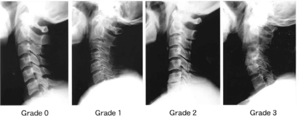
Forty - two patients out of 94 patients being managed with hemo-dialysis at Mitoyo General Hospital in 1998 have had undergone plain radiography in 1989. These 42 patients participated in this study. There were 24 male and 18 female ranged in age from 38 to 80 years old (mean, 58 years old). Duration of hemodialysis ranged from 9 to 25 years (mean, 15.3 years) at the first radiological examination. Distribution of age and duration of hemodialysis were not statistically significant difference between male and female. Nephropathy was caused by glomeruloneohritis in 38 patients and by diabetes mellitus in 4 patients. All patients underwent antero-posterior and lateral views of plain radiography of the cervical spine in 1989 for the first examination. Nine years after the initial radi-ography, all patients underwent follow - up plain radiography of the cervical spine for the second examination in 1998. Radiological findings of the lateral view were graded as follows. Normal findings without any disc space narrowing, erosive changes and subluxa-tion was defined as grade 0. Erosive changes in anterior rim of the vertebrae and endplate without remarkable disc space narrowing were defined as grade 1. Remarkable disc space narrowing with minimal osteophyte formation was defined as grade 2. Spinal fusion and/or subluxation of the vertebrae was defined as grade 3 (Fig. 1). The grade of the finding was determined in consultation by two orthopedic surgeons. All patients were graded according to their findings at the first and the second examination. The relationships between the duration of hemodialysis and the grade and between the age at the onset of hemodialysis and the grade were evaluated. Also radiological changes at each disc level from C2 - 3 to C6 - 7 were observed at the first and the second examination. Clinical symptom
ORIGINAL
Radiographic changes of cervical destructive
spondyloarthropathy in long-term hemodialysis patients : A
9-year longitudinal observational study
Akihiro Nagamachi1), Mitsuhiko Takahashi2), Noriaki Mima2), Keisuke Adachi2), Kazumasa Inoue2), Subash C. Jha1),
Akihiro Nitta1), Masatoshi Morimoto1), Tomoya Takasago1), Toshiyuki Iwame1), Keizo Wada1), Fumitake Tezuka1),
Kazuta Yamashita1), Humio Hayashi1), Ryo Miyagi1), Toshihiko Nishisyo1), Ichiro Tonogai1), Tomohiro Goto1),
Yoichiro Takata1), Toshinori Sakai1), Kosaku Higashino1), Takashi Chikawa3), and Koichi Sairyo1)
1)
Department of Orthopedic Surgery, Tokushima University, Tokushima, Japan,2)Department of Orthopedic Surgery, Mitoyo General
Hos-pital, Kanonji, Japan,3)Department of Orthopedic Surgery, Tokushima Municipal Hospital, Tokushima, Japan
Abstract : Analyses of radiographic changes and clinical symptom of destructive spondyloarthropathy (DSA) on consecutive 42 patients managed with long -term hemodialysis were performed to elucidate radiographic changes of DSA and the factors that influence to the destructive changes. Patients underwent plain radiographs of the cervical spine with 9 years interval. Grading of radiological feature from lateral view was classified into grade 0 to grade 3. Clinical symptom was evaluated using modified Japanese Orthopaedic Association scoring system for cervical myelopathy (mJOA score). Destructive changes were observed in 3 patients at the first ex-amination, and those were observed in 15 patients 9 years after the first examination. There is no statistically significant difference between the duration of hemodialysis and the grade. The mean age at the onset of hemo-dialysis, however, was significantly higher in patients of grade 2 and 3 than those of grade 1. Older patients with long -term hemodialysis had destructive changes. Destructive changes commonly observed in lower cervical spine. The average numbers of the involved disc level were 1.6 in grade 2 and 1.0 in grade 3. Clinical symptoms were varied in each grade and there was no statistically significant difference in total mJOA score among these grades. J. Med. Invest. 64 : 68-73, February, 2017
Keywords : destructive spondyloarthropathy, hemodialysis, radiological analysis, clinical symptom
Received for publication August 8, 2016 ; accepted November 11, 2016. Address correspondence and reprint requests to Akihiro Nagamachi, Department of Orthopedic Surgery, Tokushima University 3 - 18 - 15, Kuramoto, Tokushima, 770 - 8503, Japan and Fax : +81 - 88 - 633 - 0178.
at the second examination was evaluated by modified Japanese Orthopaedic Association scoring system for cervical myelopathy (Table 1). Statistical analysis was performed using unpaired student-t test, Fisher’s exact test, one way ANOVA and Fisher’s PLSD for post hoc test. For all statistical analyses, thep!0.05 level of sig-nificance was used.
RESULTS
At the first examination, 9 patients (21.4%) were classified into grade 0, 30 patients (71.4%) were classified into grade 1, 3 patients (7.1%) were classified into grade 2 and no patient was classified into grade 3. Nine years after the first examination, 1 patient (2.4%) was classified into grade 0, 26 patients (61.9%) were classified into grade 1, 7 patients (16.7%) were classified into grade 2 and 8 pa-tients (19.0%) were classified into grade 3. Patient classified into grade 2 and grade 3 were observed in 7.1% of the patients at the first examination, conversely those were observed 35.7% at the second examination (Fig. 2). The mean duration of hemodialysis of patients with grade 1, grade 2 and grade 3 was 15.5!4.2 years, 14.9!4.9 years and 15.2!4.0 years, respectively with no statistically sig-nificant difference. On the other hand, mean age at the onset of hemodialysis of patients with grade 1, grade 2 and grade 3 was 39.2!9.6 years, 48.2!11.9 years and 49.6!10.5 years, respec-tively. Mean age at the onset of hemodialysis of grade 2 and grade 3 were significantly older than that of grade 1 (Fig. 3)
A decrease in the number of patients of grade 0 and an increase of grade 1 were observed in all disc levels during nine years. Ra-diological changes of grade 2 and grade 3 significantly increased in C5 - 6 and C6 - 7 disc levels (Fig. 4). The average numbers of the involved disc level were 1.6 in grade 2 and 1.0 in grade 3 at the second examination.
Of 8 patients in grade 3, 2 patients had grade 2 changes in an adjacent disc level.
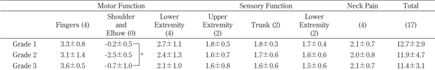
Clinical symptom was varied in each grade. There was no statisti-cally significant difference among all grade in total mJOA score, how-ever, there was statistically significant difference between grade 1 and grade 3 in shoulder and elbow function scores (Table 2).
A CASE PRESENTATION
Figure 5 shows radiological changes of a 65 years old male pa-tient who has been managed with hemodialysis during 8 years at
the first examination. He was classified into grade 1 at the first ex-amination and was classified into grade 3 at the second examina-tion. At the first examination, radiological changes of grade 1 were observed in the levels of C3 - 4, C4 - 5 and C5 - 6. At the second ex-amination, duration of hemodialysis was 17 years and the radio-logical changes of grade 3 were observed in the levels of C3 - 4 and C4 - 5. Total mJOA score was 11 point at the second examination (Fig. 5).
DISCUSSION
Since Kuntzet al. first described DSA as a new syndrome asso-ciated with long - term hemodialysis (2), many possible cause of DSA have been considered. Recently, most authors consider that the main cause of DSA is deposition ofβ2-microgloblin (3, 4, 8, 20, 21). In chronic renal failure and long - term hemodialysis pa-tients,β2-microgloblin deposit in articular, periarticular and perineu-ral tissues becauseβ2-microgloblin could not be filtered by the kidney and the column. Sebertet al. demonstrated that deposition of amyloid resulting fromβ2-microgloblin in the vertebral disc of patient with DSA (11). Miyataet al. attested β2-microgloblin modi-fied with advanced glycation end products (AGEs) is the major component of hemodialysis - associated amyloidosis (21). AGEs induce transendothelial human monocyte chemotaxis and produc-tion of TNF alpha and IL - 1β. Activation of osteoclast or chondro-clast by TNF alpha and IL - 1β promotes bone absorption, causing destructive changes of the cervical spine (22).
Maruyamaet al. described that patient’s age at the onset of hemo-dialysis was an associate risk factor for DSA (8). In the present study, we also found that duration of hemodialysis did not correlate to progress of destructive changes. Conversely, patient’s age at the onset of hemodialysis correlated to progress of destructive changes. Older patients with long - term hemodialysis had destructive changes. Although the cause of DSA is thought to be deposition of AGEs β2-microgloblin in the disc, ligament and vertebrae, it is not enough to explain the pathogenesis of DSA. If ADEsβ2-microgloblin depo-sition is the main cause of DSA, destructive changes should have occurred in younger patients. Therefor, aging is thought to be one of the most important factors of DSA.
DSA developed commonly in the lower cervical spine of which range of motion was larger. These destructive changes were lim-ited in one or sometimes two disc levels. It was thought that once the destructive changes had occurred, axial and shearing loads were concentrated to the involved level and destruction progressed Figure 1. Radiographic grading of cervical destruction on the lateral view.
Grade 0 : normal ; grade 1 : erosive changes in the anterior rim of the vertebrae and endplate ; grade 2 : disc space narrowing with minimal osteo-phyte formation ; grade 3 : spinal fusion and/or subluxation of the vertebrae.
in limited disc levels. The mechanical stress to the spine is asso-ciated with the pathogenesis of DSA. Of 8 patients with grade 3, 2 patients had grade 2 changes in an adjacent disc level in this study. This fact is important to plan the extent of spinal fusion level. Sudoet al. performed reconstructive surgery for patients with sig-nificant spinal instability and reported that progressive destructive changes causing significant instability in the mobile segments
adjacent to the previous fusion site were observed in four pa-tients (40%) who underwent circumferential fusion (23). It is im-portant to extent fusion if adjacent disc level had grade 1 or grade 2 changes.
In the present study, mJOA scores were inconsistent and low in all grades and there was no statistically significant correlation between radiological findings and clinical symptom except shoulder Table 1. Modified Japanese Orthopaedic Association scoring system for cervical myelopathy
Category
A. MOTOR FUNCTION : I. Fingers
0 = Unable to feed oneself with any tableware including chopsticks, a spoon or fork and/or unable to fasten buttons of any size 1 = Can manage to feed oneself with a spoon and/or a fork but not with chopsticks
2 = Either chopstick - feeding or writing is possible but not partial, and/or large buttons can be fastened 3 = Either chopstick - feeding or writing is clumsy but partial, and/or cuff buttons can be fastened 4 = Normal
II. Shoulder and Elbow
Evaluated by MMT score of the deltoid or biceps muscle, which is weaker - 2 = MMT 2 or below
- 1 = MMT 3 - 0.5 = MMT 4 0 = MMT 5 III. Lower Extremity
0 = Unable to stand up and walk by any means 0.5 = Able to stand up but unable to walk
1 = Unable to walk without a cane or other support on a level 1.5 = Able to walk without a support but needs support on stairs
2 = Walks independently on a level but needs support on stairs
2.5 = Walks independently when going upstairs, but needs support when going downstairs 3 = Capable of fast walking but clumsily
4 = Normal
B. SENSORY FUNCTION I. Upper Extremity
0 = Complete loss of touch and pain sensation
0.5 = 50% or below of normal sensation and/or severe pain or numbness 1 = Over 60% of normal sensation and/or moderate pain or numbness
1.5 = Subjective numbness of a slight degree without any objective sensory deficit 2 = Normal
II. Trunk
0 = Complete loss of touch and pain sensation
0.5 = 50% or below of normal sensation and/or severe pain or numbness 1 = Over 60% of normal sensation and/or moderate pain or numbness
1.5 = Subjective numbness of a slight degree without any objective sensory deficit 2 = Normal
III. Lower Extremity
0 = Complete loss of touch and pain sensation
0.5 = 50% or below of normal sensation and/or severe pain or numbness 1 = Over 60% of normal sensation and/or moderate pain or numbness
1.5 = Subjective numbness of a slight degree without any objective sensory deficit 2 = Normal
C. NECK AND SHOULDER PAIN 0 = Frequent or continuous severe pain 1 = Frequent mild or occasional severe pain 2 = Occasional mild pain
3 = None
and elbow functions. Two of 8 patients of grade 3 complained mild pain on their neck, while 4 of 27 patients in grade 1 complained continuous severe pain on their neck. Many patients had numb-ness in their limbs because of peripheral neuropathy caused by amyloid deposition including carpal tunnel syndrome and tarsal tunnel syndrome. Thus, clinical symptom did not correlate with the radiological findings. Even though total mJOA scores were low, no patient required surgery in this series. Patients with DSA oc-casionally complain severe neck pain and sometimes have radicu-lomyelopathy. Surgery would be performed when rapid neuro-logical deterioration (12) is observed. Patients with DSA have many other complications, including cardioplumonary disease, immunosuppression due to diabetes mellitus and sometimes he-patocellular carcinoma resulting from hepatitis C. Nakamuraet al. investigated the survival rate of the patients with DSA and concluded
that the survival rate was 37.4% over 10 years from the onset of hemodialysis and 0% over 20 years (24). Large amount of bleeding, surgical site infection and osteopenia are some of the major prob-lems at surgery. Kumaret al. demonstrated that successful cervical spine fusion could relieve pain and neurologic deficits in selected patients (16). However, 2 of the 11 patients who underwent surgery died during perioperative period. Itohet al. performed surgery for 17 patients with DSA and 1 patient died during the operation and 2 patients died perioperative period (15). Naitoet al. studied the outcome of 9 patients who had surgery for DSA and concluded that 5 patients had good, 2 patients had fair and 1 patient had poor results (18). They also reported that 1 patient died 46 days after the surgery due to postoperative meningitis. Because of such high mortality, patient selection based on their disability, general con-dition and prognosis is important.
CONCLUSION
During 9 years, number of patients with destructive changes increased from 3 (7.1%) to 15 (35.7%). In the present study, we found that duration of hemodialysis did not correlate to progress of destructive changes. Conversely, patient’s age at the onset of hemodialysis correlated to progress of destructive changes. De-structive changes progressed in older patients with long - term hemodialysis. Destructive changes progressed in the lower cervi-cal spine and occurred in limited number of the disc level. Mean mJOA score was low, however, there was no patient required surgery in this series. There was no statistical correlation between radiological findings and mJOA score.
CONFLICT OF INTEREST
There is nothing to declare of conflict of interest for this work.
ACKNOWLEDGEMENTS
The authors thank to Mamoru Hirohata, M.D., Toru Endo, M.D., Figure 2. Number of patients with each grade of radiographic changes
at the first and second examinations.
Grades 2 and 3 were observed in 7.1% of the patients at the first exami-nation and in 35.7% at the second examiexami-nation.
Figure 3. Grade versus duration of hemodialysis and age at initiation of hemodialysis.
Figure 4. Radiographic changes in each disc level.
Radiographic changes of grades 2 and 3 were most commonly observed in the lower cervical spine.
Table 2. Modified Japanese Orthopaedic Association scores by grade of radiographic changes at the second examination
Motor Function Sensory Function Neck Pain Total
Fingers (4) Shoulder and Elbow (0) Lower Extremity (4) Upper Extremity (2) Trunk (2) Lower Extremity (2) (4) (17) Grade 1 3.3!0.8 - 0.2!0.5 2.7!1.1 1.8!0.5 1.8!0.3 1.7!0.4 2.1!0.7 12.7!2.9 Grade 2 3.1!1.4 - 2.5!0.5 * 2.4!1.3 1.6!0.7 1.7!0.6 1.6!0.6 2.0!0.8 11.9!4.7 Grade 3 3.6!0.5 - 0.7!1.0 2.1!1.0 1.6!0.8 1.6!0.6 1.5!0.6 2.1!0.7 11.4!3.1 (Mean!SD,*P!0.05)
Figure 5. Radiographs showing the radiographic changes in a 65 - year - old man who had been on hemodialysis for 17 years. At the second ex-amination, grade 3 radiographic changes were observed in the C3 - 4 and C4 - 5 levels.
Kenji Akiyama, M.D. and Tsutomu Ishizu, M.D. for their work in the coordination of this study.
REFERENCES
1. The Japanese Society for Dialysis Therappy. The basic statis-tic of hemodialysis patients in Japan (in Japanese) : http : // docs.jsdt.or.jp/overview/pdf2015/p003.pdf
2. Kuntz D, Naveau B, Bardin T, Drueke T, Treves R, Dryll A : Destructive spondyloarthropathy in hemodialysed patients. A new syndrome. Arthritis Rheum 27 : 369 - 375, 1984 3. Bardin T, Zingraff J, Shirahama T : Hemodialysis - asociated
amyloidosis and beta- 2 microgloblin - Clinical and immuno-histochemical study - . Am J Med 83 : 419 - 424, 1987 4. Moriniere Ph, Marie A, Esper NE : Destructive
spondyloar-thropathy withβ2-microgloblin amyloid deposits in a uremic patient before chronic hemodialysis. Nephron 59 : 654 - 657, 1991
5. Alcalay M, Goupy MC, Azais I, Bontoux D : Hemodialysis is not essential for the development of destructive spondyloar-thropathy in patients with chronic renal failure. Arthritis Rheum 30 : 1182 - 1186, 1987
6. Bindi P, Lavaud S, Bernieh B, Toupance O, Chanard J : Early and late occurrence of destructive spondyloarthropathy in hemodialysed patients. Nephrol Dial Transplant 5 : 199 - 203, 1990
7. Kaplan P, Rensnick D, Murphey M, Heck L, Phalen J, Egan D, Rutsky E : Destructive noninfectious spondyloarthropathy in hemodialysis patients : A report of four cases. Radiology 162 : 241 - 244, 1987
8. Maruyama H, Gejyo F, Arakawa M : Clinical studies of de-structive spondyloarthropathy in long - term hemodialysis pa-tients. Nephron 61 : 37 - 44, 1992
9. McCarthy J, Dahlberg PJ, Krie gshauser JS : Erosive spondy-loarthropathy in long - term dialysis patients : Relationship to severe hyperparathyroidism. Mayo Clin Proc 63 : 446 - 452, 1988
10. Orzincolo C, Bedani PL, Scutellari PN, Cardona P, Trotta F, Gilli P : Destructive spondyloarthropathy and radiographic follow up in hemodialysis patients. Skeletal Radio 19 : 483 -487, 1990
11. Sebert JL, Fardellone P, Marie A : Destructive spondyloar-thropathy in hemodialyzed patients : Possible role of amyloi-dosis. Arthritis Rheum 29 : 301 - 302, 1986
12. Davidson GS, Montanera WJ, Fleming JFR, Gentili F : Amy-loid destructive spondyloarthropathy causing cord compres-sion : Related to chronic renal failure and dialysis. Neurosurgery
33 : 519 - 522, 1993
13. Deforges - Lasseur C, Combe C, Cernier A, Vital JM, Aparico M : Destructive spondyloarthropathy presenting with progres-sive paraplegia in a dialysis patient. Recovery after surgical spinal cord decompression and parathyroidectomy. Nephrol Dial Transplant 8 : 180 - 184, 1993
14. Digenis GE, Davidson G, Dombros NV, Katz A, Bookman A, Oreopoulos DG : Destructive spondyloarthropathy in a pa-tient on continuous ambulatory peritoneal dialysis for 13 years, Pert Dial Int 13 : 228 - 231, 1993
15. Ito M, Abumi K, Kaneda K, Takeda N, Satoh S, Takebayashi T : Surgical treatment of destructive spondyloarthropathy as-sociated with long term hemodialysis (in Japanese). Spine & Spinal Cord 10 : 1079 - 1085, 1997
16. Kumar A, Leventhal MR, Freedman EL, Coburn J, Delamarter R : Destructive spondyloarthropathy of the cervical spine in patients with chronic renal failure, Spine 22 : 573 - 578, 1997 17. Mikawa Y, Yamaoka T, Watanabe R : Compression of the
spinal cord due to destructive spondyloarthropathy of the atlantoaxial joints. J Bone Joint Surg [Am] 78 - A : 1911 - 1914, 1996
18. Naito M, Ogawa K, Nakamoto M, Goya T, Sugioka Y : De-structive spondyloarthrppathy during long - term hemodialy-sis. J Bone Joint Surg [Br] 74 - B : 686 - 690, 1992
19. Naidich JB, Mossey RT, McHeffey - Atkinson B : Spondyloarthropathy from long term hemodialysis. Radiology 167 : 761 -764, 1988
20. Kroner G, Stabler A, Seiderer M, Moran JE, Gurland HJ :β2 microgloblin - related amyloidosis causing atlantoaxial spon-dyloarthropathy with spinal - cord compression in haemodialy-sis patients : Detection by MRI. Nephrol Dial Transplant Suppl 2 : 91 - 95, 1991
21. Miyata T, Oda O, Inagi R :β2-microgloblin modified with advanced glycogen end products is a major compornent of hemodialysis associated amyloidosis. J Clin Invest 92 : 1243 -1251, 1993
22. Vlassara H, Brownlee M, Manogue KR, Dinarello CA, Pasagian A : Cachectin/TNF and IL - 1 induced by glucose - modified proteins : role in normal tissue remodeling. Science 240 : 1546 - 1548, 1988
23. Sudo H, Ito M, Abumi K, Kotani Y, Takeuchi T, Yasui K, Minami A : Long - term follow up of surgical outcomes in patients with cervical disorders undergoing hemodialysis. J Neurosurg Spine 5 : 313 - 319, 2006
24. Nakamura M, Taketomi E, Matsunaga S : Long term follow up of destructive spondyloarthropathy in hemodialyzed pa-tients (in Japanese). J Jpn Resea Soci Spine 25 : 70 - 72, 1999