293
Current Perspective
1. Introduction
Stem cell therapy for central nervous system (CNS) repair has great clinical potential (1). In contrast to the controversy over utilization of embryonic stem cells which are theoretically highly beneficial due to their pluripotent ability, mesenchymal stem cells (MSCs) are recognized to be a relatively effective, ethically safe, and acceptable source for cell therapy to treat various neurological diseases such as stroke (2).
Up to now, large amount of evidences accumulated on MSC behavior demonstrated that under appropriate conditions, MSCs selectively differentiate not only into mesenchymal lineages but also into endodermal and ectodermal cell lineages in vitro (3 – 7). MSCs are present in adult tissues including bone marrow and adipose tissue. However, the clinical use of bone marrow–derived stem cells (BSCs) has presented
problems including pain, morbidity, and low numbers of harvested cells. In contrast, adipose-derived stem cells (ASCs), which are derived from adipose tissue, are one of the most advantageous resources due to easier access to adipose tissue and abundance with proliferation and differentiation potential (8). ASCs have a high proliferation capacity in vitro and differentiate into cells with several neuronal and glial characteristics, including expression of neuronal and glial proteins (9, 10). Implantation of human ASCs leads to no adverse side effects such as tumorigenicity, chromosomal abnormalities, or immune rejection (11).
Although the stem nature of ASCs could be doubted, previous studies have suggested that BM-MSCs and ADSCs exhibit a virtually identical transcriptional profile for stemness-related genes (12, 13) and further-more, MSC derived neuronal cells have been shown to be electrically excitable (7). In addition, some authors have found that few cells that were capable of engrafting into the nervous tissue fused with endogenous cells and thereby acquired the phenotype of the partner host cell (14 – 16).
Therefore, ASCs have recently received attention as a promising source of cells for cell therapy, and the
Therapeutic Potential of Human Adipose-Derived Stem Cells
in Neurological Disorders
Keun-A Chang
1,†, Jun-Ho Lee
2,†, and Yoo-Hun Suh
3,*
1Department of Pharmacology, College of Medicine, Neuroscience Research Institute, Gachon University,
Incheon 405-760, Korea
2Department of Emergency Medical Technology, College of Natural Science, Daejeon University, Daejeon 300-716, Korea 3Korea Brain Research Institute (KBRI), 425 Jungang-daero, Jung-gu, Daegu 700-010, Korea
Received June 16, 2014; Accepted September 30, 2014
Abstract. Stem cell therapy has been noted as a novel strategy to various diseases including
neurological disorders such as Alzheimer’s disease, Parkinson’s disease, stroke, amyotrophic lateral sclerosis, and Huntington’s disease that have no effective treatment available to date. The adipose-derived stem cells (ASCs), mesenchymal stem cells (MSCs) isolated from adipose tissue, are well known for their pluripotency with the ability to differentiate into various types of cells and immuno-modulatory property. These biological features make ASCs a promising source for regenerative cell therapy in neurological disorders. Here we discuss the recent progress of regenerative therapies in various neurological disorders utilizing ASCs.
Keywords: neurological disorder, adipose-derived stem cell (ASC), Alzheimer’s disease (AD),
Parkinson’s disease (PD), Huntington’s disease (HD)
†These authors contributed equally on this study.
*Corresponding author. [email protected] Published online in J-STAGE on November 18, 2014 doi: 10.1254/jphs.14R10CP
therapeutic efficacy of ASCs has been assessed in various animal models with specific neurological dis-orders (Table 1).
2. Therapeutic potential of ASCs for Alzheimer’s disease (AD)
AD is the most common neurodegenerative disorder characterized by the accumulation of amyloid plaques and neurofibrillary tangles accompanied by cognitive dysfunction. There are no drug treatments available that can provide a cure for AD. However, medicines have been developed that can improve symptoms or tempo-rarily slow down their progression in some people. There are two main types of medication used to treat AD: cholinesterase inhibitors and NMDA-receptor
antagonists. Cholinesterase inhibitors include donepezil hydrochloride, rivastigmine, and galantamine. The NMDA-receptor antagonist is memantine (17 – 19).
Therapeutic potential of intracerebral (i.c.) or intra-venous (i.v.) injection of human ASCs was previously reported in an AD mouse model, Tg2576 transgenic (Tg) mice, by our group. Our study showed that intra venously transplanted ASCs passed through the blood brain barrier (BBB) and migrated into the brains of Tg mice (20). I.v. or i.c. injected ASCs significantly improved learning and memory and restored neuro pathology including amyloid beta deposition in Tg mice (20). In addition, elevating endogenous neurogenesis and synaptic and dendritic stability were shown in ASCs-transplanted Tg mouse brains (20). However, inter leukin-10 (IL-10) and vas cular endothelial growth factor (VEGF) were
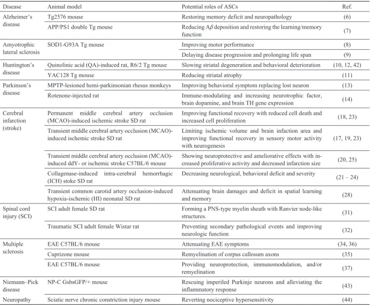
up-Table 1. Applications of adipose-derived stem cells (ASCs) in the treatment of neurological diseases
Disease Animal model Potential roles of ASCs Ref.
Alzheimer’s
disease Tg2576 mouseAPP/PS1 double Tg mouse Restoring memory deficit and neuropathologyReducing Ab deposition and restoring the learning/memory (6)
function (7)
Amyotrophic
lateral sclerosis SOD1-G93A Tg mouse Improving motor performanceDelaying disease progression and prolonging life span (8)(9) Huntington’s
disease Quinolinic acid (QA)-induced rat, R6/2 Tg mouseYAC128 Tg mouse Slowing striatal degeneration and behavioral deteriorationReducing striatal atrophy (10, 12, 42)(11) Parkinson’s
disease MPTP-lesioned hemi-parkinsonian rhesus monkeysRotenone-injected rat Improving behavioral symptom replacing lost neuronImmune-modulating and increasing neurotrophic factor, (13) brain dopamine, and brain TH gene expression (14) Cerebral
infarction (stroke)
Permanent middle cerebral artery occlusion
(MCAO)-induced ischemic stroke SD rat Improving functional recovery with reduced cell death and increased cell proliferation (18, 23) Transient middle cerebral artery occlusion
(MCAO)-induced ischemic stroke SD rat Limiting ischemic volume and brain infaction area and improving functional recovery in sensory motor activity
with neurogenesis (17, 19, 23)
Transient middle cerebral artery occlusion
(MCAO)-induced ddY- or ischemic stroke C57BL/6 mouse Showing neuroprotective and ameliorative effects with in-creased proliferative activity and decreased infarction size (20, 25) Collagenase-induced intra-cerebral hemorrhagic
(ICH) stoke SD rat Decreasing neurological, behavioral deficit and severity (21 – 24) Transient common carotid artery occlusion-induced
hypoxia-ischemic (HI) neonatal SD rat Attenuating brain damages and deficit in spatial learning and memory (28) Spinal cord
injury (SCI) SCI adult female SD rat Forming a PNS-type myelin sheath with Ranvier node-like structures. (31) Traumatic SCI adult female Wistar rat Preventing secondary pathological events and improving
neurologic function (32)
Multiple
sclerosis EAE C57BL/6 mouseCuprizone mouse Attenuating EAE symptomsRemyelination of corpus callosum axons (34, 36)(35) EAE C57BL/6 mouse Providing neuroprotection, immunomodulation, and/or
remyelination (37)
Niemann–Pick
disease NP-C GsbsGFP/+ mouse Rescuing imperiled Purkinje neurons and alleviating the inflammatory response (43) Neuropathy Sciatic nerve chronic constriction injury mouse Reverting nociceptive hypersensitivity (44)
regulated in ASCs-transplanted Tg mouse brains (6). The therapeutic effects of i.c. ASCs transplantation were also evaluated in an APP/PS1 double Tg AD mouse model (21). The transplantation of ASCs reduced amyloid beta (Ab) deposition and restored the learning and memory function in APP/PS1 double Tg mice (21).
These studies suggested that activated microglia might be involved in the mechanisms to ameliorate the neuro-pathological deficits in AD (20, 21). More activated microglia were detected in both regions of the hippo-campus and cortex after ASCs transplantation, exhibiting decreased expression levels of pro-inflammatory factors and elevated expression levels of Ab-degrading enzymes as well as neprilysin (20, 21). The mechanisms behind the role of ASC in microglial activation are largely unknown. However, activation of microglia by ASC transplantation may act as a natural defense mechanism to prevent Ab accumulation or reduce Ab deposits. Another study has also shown a similar set of results and the BM-MSCs were able to reduce the inflammatory response and also restore defective microglial function (22). This study suggested that activation of microglia with conversion to an alternative phenotype results in a feed-forward loop whereby significantly increased levels of microglial cells correlate with enhanced Ab-clearance following BM-MSC transplantation.
3. Therapeutic potential of ASCs for amyotrophic lateral sclerosis (ALS)
ALS is a neurodegenerative disease that selectively affects motor neurons in the cortex, brain stem, and spinal cord. The precise pathogenic mechanism remains unknown, and there is no effective therapy.
The efficacy of the systemic administration of ASCs was assessed in an ALS mouse model, superoxide- dismutase 1 mutant [SOD1-G93A] Tg mice (23). Clinical and neurophysiological tests showed that the administra-tion of ASCs to Tg mice at the clinical onset significantly delayed motor deterioration for several weeks (23). In ASCs-treated Tg mice, the number of lumbar motor neurons and levels of glial-derived neurotrophic factor (GDNF) and basic fibroblast growth factor (bFGF) were increased (23).
A recent study from Kim et al. suggest that transplan-tation of ASCs in [SOD1-G93A] Tg mice delays disease progression and prolongs life span of Tg mice through neuroprotective effects by production of cytokines/ growth factors (24). Human ASCs were intravenously (i.v.) and intracerebroventricularly (i.c.v.) transplanted into Tg mice before the appearance of clinical symptoms (24). In Tg mice transplanted with ASCs via the i.c.v. route, delayed clinical symptoms were shown by
behavior assessments such as the rotarod test, paw grip endurance, and reflex index; and the survival of animals was extended (24). In cultured neuronal cells and in the ALS spinal cord, ASCs secreted high levels of neuro-trophic factors such as nerve growth factor (NGF), brain-derived neurotropic factor (BDNF), insulin-like growth factor 1 (IGF-1), and VEGF and reduced the cytotoxicity by these factors (24).
4. Therapeutic potential of ASCs for Huntington’s disease (HD)
HD is a late-onset neurodegenerative disease caused by abnormal expansion of CAG repeats in the huntingtin gene, which is characterized by a progressive loss of medium spiny neurons in the basal ganglia. Several studies have examined the potential of MSCs including ASCs for transplantation into HD models as neuro-protective treatment or cell replacement therapies.
The effects of human ASCs on HD pathology was investigated by Lee et al. (25) in cell culture experiments and in two different rodent HD models, QA model and R6/2 Tg mice. In vitro study showed that human ASCs secreted multiple growth factors, including epidermal growth factor (EGF), BDNF, NGF, IGF-1, and ciliary neurotrophic factor (CNTF) (25). Transplan-tation of ASCs into the QA model revealed significant improvement in the apomorphine-induced rotation test over 4 weeks, reduced lesion volume, and a lower number of apoptotic striatal cells compared to control animals (25). In R6/2 Tg mice, transplantation of ASCs improved rotarod performance and limb clasping, increased survival, attenuated the loss of striatal neurons, and reduced the huntingtin aggregates (25).
In the same group, transplantation of human ASCs was compared with normal human ASCs in another HD animal model, YAC128 Tg mice, which showed slow disease progression for 12 months (26). An in vitro study showed that cultured HD ASCs express multiple growth factors (BDNF, HGF, IGF, LIF, NGF, and VEGF) with the same cell surface markers (CD13, CD29, CD31, CD34, and CD44) as those of normal ASCs (26). There was no significant difference in rotarod performance and body weight in Tg mice transplanted with either normal ASCs or HD ASCs compared with Tg control mice (26). Normal ASC transplantation reduced the striatal atrophy, while HD ASCs failed to prevent it (26). Transplantation of normal ASCs after onset of disease phenotype main-tained the rotarod performance for 4 weeks, but no significant difference in striatal atrophy was observed. In the same case, transplantations of HD ASCs were not effective (26).
HD model and to further assess the possibility of the paracrine effects of human ASCs via secreted multiple growth factors, the same researchers investigated effects of cell-free extracts of ASCs (ASCs-E) on the R6/2 HD mouse model (27). Injection of ASCs-E improved the performance in the rotarod test, ameliorated the striatal atrophy and reduced mutant huntingtin aggregation in the striatum in the R6/2 HD mouse model (27). There-fore, ASCs-E might slow disease progression in an animal model of HD and could be a potential resource for treatment of HD (27).
5. Therapeutic potential of ASCs for Parkinson’s disease (PD)
PD is the second most common neurodegenerative disorder characterized by the loss of neurons in the substantia nigra pars compacta (SNpc) in middle-aged and elderly people.
A recent study investigated the therapeutic effects of the combined transplantation of neuronal-primed ASCs derived from rhesus monkey and adenovirus containing Neurturin (NTN) and tyrosine hydroxylase (TH) (Ad-NTN-TH) into methyl-4-phenyl-1,2,3,6-tetrahydropyri-dine (MPTP)-lesioned hemi-parkinsonin rhesus monkeys (28). An in vitro study with the use of LIM homeobox transcription factor 1, alpha (LMX1A) and NTN showed that NTN-conditioned medium protected dopaminergic neurons against 1-methyl-4-phenylpyridinium (MPP+) toxicity and the LMX1A- and NTN-infected ASCs showed a dopaminergic differentiation with secreting the dopamine (28). ASCs combined with Ad-NTN-TH were implanted into the striatum and SNpc of the rhesus monkey PD model (28). Transplantation of ASCs improved parkinsonian behavior as such in tremor recovery and motility in combination-transplanted monkeys (28). Single-photon emission computed tomo-graphy analysis showed that the transplantation of combined ASCs with Ad-NTN-TH increased dopamine transporter uptake at the striatum, but not the neuronal-primed ASC-transplantation (28). In postmortem analysis, neuronal-primed ASCs could replace lost neurons and reconstruct the nigrostriatal pathway in the brain (28). The grafting of ASCs combined with Ad-NTN-TH had more neuro protective effects compared with Ad-NTN-TH or ASCs alone (28).
Another group performed a biomarker study in a rotenone-induced PD rat model treated with ASCs (29). ASCs treatment restored the increased serum trans-forming growth factor b (TGF-b) and monocyte chemo-attractant protein 1 (MCP-1) levels and increased serum BDNF, brain dopamine, and brain TH gene expression levels in a PD rat model (29). Their study suggests that
ASCs infusion might be attributed to their immuno-modulatory, anti-inflammatory, and neurotrophic effects.
6. Therapeutic potential of ASCs for stroke
Stroke is known to be the third most frequent cause of mortality in industrial countries (30) with ischemic stroke being the most prevalent. Considering the compli-cations of thrombolytic therapy in acute ischemic stroke, development of an alternative therapeutic intervention is necessary and utilization of various kinds of stem cells including MSCs have been explored to treat ischemic stroke patients such as in cytotherapy. Among the MSCs, ASC is one of the most advantageous resources (31). Thus, a number of studies have demonstrated its positive effects on cerebrovascular insults such as hemorrhagic and mostly ischemic stroke (32 – 44). Although none has been proven practically efficient in human stroke patients to this point, numbers of experi-mental trials in vitro or in vivo animal models regarding ASC-associated therapy have been introduced as a promising therapeutic option against stroke.
6.1. Clinical potential for transplantation of ASCs against stroke
Intravenously transplanted autologous ASCs in tran-sient or permanent middle cerebral artery occlusion (MCAO)-induced ischemic stroke adult Sprague-Dawley (SD) rats demonstrated positive effects in infarct size, apoptosis, oxidative stress, and inflammatory response with enhanced signatures of neurogenesis, oligodendro-genesis, angiooligodendro-genesis, or synaptogenesis in addition to neurological functional recovery against experimental ischemic stroke (32 – 34), while no reduction in infarct volume or any migration/implantation of cells into the lesion were observed in certain cases (33, 34). Similarly, i.v. administration of allogenic ASCs into age-matched ischemic stroke C57BL/6J mice generated by transient MCAO attenuated ischemic damage such as infarct size and brain swelling and enhanced motor functional recovery with expression of some growth factors such as hepatocyte growth factor (HGF) and angiopoietin-1 in ischemic brain tissue, although ASCs were not fully incorporated into the infarct area (35). Despite ischemia is accountable for most cases of stroke, the thera peutic potential of ASCs in hemorrhagic stroke has been also demonstrated. Intraventricular (i.v.) injection of either human or murine ASCs as well as intraperitoneal (i.p.) admini stration of cell-free extract of human ASCs into rats inflicted with experimental intracerebral hemorrhage (ICH) resulted in the induction of neuronal differentia-tion and funcdifferentia-tional improvement represented by a considerable reduction in neurological or behavioral
deficit and severity in addition to a decrease in brain water content, hemorrhagic volume, and parenchymal atrophy with reduced apoptosis and cerebral inflam-mation (36 – 39).
However, due to the low survival rate and potential risk for tumorigenicity elicited by the implantation of stem cells, utilization of conditioned medium of ASCs (ASCs-CM) might provide an alternative way to over-come these limitations, and its application has also been attempted as ASC therapy.
6.2. Clinical potential for administration of ASCs-CM against stroke
The neuroprotective effects of human and murine ASCs-CM were observed in vitro. Pretreatment with ASCs-CM collected from the cultures of ASCs originally isolated from human subcutaneous adipose tissue or the fat pad of female C57BL/6-Tg mice significantly reduced glutamate-induced excitotoxicity in the SH-SY5Y or PC12 cells (41, 43). Related to the glutamate-induced excitotoxicity, rat cortical neurons were also co-cultured with human ASCs-CM or ASCs that were separated from neurons with porous membrane to see the effects of ASCs-CM. The neuroprotective effects were verified against glutamate excitotoxicity supported by the data showing inhibition of neuronal cell damage, apoptosis, and glutamate-induced energy depletion as well as the promotion of nerve regeneration and repair (42). A range of in vivo models employing ASCs-CM have also been introduced. Pretreatment of murine or human ASCs-CM by i.c.v. administration before MCAO demonstrated significant reduction in infarct area and volume. In addition, rapid treatment of ASCs-CM immediately after MCAO was also effective, although administration 2 h after MCAO was not (41). I.v. administration of concentrated ASCs-CM collected from SD rat into 7-day-old neonatal SD rat either 1 h before or 24 h after induction of hypoxia-ischemia significantly protected hippocampal and cortical volume loss and demonstrated significant behavioral and learning functionality (44). Although treatment of the initial phase of the stroke is essential, increased intracerebral pressure, which could worsen the consequences of the stroke and increase lethality, is likely due to the infusion of ASCs-CM at the acute phase of the stroke. Continuous infusion of ASCs-CM collected from human ASCs in a three- dimensional spheroid form was administered in the subchronic phase of the SD rat stroke model induced by standard MCAO, and significant therapeutic effects against stroke were also observed (40). Accordingly, utilization of ASCs-CM appears to exert similar amelio-rative effects on both in vitro and in vivo stroke models. More discussions in depth for clinical feasibility and
plausible approaches of ASC-associated therapy to treat chronic stroke were also proposed (45).
7. Therapeutic potential of ASCs for other neuronal disorders
7.1. Spinal cord injury (SCI)
Therapeutic promise for SCI has also been assessed using ASCs. SCI typically results from sustained trauma to the spinal cord, resulting in loss of neurologic function at the level of the injury. When the induced ASCs were engrafted to SCI lesions, they formed a peripheral nervous system (PNS)-type myelin sheath on CNS axons (46). In in vitro study, ASCs expressed Schwann cells (SCs) markers such as A2B5, GFAP, O4, p75, S100, Sox10, Krox-20, and L1, indicating the possibility to differentiate into SCs (46). In addition, the ASCs induced into SCI expressed several neurotrophic factors in repaired tissue (46). Especially, the secondary damage following SCI exacerbates the injury and retards repair mechanisms. ASCs have a strong, long-lasting immuno-suppressive capacity via soluble factors. Direct injection of ASC extracts (ASC-E) mixed with matrigel into the spinal cord immediately after SCI also resulted in reduced apoptotic cell death, astrogliosis, and hypo- myelination but did not reduce the extent of microglia infiltration (47). In addition, ASC-E injected animal showed significant functional improvement of hind limbs (47).
7.2. Multiple sclerosis
Multiple sclerosis (MS), also known as disseminated sclerosis or encephalomyelitis disseminate, is an inflam-matory disease (48). Previous studies have demonstrated the potential of stem cell–based MS therapy in auto-immune and cuprizone models of MS (49 – 51). I.v. administration of ASCs before disease onset significantly reduces the severity of experimental autoimmune encephalomyelitis (EAE) by immune modulation and decreases spinal cord inflammation and demyelination in MS animal model (49). Combined therapy of i.v. transplantation of ASCs with 17b-estradiol (E2) showed the enhanced efficacy on remyelination of corpus callosum axons in a cuprizone mouse model of MS compared with ASCs monotherapy (50). A more recent study demonstrated that therapeutic efficacy of human ASCs from older donors was reduced compared with that of cells from younger donors for disease prevention. ASCs from older donors failed to ameliorate the neuro-degeneration associated with EAE and increased CNS inflammation and demyelination (51). Therapeutic effects of ASCs were compared in two independent routes of injection, i.p. and i.v. (52). ASCs transplantation in the
i.p. or i.v. route showed immunomodulative and neuro-protective effects; however, i.p. injection of ASCs has an enhanced efficacy in maintaining the splenic CD4 + CD25 + FOXP3 + T cell population and increase of IL-4 secretion (52).
8. Conclusion
To explain mechanisms underlying the ameliorative and neuroprotective effects of ASC therapy against neurological disorders, researchers introduced stacks of mediators mostly involved in angiogenesis, synapto-genesis, gliosynapto-genesis, and/or neurogenesis. The mediators expressed or secreted from ASCs include various growth factors containing neurotrophic factors such as NGF, BDNF, GDNF, VEGF, HGF, IGF-1, and others such as chemotactic factors (SDF-1 and CXCR4) or a synaptic vesicle protein, synaptophysin, along with diverse neuro-nal and glial cell proteins and MSC markers (32, 33, 35,
36, 43, 53).
In addition, transplantation of ASCs ameliorates the increased levels of pro-inflammatory cytokine expression, such as TGF-b and IL-1b; secretes anti- inflammatory cytokine, IL-10; and is thought to partici-pate in immunomodulation (54, 55) (Fig. 1). Further-more, in line with the positive consequences obtained from ASCs-CM approaches, recent findings underline the importance of the “paracrine effect” of stem cell treatment (56). Although we discussed current clinical applications of ASCs in neurological disorders in this review, the potential relevance of ASCs or even other MSCs for the treatment of neuropsychiatric disorders such as schizophrenia, autism or depression is plausible. However, it seems to be too immature to discuss in this review since the evidences related to clinical applications utilizing ASCs are rare and even those are suggestive and currently in trial stage (57 – 61).
Altogether, ASC-associated therapy is very likely to
Fig. 1. Paracrine and immunomodulatory effects as possible mechanisms of roles of adipose-derive adult stem cells (ASCs) in
the treatment of neurological diseases. A: Neurological diseases are associated with immune alterations and neuroinflammation including pro-inflammatory cytokines (i.e., IL-1b and TNFa) overproduction. B: Intracerebrally or intravenously injected ASCs are thought to modulate a hostile microenvironment of diseases through secretion of large amounts of several bioactive molecules (paracrine activity) such as VEGF and IL-10. In addition, transplanted hASCs might benefit the brain by inducing proliferation of endogenous early-stage neurons and surrounding cells through increasing neurotropic factors such as BDNF, NGF, and VEGF, suggesting the increase of neurogenesis and dendrite and synaptic stability. Therefore, these molecules derived from transplanted ASCs attribute to their immunomodulatory, anti-inflammatory, and neurotrophic effects; neurogenesis; or synaptogenesis.
be a promising clinical option for the treatment of diverse neurological disorder patients. To optimize and maximize the efficiency with safety and expediency in ASC therapy, future trials need to sharpen our current knowledge and perspectives for the clinical utilization of ASC or ASC-derived resources, including ASCs-CM.
Acknowledgments
This work was supported by the Gachon University research fund of 2014 (GCU 2014-5106) and a research grant of Daejeon University in 2012, the Republic of Korea. The authors declare no competing fi-nancial interests.
References
1 Gage FH. Mammalian neural stem cells. Science. 2000;287: 1433–1438.
2 Mezey E. The therapeutic potential of bone marrow-derived stromal cells. J Cell Biochem. 2011;112:2683–2687.
3 Lu P, Blesch A, Tuszynski MH. Induction of bone marrow stromal cells to neurons: differentiation, transdifferentiation, or artifact? J Neurosci Res. 2004;77:174–191.
4 Ferrari G, Cusella-De Angelis G, Coletta M, Paolucci E, Stornaiuolo A, Cossu G, et al. Muscle regeneration by bone marrow-derived myogenic progenitors. Science. 1998;279:1528– 1530.
5 Pittenger MF, Mackay AM, Beck SC, Jaiswal RK, Douglas R, Mosca JD, et al. Multilineage potential of adult human mesenchymal stem cells. Science. 1999;284:143–147.
6 Bossolasco P, Cova L, Calzarossa C, Rimoldi SG, Borsotti C, Deliliers GL, et al. Neuro-glial differentiation of human bone marrow stem cells in vitro. Exp Neurol. 2005;193:312–325. 7 Wislet-Gendebien S, Hans G, Leprince P, Rigo JM, Moonen G,
Rogister B. Plasticity of cultured mesenchymal stem cells: switch from nestin-positive to excitable neuron-like phenotype. Stem Cells. 2005;23:392–402.
8 Parker AM, Katz AJ. Adipose-derived stem cells for the regenera-tion of damaged tissues. Expert Opin Biol Ther. 2006;6: 567–578.
9 Zavan B, Vindigni V, Gardin C, D’Avella D, Della Puppa A, Abatangelo G, et al. Neural potential of adipose stem cells. Discov Med. 2010;10:37–43.
10 Deng J, Petersen BE, Steindler DA, Jorgensen ML, Laywell ED. Mesenchymal stem cells spontaneously express neural proteins in culture and are neurogenic after transplantation. Stem Cells. 2006;24:1054–1064.
11 Ra JC, Shin IS, Kim SH, Kang SK, Kang BC, Lee HY, et al. Safety of intravenous infusion of human adipose tissue-derived mesenchymal stem cells in animals and humans. Stem Cells Dev. 2011;20:1297–1308.
12 Peroni D, Scambi I, Pasini A, Lisi V, Bifari F, Krampera M, et al. Stem molecular signature of adipose-derived stromal cells. Exp Cell Res. 2008;314:603–615.
13 Case J, Horvath TL, Ballas CB, March KL, Srour EF. In vitro clonal analysis of murine pluripotent stem cells isolated from skeletal muscle and adipose stromal cells. Exp Hematol. 2008; 36:224–234.
14 Alvarez-Dolado M, Pardal R, Garcia-Verdugo JM, Fike JR,
Lee HO, Pfeffer K, et al. Fusion of bone-marrow-derived cells with Purkinje neurons, cardiomyocytes and hepatocytes. Nature. 2003;425:968–973.
15 Weimann JM, Charlton CA, Brazelton TR, Hackman RC, Blau HM. Contribution of transplanted bone marrow cells to Purkinje neurons in human adult brains. Proc Natl Acad Sci U S A. 2003;100:2088–2093.
16 Weimann JM, Johansson CB, Trejo A, Blau HM. Stable repro-grammed heterokaryons form spontaneously in Purkinje neurons after bone marrow transplant. Nat Cell Biol. 2003;5:959–966. 17 Ago Y, Koda K, Takuma K, Matsuda T. Pharmacological aspects
of the acetylcholinesterase inhibitor galantamine. J Pharmacol Sci. 2011;116:6–17.
18 Oda T, Kume T, Katsuki H, Niidome T, Sugimoto H, Akaike A. Donepezil potentiates nerve growth factor-induced neurite outgrowth in PC12 cells. J Pharmacol Sci. 2007;104:349–354. 19 Zemek F, Drtinova L, Nepovimova E, Sepsova V, Korabecny
J, Klimes J, et al. Outcomes of Alzheimer’s disease therapy with acetylcholinesterase inhibitors and memantine. Expert Opin Drug Saf. 2014;13:759–774.
20 Kim S, Chang KA, Kim J, Park HG, Ra JC, Kim HS, et al. The preventive and therapeutic effects of intravenous human adipose-derived stem cells in Alzheimer’s disease mice. PLoS One. 2012;7:e45757.
21 Ma T, Gong K, Ao Q, Yan Y, Song B, Huang H, et al. Intra-cerebral transplantation of adipose-derived mesenchymal stem cells alternatively activates microglia and ameliorates neuropathological deficits in Alzheimer’s disease mice. Cell transplantation. 2013;22 Suppl 1:113–126.
22 Lee JK, Jin HK, Bae JS. Bone marrow-derived mesenchymal stem cells reduce brain amyloid-beta deposition and accelerate the activation of microglia in an acutely induced Alzheimer’s disease mouse model. Neurosci Lett. 2009;450:136–141. 23 Marconi S, Bonaconsa M, Scambi I, Squintani GM, Rui W,
Turano E, et al. Systemic treatment with adipose-derived mesen-chymal stem cells ameliorates clinical and pathological features in the amyotrophic lateral sclerosis murine model. Neuroscience. 2013;248C:333–343.
24 Kim KS, Lee HJ, An J, Kim YB, Ra JC, Lim I, et al. Transplanta-tion of human adipose tissue-derived stem cells delays clinical onset and prolongs life span in ALS mouse model. Cell Transplant. (In press)
25 Lee ST, Chu K, Jung KH, Im WS, Park JE, Lim HC, et al. Slowed progression in models of Huntington disease by adipose stem cell transplantation. Ann Neurol. 2009;66:671–681.
26 Im W, Lee ST, Park JE, Oh HJ, Shim J, Lim J, et al. Transplanta-tion of patient-derived adipose stem cells in YAC128 Huntington’s disease transgenic mice. PLoS Curr. 2010;2. doi: 10.1371/currents.RRN1183.
27 Im W, Ban J, Lim J, Lee M, Lee ST, Chu K, et al. Extracts of adipose derived stem cells slows progression in the R6/2 model of Huntington’s disease. PLoS One. 2013;8:e59438.
28 Zhou Y, Sun M, Li H, Yan M, He Z, Wang W, et al. Recovery of behavioral symptoms in hemi-parkinsonian rhesus monkeys through combined gene and stem cell therapy. Cytotherapy. 2013;15:467–480.
29 Ahmed H, Salem A, Atta H, Ghazy M, Aglan H. Do adipose tissue-derived mesenchymal stem cells ameliorate Parkinson’s disease in rat model? Hum Exp Toxicol. 2014. (In press) 30 Bacigaluppi M, Pluchino S, Martino G, Kilic E, Hermann DM.
Neural stem/precursor cells for the treatment of ischemic stroke. J Neurol Sci. 2008;265:73–77.
31 Gutierrez-Fernandez M, Rodriguez-Frutos B, Otero-Ortega L, Ramos-Cejudo J, Fuentes B, Diez-Tejedor E. Adipose tissue- derived stem cells in stroke treatment: from bench to bedside. Discov Med. 2013 Aug;16:37–43.
32 Leu S, Lin YC, Yuen CM, Yen CH, Kao YH, Sun CK, et al. Adipose-derived mesenchymal stem cells markedly attenuate brain infarct size and improve neurological function in rats. J Transl Med. 2010;8:63.
33 Gutierrez-Fernandez M, Rodriguez-Frutos B, Ramos-Cejudo J, Teresa Vallejo-Cremades M, Fuentes B, Cerdan S, et al. Effects of intravenous administration of allogenic bone marrow- and adipose tissue-derived mesenchymal stem cells on functional recovery and brain repair markers in experimental ischemic stroke. Stem Cell Res Ther. 2013;4:11.
34 Jiang W, Liang G, Li X, Li Z, Gao X, Feng S, et al. Intracarotid transplantation of autologous adipose-derived mesenchymal stem cells significantly improves neurological deficits in rats after MCAo. J Mater Sci Mater Med. 2014;25:1357–1366. 35 Ikegame Y, Yamashita K, Hayashi S, Mizuno H, Tawada M, You
F, et al. Comparison of mesenchymal stem cells from adipose tissue and bone marrow for ischemic stroke therapy. Cytotherapy. 2011;13:675–685.
36 Yang KL, Lee JT, Pang CY, Lee TY, Chen SP, Liew HK, et al. Human adipose-derived stem cells for the treatment of intra-cerebral hemorrhage in rats via femoral intravenous injection. Cell Mol Biol Lett. 2012;17:376–392.
37 Chen J, Tang YX, Liu YM, Chen J, Hu XQ, Liu N, et al. Trans-plantation of adipose-derived stem cells is associated with neural differentiation and functional improvement in a rat model of intracerebral hemorrhage. CNS Neurosci Ther. 2012;18:847–854. 38 Jeon D, Chu K, Lee ST, Jung KH, Ban JJ, Park DK, et al.
Neuroprotective effect of a cell-free extract derived from human adipose stem cells in experimental stroke models. Neurobiol Dis. 2013;54:414–420.
39 Kim JM, Lee ST, Chu K, Jung KH, Song EC, Kim SJ, et al. Systemic transplantation of human adipose stem cells attenuated cerebral inflammation and degeneration in a hemorrhagic stroke model. Brain Res. 2007;1183:43–50.
40 Cho YJ, Song HS, Bhang S, Lee S, Kang BG, Lee JC, et al. Therapeutic effects of human adipose stem cell-conditioned medium on stroke. J Neurosci Res. 2012;90:1794–1802.
41 Egashira Y, Sugitani S, Suzuki Y, Mishiro K, Tsuruma K, Shimazawa M, et al. The conditioned medium of murine and human adipose-derived stem cells exerts neuroprotective effects against experimental stroke model. Brain Res. 2012;1461: 87–95.
42 Hao P, Liang Z, Piao H, Ji X, Wang Y, Liu Y, et al. Conditioned medium of human adipose-derived mesenchymal stem cells mediates protection in neurons following glutamate excito-toxicity by regulating energy metabolism and GAP-43 expres-sion. Metab Brain Dis. 2014;29:193–205.
43 Lu S, Lu C, Han Q, Li J, Du Z, Liao L, et al. Adipose-derived mesenchymal stem cells protect PC12 cells from glutamate excitotoxicity-induced apoptosis by upregulation of XIAP through PI3-K/Akt activation. Toxicology. 2011;279:189–195. 44 Wei X, Du Z, Zhao L, Feng D, Wei G, He Y, et al. IFATS
collec-tion: The conditioned media of adipose stromal cells protect against hypoxia-ischemia-induced brain damage in neonatal
rats. Stem Cells. 2009;27:478–488.
45 Chan TM, Harn HJ, Lin HP, Chiu SC, Lin PC, Wang HI, et al. The Use of ADSCs as a Treatment for Chronic Stroke. Cell transplantation. 2014;23:541–547.
46 Chi GF, Kim MR, Kim DW, Jiang MH, Son Y. Schwann cells differentiated from spheroid-forming cells of rat subcutaneous fat tissue myelinate axons in the spinal cord injury. Exp Neurol. 2010;222:304–317.
47 Kang SK, Yeo JE, Kang KS, Phinney DG. Cytoplasmic extracts from adipose tissue stromal cells alleviates secondary damage by modulating apoptosis and promotes functional recovery follow-ing spinal cord injury. Brain Pathol. 2007;17:263–275.
48 Tajiri N, Acosta SA, Shahaduzzaman M, Ishikawa H, Shinozuka K, Pabon M, et al. Intravenous transplants of human adipose- derived stem cell protect the brain from traumatic brain injury-induced neurodegeneration and motor and cognitive impair-ments: cell graft biodistribution and soluble factors in young and aged rats. J Neurosci. 2014;34:313–326.
49 Constantin G, Marconi S, Rossi B, Angiari S, Calderan L, Anghileri E, et al. Adipose-derived mesenchymal stem cells ameliorate chronic experimental autoimmune encephalomyelitis. Stem Cells. 2009;27:2624–2635.
50 Ragerdi Kashani I, Hedayatpour A, Pasbakhsh P, Kafami L, Atlasi N, Pirhajati Mahabadi V, et al. 17beta-Estradiol enhances the efficacy of adipose-derived mesenchymal stem cells on remyelination in mouse model of multiple sclerosis. Acta Med Iran. 2012;50:789–797.
51 Yousefi F, Ebtekar M, Soleimani M, Soudi S, Hashemi SM. Comparison of in vivo immunomodulatory effects of intravenous and intraperitoneal administration of adipose-tissue mesenchy-mal stem cells in experimental autoimmune encephalomyelitis (EAE). Int Immunopharmacol. 2013;17:608–616.
52 Scruggs BA, Semon JA, Zhang X, Zhang S, Bowles AC, Pandey AC, et al. Age of the donor reduces the ability of human adipose-derived stem cells to alleviate symptoms in the experi-mental autoimmune encephalomyelitis mouse model. Stem Cells Transl Med. 2013;2:797–807.
53 Wei X, Zhao L, Zhong J, Gu H, Feng D, Johnstone BH, et al. Adipose stromal cells-secreted neuroprotective media against neuronal apoptosis. Neurosci Lett. 2009;462:76–79.
54 Kalbermatten DF, Schaakxs D, Kingham PJ, Wiberg M. Neuro-trophic activity of human adipose stem cells isolated from deep and superficial layers of abdominal fat. Cell Tissue Res. 2011;344:251–260.
55 Rehman J, Traktuev D, Li J, Merfeld-Clauss S, Temm-Grove CJ, Bovenkerk JE, et al. Secretion of angiogenic and antiapoptotic factors by human adipose stromal cells. Circulation. 2004;109: 1292–1298.
56 Martino G, Pluchino S. The therapeutic potential of neural stem cells. Nat Rev Neurosci. 2006;7:395–406.
57 Hemmrich K, Kappel BA, Paul NE, Goy D, Luckhaus C, Pallua N, et al. Antipsychotic drugs increase adipose stem cell differentiation--implications for treatment with antipsychotic drugs. J Clin Psychopharmacol. 2011;31:663–665.
58 Ng TK, Fortino VR, Pelaez D, Cheung HS. Progress of mesen-chymal stem cell therapy for neural and retinal diseases. World J Stem Cells. 2014;6:111–119.
59 Siniscalco D, Bradstreet JJ, Sych N, Antonucci N. Mesenchymal stem cells in treating autism: Novel insights. World J Stem Cells. 2014;6:173–178.
60 Tfilin M, Sudai E, Merenlender A, Gispan I, Yadid G, Turgeman G. Mesenchymal stem cells increase hippocampal neurogenesis and counteract depressive-like behavior. Mol Psychiatry. 2010;15:1164–1175.
61 Barzilay R, Ganz J, Sadan O, Ben-Zur T, Bren Z, Hinden N, et al. Mesenchymal stem cells protect from sub-chronic phencyclidine insult in vivo and counteract changes in astrocyte gene expres-sion in vitro. Eur Neuropsychopharmacol. 2013;23:1115–1123.