INTRODUCTION
Sex differentiation is an elaborated cascade which requires participation of many different genes (1-3). In male differentiation, indifferent gonad (bipotent gonad) is differentiated into fetal testis after the
ex-pression of SRY (sex-determining region on Y). During
¨
sex differentiation for males, Mullerian inhibitory substance (MIS) is expressed by Sertoli cells of fetal
¨
testis and induces regression of the Mullerian duct that forms the anlagen of the uterus, oviducts, and upper part of the vagina (4). For sex differentiation for females, MIS is not produced by the ovaries during fetal de-velopment (5). However, it is produced by granulosa cells of developing follicles in postnatal ovaries (5). Recent studies have revealed that the MIS expression is regulated by several transcription factors (6-9). In fetal Sertoli cells, SF-1up-regulates the expression of
ORIGINAL
Roles of estrogen receptorα(ERα) in the regulation of the
¨
human Mullerian inhibitory substance (MIS) promoter
Gang Chen
1,3), Toshikatsu Shinka
1,3), Keigo Kinoshita
1,3), Hong-Tao Yan
1,3), Teruaki Iwamoto
2,3),
and Yutaka Nakahori
1,3)1)
Department of Human Genetics and Public Health, Graduate School of Proteomics, Faculty of Medicine, The University of Tokushima, Tokushima, Japan;2)
Department of Urology, St. Marianna Medical University School of Medicine, Kanagawa, Japan ; and3)
Core Research for Evolutional Science and Technology (CREST), Saitama, Japan
Abstract : Sex differentiation consists of multi-step pathway that involves expression of many
¨
different genes. Mullerian duct inhibitory substance (MIS) has a key role for regression of the
¨
Mullerian duct during male sex differentiation. Recently, endocrine disruptors (EDs), which often have estrogen-like activities, have caused concern over worldwide. It has been reported that estrogen regulates the MIS expression. Therefore, we tested whether ERαand ERβinfluence the MIS promoter activity in the NT2/D1 cell line which expresses many sex differentiation-related genes such as SRY, SOX9, and DAX-1. RT-PCR analysis revealed that the NT2/D1 cells express both ERα and ERβ in addition to MIS. Under the low concentration of 17β-estradiol (E2), the over-expression of exogenous ERα increased the MIS promoter activity 3.3-fold compared with the control. However, as E2 concentration was increased, the MIS promoter activity was decreased. For ERβ, we could not observe alterations of the MIS promoter activity. Furthermore, the over-expression of the exogenous SF-1 inhibited the activation of the MIS promoter with ERα. Although it remains unclear whether the effects of ERα on the MIS promoter are mediated through the genomic or the no-genomic actions, the present results suggest that ERα up-regulates the MIS promoter activity in the NT2/D1 cells under low concentrations of E2, and that the two ERs may work in different manners for the MIS promoter activation. The present findings may be useful to understand the molecular mechanisms by which EDs or estrogens affect the MIS expression. J. Med. Invest. 50 : 192-198, 2003
Keywords : sex differentiation, MIS, promoter, estrogen, estrogen receptor
Received for publication June 30, 2003 ; accepted July 31, 2003. Address correspondence and reprint requests to Prof. Yutaka Nakahori, Department of Human Genetics and Public Health, Graduate School of Proteomics, Faculty of Medicine, The University of Tokushima, Kuramoto-cho, Tokushima 770-8503, Tokushima, Japan and Fax : +81-88-633-7453.
The Journal of Medical Investigation Vol. 50 2003 192
MIS through its binding to the two binding sites within the proximal MIS promoter (6). Situated there are the binding sites of SOX9 and GATA4, which are presumed to have important roles for male sex-differentiation, within the proximal MIS promoter (6). SOX9 and GATA4 activate the MIS gene expression through their binding to the MIS promoter and synergistic interaction with SF-1 (6-9). WT-1also activates MIS gene by interaction with SF-1, while DAX-1inhibits the MIS expression by interaction with SF-1 (9, 10).
In the last few years, growing attention has focused on estrogen-like activity exerted by the endocrine disruptors (EDs), which includes pharmacological compounds, pesticides and industrial by-products whose environmental levels have been suggested to increase health risks (11). Some of these compounds can bind to ERs as either agonist or antagonists of the steroid hormone (12). Estrogens are known to be important for the development and the function of the reproductive system (13, 14). It is possible that estrogen-mimics perturb the function of the reproductive system. For example, the clinical use of diethylstilbestrol (DES) by pregnant women has resulted in the presence of
¨
the Mullerian duct remnants of in live fetus (15). The prenatal exposure to diethylstilbestrol delayed the
¨
onset of Mullerian duct formation in fetal male mice (16). The expression of MIS was up-regulated in the female mice without both estrogen receptorαand β, suggesting that estrogen inhibits the expression of MIS (17). However, it was also reported that the ex-pression of MIS was up -regulated by estrogen (16). Therefore, we set out to determine what has on estrogen affects the MIS promoter activity.
Here, we show that ERαand ERβhave different effects on the MIS promoter activity in the NT2/D1 cells, which was derived from a human testicular em-bryonal cell carcinoma. Furthermore, we also show that the ERα-E 2 complex repressed the MIS promoter activity mediated with SF-1 in those cells.
MATERIALS AND METHODS
Preparation of the human MIS promoter-firefly luciferase reporter construct
Human MIS promoter a 273 bp fragment was ampli-fied using a PCR technique using normal male genomic DNA as a template (upstream primer : 5’-CTCGAGGG ACAGAAAGGGCTCTTTGA-3’. downstream primer : 5’-AGATCTCGTGGGTGCTGCCAGGGGCT-3’), and was cloned into pGL3-basic using Bgl Ⅱand XhoⅠsites. ERα-pSG5 and SF-1-pBluescript, which were used
for over-expression of ERαand SF-1, were from Dr. Jean-Marc. Vanacker (Lyon, Cedex, France). To insert into the pCXN2 vector, SF-1 cDNA was modified with a PCR technique using SF-1-pBluescript as a template. pCXN2 empty vector was obtained from Dr. Miyazaki (Osaka, Japan)(18). Authenticity of all constructs was confirmed by sequencing. Those constructs were shown to work well in the NT2/D1cells with RT-PCR analysis. Transfection of plasmid DNA and dual luciferase assay The NT2/D1cells and the human fetal fibroblast cells (ATCC) were cultured in Dulbecco’s modified Eagle’s medium (DMEM), which contained no phenol red and was supplemented with 10% fetal calf serum, penicillin-streptomycin at 37℃ in a 5% CO2. then, 100 ng MIS -273-pGL 3 basic reporter plasmid, 10ng pRL-TK Renilla luciferase plasmid and 50 ng of ERα-pSG5 or/ and 50 ng of SF-1-pCXN2 were co-transfected into the cells using FuGENE6 transfection reagent (Roche) according to the manufacturer’s method. Dual luciferase assay was performed by at least three separate transfections in triplicate using picha gene dual luciferase Assay Kit (Toyo Ink) according to the manufacturer’s instructions. RT-PCR
Total RNA from the NT 2/D 1 cells were isolated by TRIzol Reagent (invitrogen) according to the manufacturer’s method. Then 2.5µg of the total RNA was subjected to reverse transcription. The condition for PCR was as follows : initial denaturation at 94℃ for 30 secs, annealing at 55℃ (ERα, ERβ) or 63℃ (MTS) for 30 secs and extension at 72℃ for 1min. The PCR was carried out in 35 cycles. The final extension step was at 72℃ for 10 min. The reaction mixture consisted of 2.0µl of cDNA, 10×PCR buffer, 0.8 mM dNTPs, primers and 0.5 units of Taq polymerase in a total volume of 20µl. The PCR products were loaded on 2.5% agarose gel with 1×TBE as buffer and were visualized with ethidium bromide staining. The primers used for MIS were as follows : the forward primer : 5’-GCAA CACCGGTGACAGGCAG -3’, and the reverse primer : 5’-CAGCCCTCGTCACAGTGACC-3’. The primers used for ERαwere as follows : the forward primer : 5’-ACTGTGCAGTGTGCAATGAC-3’, and the reverse primer: 5’-CATCATCTCTCTGGCGCTTG-3’. The primers used for ERβwere as follows : the forward primer : 5’-ATCGCTAGAACACACCTTAC-3’, and the reverse primer :5’-CACTTCGTAACACTTCCGAA-3’. All primer sets for amplification of cDNA were generated to encompass at least one intron in each gene.
RESULTS
NT2/D1 cell line expresses ER
α
and ERβ
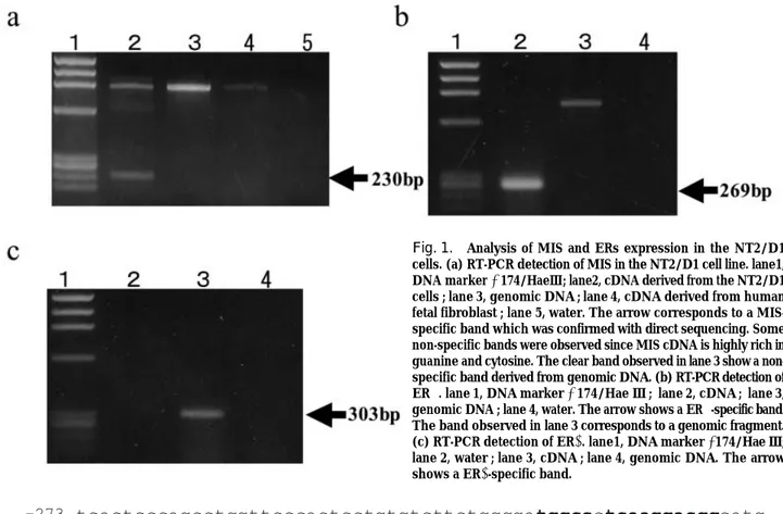
Some studies reported that the NT2/D1cell line expresses the genes participating in sex-determination or sex-differentiation such as SRY, SOX9 and DAX-1 (6,19). To elucidate whether MIS is expressed in this cell line, we carried out RT-PCR analysis using a MIS-specific primer set. As a result, we found that MIS was expressed at a low level in the NT2/D1 cell line (Fig.1a). Therefore, we used this cell line for molecular analysis for effects of estrogen on the MIS expression. When we tested whether ERαand ERβare expressed in the NT 2/D 1 cell line using RT-PCR, we found that both genes were expressed in this cell line (Fig.1b, 1c).
To clarify whether estrogen has some effects on the expression of MIS, we carried out dual luciferase reporter assay to assess the MIS promoter activity under different
concentrations ofβ-estradiol (E2). It was reported that a 273 bp DNA fragment upstream from the translation initiation codon of the MIS was important for MIS expression (6). This fragment contains a SOX9 binding site and two SF-1binding sites, and those transcription factors were suggested important for MIS expression (6). Therefore, we generated a firefly luciferase reporter construct that possesses the 273 bp DNA fragment of the MIS promoter, designed as pGL3 -MIS 273, according to a previous study (6). The sequence of the human proximal MIS promoter is shown in Fig.2. When we cultured the NT2/D1 cells using the me-dium containing the fetal calf serum stripped for lipid-soluble hormones, those cells could not growth. There-fore, in the present study, we cultured cells in the medium that was not stripped for lipid-soluble hormones. The concentration of E2 in the fetal calf serum was approx-imately 22.84 pg/ml(0.08 nM), according to the certificate
Fig. 1. Analysis of MIS and ERs expression in the NT2/D1 cells. (a) RT-PCR detection of MIS in the NT2/D1 cell line. lane1, DNA markerφ174/HaeⅢ; lane2, cDNA derived from the NT2/D1 cells ; lane 3, genomic DNA ; lane 4, cDNA derived from human fetal fibroblast ; lane 5, water. The arrow corresponds to a MIS-specific band which was confirmed with direct sequencing. Some non-specific bands were observed since MIS cDNA is highly rich in guanine and cytosine. The clear band observed in lane 3 show a non-specific band derived from genomic DNA. (b) RT-PCR detection of ERα. lane 1, DNA markerφ174/Hae Ⅲ ; lane 2, cDNA ; lane 3, genomic DNA ; lane 4, water. The arrow shows a ERα-specific band. The band observed in lane 3 corresponds to a genomic fragment. (c) RT-PCR detection of ERβ. lane1, DNA markerφ174/Hae Ⅲ; lane 2, water ; lane 3, cDNA ; lane 4, genomic DNA. The arrow shows a ERβ-specific band.
Fig. 2. The sequence of the human MIS proximal promoter. Bold letters correspond to the binding sequences of the transcription factors. Ⅰ, ERE-like motives ; Ⅱ, SF-1 binding sites ; Ⅲ, SOX binding site ; Ⅳ, TATA box
G. Chen et al. Effects of ERα on the MIS promoter 194
of product.
When the amount of E2 in the culture medium was increased, no apparent dose-dependent effects of E2 on the MIS promoter was observed (Fig. 3). However, we could not rule out the possibility that the amount of ERs expressed in the NT2/D1 cells was inadequate to exhibit their functions. Semi-quantitative RT-PCR analysis of the two ERs under the different concentrations of E2 could not reveal significant alterations of expression of both ERs (data not shown).
Over-expression of ER
α
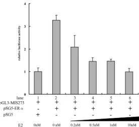
increases the MIS promoter activityTo over-express ERαor ERβin the NT2/D1cells, ERα-pSG5 and ERβ-pCXN2 were transfected into the NT2/D1 cells. When ERαor ERβwere over-expressed in the NT2/D1 cells under several concentrations of E2, the two ERs were completely different in the dose-responsiveness to E2 for the MIS promoter activity (Figs. 4, 5).
For ERα, the MIS promoter activity was increased 3.3-fold compared with the controls under no additional E2 to the medium (Fig.4). As the E2 concentration was increased, the MIS promoter activity was decreased (Fig. 4). When E2 was added in the medium up to final concentrations of 10 nM, the activity of MIS proximal promoter became almost one-third compared with no additional E2 (Fig.4). To confirm that ERαhas transcriptional activity in the NT2/D1 cells, the reporter construct, which contains a firefly luciferase gene ligated with a thymidine kinase minimum promoter and es-trogen responsive element (ERE), was co-transfected into the NT2/D1cells with the ERαover-expression vector. As a result, the reporter gene activity was
in-creased according to the concentrations of E2, suggesting that the exogenous ERαcan work in the NT2/D1 cells (data not shown).
In contrast to ERα, for ERβ, the response of the MIS promoter to E2, which was observed for ERα, was not detected, and the promoter activity was similar to the controls, suggesting that the type of ERs is crucial for regulation of the MIS promoter activity (Fig.5). ERαaffects SF-1-dependent MIS proximal promoter activity
When SF-1 and ERα were simultaneously over-expressed in the NT2/D1 cells, it became clear that the MIS promoter activity was similar to that observed for SF-1alone which increased the promoter activity
Fig. 3. The MIS promoter activity in the NT2/D1cells under the different concentrations of E2. The lanes and E2 concentrations in each sample are below.
“+” indicates addition of pGL3-MIS 273. Fold activation was compared with pGL3-MIS 273 alone, lane 1. Errors bars show the standard error.
Fig. 4. The effects of over- expression of the ERαon the MIS promoter activity.
The lanes and expression constructs in each sample are below. “+” and “-”indicate addition and omission, respectively. pSG 5 is an empty expression vector. Fold activation was compared with lane 1.Errors bars show the standard error.
Fig. 5. The effects of over- expression of the ERβon the MIS promoter activity. The lanes and expression constructs in each sample are below. “+” and “-” indicate addition and omission, respectively. pCXN 2 is a empty expression vector. Fold activation was compared with lane 1. Errors bars show the standard error. The Journal of Medical Investigation Vol. 50 2003 195
by 2.5 -fold compared with the control (Fig.6). This result suggested that SF-1inhibits the MIS promoter activity mediated with ERα(Fig.6). Furthermore, the MIS promoter activity mediated with SF-1was repressed in a E2 dose-dependent manner (Fig.6).
DISCUSSION
One of major targets for EDs is thought to be a sex differentiation system during fetal stage (11). MIS has
¨
important roles for regression of the Mullerian duct during male sex differentiation (1-4). Therefore, we addressed whether E2 affects the MIS expression in the promoter level using the NT2/D1 cells as a model of the pre-mature Sertoli cell.
ERs are known to be different in their tissue distributions and presumed functions (20). In the present study, over-expression of two ERs in the NT2/D1 cells demon-strated that ERαbut not ERβup-regulated the MIS promoter activity. Furthermore, we showed that the MIS promoter activity mediated with ERαwas repressed in an E2 dose-dependent manner.
It was reported that DES-exposed fetus exhibits a
¨
delay in the formation and regression of the Mullerian duct (15, 16). Therefore, it may be possible that one of the effects of E2 on the sex differentiation may be to change the timing of the initiation for formation and
¨
regression of the Mullerian duct (16).
It was reported that SF-1is expressed in the fetal Sertoli cells and has important roles for expression of MIS (6-9, 21). The present results suggested that the
MIS expression mediated with SF-1may be influenced by E2 through ERα. However, in the fragment of the MIS promoter used in the present study, which contained a 273 bp region upstream from the translation start codon of the MIS gene, there is no complete estrogen receptor response element (6). However, there are two SF-1 binding sites that contain ERE half-site like motives and four sequences similar to ERE half-sites (Fig.2). For examples, Jena-Marc Vanacker et al reported that ERαbut not ERβbinds to SF-1 response elements and activates the osteopontin gene promoter (22). Indeed, since there are two SF-1 response elements, which contain similar sequences with half motives of ERE, in the MIS-273 bp promoter, ERαmay bind to them.
There are some studies that reported that ligand-free and ligand-bound nuclear receptors have different functions for the transcription of their target genes (23, 24). The findings of present study suggested that ERα up-regulated transcriptional activity of the human MIS promoter under low concentrations of E2, and that it was decreased as the concentration of E2 was increased. Although we could not remove estrogens completely because the NT2/D1 could not survive without lipid-soluble hormones, it is possible that ligand-unbound ERαup-regulated transcriptional activity of the human MIS promoter.
For ERβ, Shapiro et al. has reported that E2-unbound ERβinduced the high constitutive activity of 5.5 kb rat vasopressin promoter and that the E2-bound ERβ inhibited the high constitutive activity (25). They re-ported that the constitutive activity of ERβmay be due to transcription from an AP1-like sequence, which was not found in the MIS promoter region analyzed in the present study (25). In present study, over-expression of ERβdid not show the response of the MIS promoter to E2, which was observed for ERα.
Some hypotheses appear to explain that SF-1 an-tagonizes the effect of ERαon the MIS promoter. First, the SF1-response elements may be important for both ERαto bind to the human MIS promoter, and SF-1 may compete with ERαfor its binding sites (22). Second, since ERαcan physically interact with SF-1 (26), this interaction may lead to block access of ERαto the MIS promoter.
In the present study, when SF-1and ERαwere simul-taneously over-expressed in the NT2/D1 cells, the MIS promoter activity was decreased as the E2 concen-tration was increased. Liganded ERαmay be different from the un-liganded one in the interaction with SF-1or the affinity to the MIS promoter. We may also have to pay attention to the possibility that ERαexhibits non-genomic functions in a ERE-independent manner
Fig. 6. SF-1inhibits ERα-mediated activation of the MIS promoter and E2 affects the MIS promoter activity mediated with SF-1 through ERα. The lanes and expression constructs in each sample are below. “+” and “-” indicate addition and omission, respectively. There is a significant difference (p<0.05) between the results in lane 2 and lane 6 by unpaired Student’s t test. There is also a significant difference (p<0.01) between the results lane 2 and lane 7.
G. Chen et al. Effects of ERα on the MIS promoter 196
to affect the functions of SF-1 (27, 28). To test these hypotheses, future studies are required.
In conclusion, although it remains unclear whether the effects of ERαon the MIS promoter are mediated through the genomic or non-genomic actions, the present results may suggest that ERαup-regulates the MIS promoter activity under low concentrations of E2 and that it inhibits the effects of SF-1 on the MIS promoter. The present findings may be useful to under-stand the molecular mechanisms by which EDs or DES affects the MIS expression.
ACKNOWLEDGEMENT
We are grateful to Miss Unemi and Miss Tsuji, Uni-versity of Tokushima for technical assistance. Our study was supported in part by grants from Core Research for Evolutional Science and Technology (CREST) and the Ministry of Health, Labour and Welfare.
REFERENCES
1. Koopman P, Bullejos M, Bowles J : Regulation of male sexual development by Sry and Sox9. J Exp Zool 290 : 463 -74, 2001
2. Cotinot C, Pailhoux E, Jaubert F, Fellous M : Molecular genetics of sex determination. Semin Reprod Med 20 : 157-68, 2002
3. Veitia RA, Salas-Cortes L, Ottolenghi C, Pailhoux E, Cotinot C, Fellous M : Testis determination in mammals : more questions than answers. Mol Cell Endocrinol 179 : 3 -16, 2001
4. Lee MM, Donahoe PK. : Mullerian inhibiting substance : a gonadal hormone with multiple functions. Endocr Rev 14 : 152- 64, 1993 5. Hirobe S, He WW, Lee MM, Donahoe PK :
Mulle-rian inhibiting substance messenger ribonucleic acid expression in granulosa and Sertoli cells coincides with their mitotic activity. Endocrinology 131 : 854-62, 1992
6. De Santa Barbara P, Bonneaud N, Boizet B, Desclozeaux M, Moniot B, Sudbeck P, Scherer G, Poulat F, Berta P : Direct interaction of SRY-related protein SOX9 and steroidogenic factor 1 regulates transcription of the human anti-Mullerian hormone gene. Mol Cell Biol 18 : 6653-65, 1998 7. Watanabe K, Clarke TR, Lane AH, Wang X, Donahoe PK : Endogenous expression of Mullerian inhibiting substance in early postnatal rat sertoli
cells requires multiple steroidogenic factor-1 and GATA-4-binding sites. Proc Natl Acad Sci USA 97 : 1624-9, 2000
8. Tremblay JJ, Viger RS : Transcription factor GATA-4 enhances Mullerian inhibiting SF-1. Mol En-docrinol 13 : 1388-401, 1999
9. Tremblay JJ, Viger RS : Nuclear receptor Dax-1 represses the transcriptional cooperation between GATA-4 and SF-1in Sertoli cells. Biol Reprod 64: 1191-9, 2001
10. Nachtigal MW, Hirokawa Y, Enyeart-VanHouten DL, Flanagan JN, Hammer GD, Ingraham HA. Wilms’ tumor1and Dax-1modulate the orphan nuclear receptor SF-1in sex-specific gene expression. Cell 93 : 445-54, 1998
11. Mori C : Possible effects of endocrine disruptors on male reproductive function. Kaibogaku Zasshi, 76 (in Japanese) : 361-8, 2001
12. Sonnenschein C, Soto AM : An updated review of environmental estrogen and androgen mimics and antagonists. J Steroid Biochem Mol Biol 65 : 143-50, 1998
13. Simpson ER : Genetic mutations resulting in loss of aromatase activity in humans and mice. J Soc Gynecol Invest 7 (Suppl.1) : S18-21, 2000 14. Eddy EM, Washburn TF, Bunch DO, Goulding
EH, Gladen BC, Lubahn DB, Korach KS : Targeted disruption of the estrogen receptor gene in male mice causes alteration of spermatogenesis and infertility. Endocrinology 137 : 4796-805, 1996 15. Gill WB, Schumacher GF, Bibbo M : Structural and functional abnormalities in the sex organ of male offspring of mothers treated with diethylstilbestrol (DES). J Reprod Med 16 : 147-153, 1976 16. Visser JA, McLuskey A, Verhoef-Post M, Kramer P,
Grootegoed JA, Themmen AP: Effect of prenatal exposure to diethylstilbestrol on Mullerian duct development in fetal male mice. Endocrinology, 139 : 4244-51, 1998
17. Couse JF, Hewitt SC, Bunch DO, Sar M, Walker VR, Davis BJ, Korach KS: Postnatal sex reversal of the ovaries in mice lacking estrogen receptors alpha and beta. Science 286 : 2328 -31, 1999 18. Miyazaki J, Takaki S, Araki K, Tashiro F, Tominaga
A, Takatsu K, Yamamura K. Expression vector system based on the chicken beta-actin promoter directs efficient production of interleukin-5.Gene 15 : 269 -77, 1989
19. Clepet C, Schafer AJ, Sinclair AH, Palmer MS, Lovell-Badge R, Goodfellow PN : The human SRY transcript. Hum Mol Genet 2 : 2007-12, 1993 20. Denger S, Reid G, Brand H, Kos M, Gannon F :
Tissue-specific expression of human ERalpha and ERbeta in the male. Mol Cell Endocrinol 178 : 155-60, 2001
21. Hatano O, Takayama K, Imai T, Waterman MR, Takakusu A, Omura T, Morohashi K : Sex-dependent expression of a transcription factor, Ad4 BP, regulating steroidogenic P-450 genes in the gonads during prenatal and postnatal rat development. Developmen 120 : 2787-97,1994
22. Vanacker JM, Pettersson K, Gustafsson JA, Laudet V : Transcriptional targets shared by estrogen receptor-related receptors (ERRs) and estrogen receptor (ER) alpha, but not by ERbeta. EMBO J 18 : 4270-9, 1999
23. Weston AD, Blumberg B, Underhill TM : Active repression by unliganded retinoid receptors in development : less is sometimes more. J Cell Biol 161 : 223-8, 2003
24. Apriletti JW, Ribeiro RC, Wagner RL, Feng W, Webb P, Kushner PJ, West BL, Nilsson S, Scanlan
TS, Fletterick RJ, Baxter JD : Molecular and structural biology of thyroid hormone receptors. Clin Exp Pharmacol Physiol Suppl 25 : S 2-11, 1998
25. Shapiro RA, Xu C, Dorsa DM : Differential transcrip-tional regulation of rat vasopressin gene expression by estrogen receptor alpha and beta. Endocrinology 141 : 4056-64, 2000
26. Drean YL, Liu D, Wong AO, Xiong F, Hew CL: Steroidogenic factor 1 and estradiol receptor act in synergism to regulate the expression of the salmon gonadotropinⅡbeta subunit gene. Mol Endocrinol 10 : 217-29, 1996
27. Coleman KM, Smith CL : Intracellular signaling pathways : nongenomic actions of estrogens and ligand-independent activation of estrogen receptors. Front Biosci 6 : 1379-91, 2001
28. Moggs JG, Orphanides G : Estrogen receptors : orchestrators of pleiotropic cellular responses. EMBO Rep 2 : 775-81, 2001
G. Chen et al. Effects of ERα on the MIS promoter 198