Author(s) TANABE, Kumiko; NISHIMURA, Kazumi; DOHI, Shuji;KOZAWA, Osamu
Citation [Brain Research] vol.[1274] p.[11]-[20]
Issue Date 2009-06-05
Rights Elsevier Science Bv
Version 著者最終稿 (author final version) postprint
URL http://hdl.handle.net/20.500.12099/34172
Mechanisms of interleukin-1 -induced GDNF release from rat glioma cells
Kumiko Tanabea,*, Kazumi Nishimuraa, Shuji Dohia and Osamu Kozawab
Departments of aAnesthesiology and Pain Medicine, and bPharmacology, Gifu University Graduate School of Medicine, Gifu 501-1194, Japan
34 pages in the article
Correspondence: Kumiko Tanabe
Department of Anesthesiology and Pain Medicine, Gifu University Graduate School of Medicine, Gifu 501-1194, Japan
Fax: +81-58-230-6405
E-mail: kumiko-t@m2.gyao.ne.jp
Abstract
Glial cell line-derived neurotrophic factor (GDNF) is highly expressed both in neurons and astrocytes in injured tissues. Astrocytes support neurons by releasing neurotrophic factors including GDNF. It has been reported that various agents including cytokines such as interleukin (IL)-1 induce GDNF mRNA expression and the release in astrocytes. However, the mechanism behind the GDNF synthesis and release remains unclear. Herein, we investigated the mechanisms of the IL-1 -induced GDNF release from rat C6 glioma cells. IL-1 time dependently stimulated GDNF release from C6 cells. IL-1 induced the phosphorylation of inhibitor kappa B (I B), p38 mitogen-activated protein (MAP) kinase, p44/p42 MAP kinase, stress-activated protein kinase/c-Jun N-terminal kinase (SAPK/JNK) and signal transducer and activator of transcription (STAT) 3. The IL-1 -stimulated levels of GDNF were suppressed by wedelolactone, an inhibitor of I B kinase, SB203580, an inhibitor of p38 MAP kinase, PD98059, an inhibitor of MAP kinase kinase 1/2 or Janus family of tyrosine kinase (JAK) inhibitor , an inhibitor of upstream kinase of STAT3. On the contrary, SP600125, an inhibitor of SAPK/JNK, failed to reduce the IL-1 -effect. These results strongly suggest that IL-1 stimulates GDNF release through the pathways of I B-nuclear factor kappa B, p38 MAP kinase, p44/p42 MAP kinase and JAK-STAT3, but not through the SAPK/JNK pathway in glioma cells.
Abbreviations
CNS, central nervous system; DMEM, Dulbecco’s modified Eagle’s medium; ELISA, enzyme-linked immunosolvent assay; GDNF, glial cell line-derived neurotrophic factor; I B, inhibitor kappa B; IKK, I B kinase; IL, interleukin; JAK, Janus family of tyrosine kinase; LPS, lypopolysaccharide; MAP, mitogen-activated protein; MEK, MAP kinase kinase; NF B, nuclear factor kappa B; PAGE, polyacrylamide gel electrophoresis; SAPK/JNK, stress-activated protein kinase/c-Jun N-terminal kinase; SDS, sodium dodecyl sulfate; STAT, signal transducer and activator of transcription; TNF, tumor necrosis factor
1. Introduction
Glial cell line-derived neurotrophic factor (GDNF)-family ligands are synthesized and released in many tissues in response to a variety of agonists (Airaksinen and Saarma, 2002; Saavedra et al., 2008). In the central nervous system (CNS), it has been first reported that GDNF has ability to promote dopamine uptake and cell survival of embryonic mesencephalic dopaminergic neurons (Lin et al., 1993). Later, GDNF has been shown to play important roles in the development, differentiation, neurodegenerative disease, protection of brain cells against ischemia, cognition, neuronal plasticity, drug abuse and neuropathic pain (Airaksinen and Saarma, 2002). The GDNF mRNA expression is elevated in neurons and astrocytes in injured tissue (Saavedra et al., 2008). Astrocytes protect neurons by secreting neurotrophic factors including GDNF (Villegas et al., 2003). Therefore, GDNF release would be expected to have therapeutic potential for neurodegenerative disease, traumatic, ischemic, inflammatory brain lesion and others.
As the mechanisms for the GDNF synthesis and release from astrocytes, it has been reported that tumor necrosis factor (TNF)- or lypopolysaccharide (LPS) induces GDNF mRNA expression and elevation of protein level through TNF- receptor in mouse astrocytes (Kuno et al., 2006) and that LPS stimulates GDNF mRNA expression through not either nuclear factor kappa B (NF B) pathway or mitogen-activated protein (MAP) kinase pathway in rat astrocytes (Tanaka et al., 2008). Various agents such as fibroblast growth factor, TNF- , interleukin (IL)-1 , IL-6, serotonin and amitriptyline are reportedly stimulate GDNF release from rat C6 glioma cells (Verity et al., 1998; Hisaoka et al., 2001, 2004, 2008; Tsuchioka et al., 2008). In these cells, it has been reported that p44/p42 MAP kinase activation and cyclic AMP responsive element binding protein phosphorylation are involved in amitriptyline-induced GDNF release (Hisaoka et al., 2001, 2008) and that serotonin-induced GDNF mRNA expression is
regulated by p44/p42 MAP kinase activation via fibroblast growth factor receptor (Tsuchioka et al., 2008). However, the mechanism of GDNF release from astrocytes is not precisely clarified. It has been shown that GDNF is highly produced and released by C6 glioma cells, which are considered astrocyte-like cells (Hisaoka et al., 2001, 2004, 2008; Tsuchioka et al., 2008), and C6 cells are widely used for investigating the mechanisms of GDNF synthesis or release (Saavedra et al., 2008). Therefore, we used C6 glioma cells for the present study.
The pro-inflammatory cytokines including ILs, interferons and TNFs, are secreted from a variety of cells in response to infection, activated lymphocyte products, microbial toxins and inflammatory stimuli (Merrill and Benveniste, 1996; Rothwell and Luheshi, 2000; Viviani et al., 2004). In the CNS, cytokines act as mediator of neuroimmune responses to neurodegeneration and the other diseases (Merrill and Benveniste, 1996; Rothwell and Luheshi, 2000; Viviani et al., 2004). IL-1 expression is at low levels in the CNS in healthy humans, whereas it is elevated in neurons, microglia and astrocytes after insult (Merrill and Benveniste, 1996; Rothwell and Luheshi, 2000; Viviani et al., 2004). It has been reported that damaged dopaminergic neurons induce GDNF release via IL-1 from astrocytes in rat neuron-glia coculture (Saavedra et al., 2007). However, the mechanism behind the IL-1 -induced GDNF release remains unclear.
IL-1 binds its receptor, which associates with IL-1 receptor-accessory proteins to initiate an intracellular signaling. The complex of receptor and IL-1 receptor-accessory protein recruits and activates IL-1 receptor associated kinase. The inhibitor kappa B (I B) and MAP kinase superfamily, such as p38 MAP kinase, p44/p42 MAP kinase and stress-activated protein kinase/c-Jun N-terminal kinase (SAPK/JNK) are then activated by IL-1 (Rothwell and Luheshi, 2000; Boutin et al., 2003; Viviani et al., 2004). I B is phosphorylated and degradated by I B kinase (IKK), subsequently NF B is freed from I B and translocates into the nucleus
(Rothwell and Luheshi, 2000; Hayden and Ghosh, 2004). In addition, the Janus family of tyrosine kinase (JAK)-signal transducer and activator of transcription (STAT) pathway is recognized to have important role in cytokine signaling such as interferons and ILs. Activation of JAK-STAT pathway leads to a rapid signaling from the cell surface to the nucleus (Imada and Leonard, 2000). Seven STAT proteins have been identified in mammalian cells (Imada and Leonard, 2000). Among them, it has been reported that STAT3 is phosphorylated and activated by IL-1 (Ng et al., 2001). In the CNS, STAT3 has important roles in post-ischemic brain damage (Yi et al., 2007).
In the present study, we investigated the involvement of I B-NF B pathway, three MAP kinases and JAK-STAT pathway in the IL-1 -induced GDNF release from rat C6 glioma cells.
2. Results
2.1. Effect of IL-1 on GDNF release from C6 cells
It has been reported that IL-1 stimulated GDNF release from rat C6 glioma cells (Verity et al., 1998). We confirmed that when the cells were exposed to IL-1 for 12 h, GDNF levels were significantly increased compared to those in the non-treated cells. The stimulatory effect was observed even 24 h after exposure to IL-1 (Fig. 1A). The net increase of GDNF by IL-1 was observed time dependently until 24 h after the stimulation. The effect of IL-1 on GDNF release was concentration-dependent in the range between 1 and 50 ng/ml, statistical significance being reached at concentrations over 1 ng/ml (Fig. 1B). We examined the effect of cycloheximide, an inhibitor of protein synthesis (Obrig et al., 1971), on the IL-1 -stimulated GDNF release. We found that the IL-1 -stimulated GDNF release was significantly reduced by 30 M cycloheximide (Table 1), suggesting that IL-1 stimulates de novo synthesis of GDNF protein.
2.2. Effect of wedelolactone on the IL-1 -induced GDNF release from C6 cells
It is well known that cytokines activate the I B-NF B pathway (Hayden and Ghosh, 2004). NF B binds to its consensus sequence on a target gene promoting transcription and upregulation of gene expression within the nucleus (Hayden and Ghosh, 2004). Since it has been reported that IL-1 induces I B phosphorylation and its degradation in C6 cells, we confirmed that IL-1 significantly phosphorylated and degradated I B in a time dependent manner (data not shown). We next examined whether I B phosphorylation is involved in the IL-1 -induced GDNF release. Wedelolactone, an inhibitor of IKK (Kobori et al., 2004), which by itself had little effect on the GDNF levels, significantly suppressed the IL-1 -induced GDNF release. The suppressive effect was concentration-dependent in the range between 1 and 50 M (Fig.
2A). We found that wedelolactone suppressed both the IL-1 -induced phosphorylation and degradation of I B at 50 M (Fig. 2B).
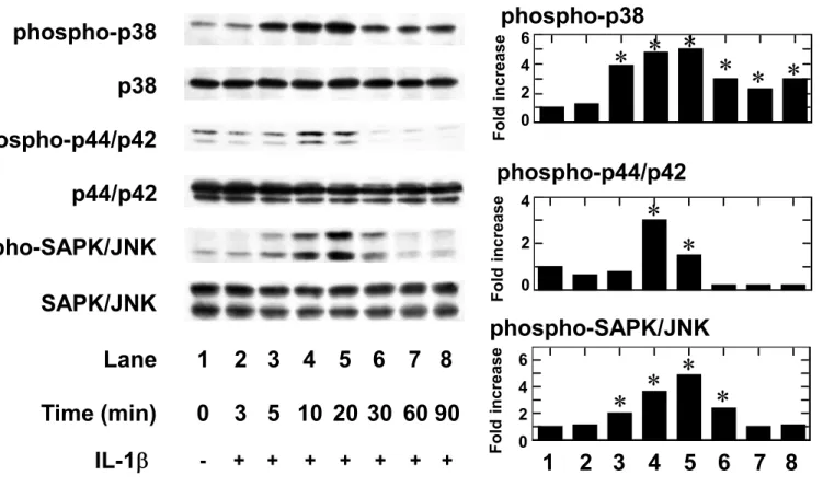
2.3. Effects of IL-1 on phosphorylation of the MAP kinase superfamily in C6 cells It is generally recognized that the MAP kinase superfamily members such as p38 MAP kinase, p44/p42 MAP kinase and SAPK/JNK are central elements used by mammalian cells to transduce the various messages of variety of agonists (Johnson and Lapadat, 2002). To investigate whether IL-1 stimulates GDNF release through activation of the MAP kinase superfamily in C6 glioma cells, we first examined the effects of IL-1 on the phosphorylation of three MAP kinases. IL-1 significantly induced the phosphorylation of p38 MAP kinase, p44/p42 MAP kinase and SAPK/JNK in a time dependent manner. The IL-1 -induced phosphorylation of p38 MAP kinase and SAPK/JNK were observed at 5 min, reached its peak at 20 min after the stimulation and decreased thereafter (Fig. 3). On the other hand, the IL-1 -induced phosphorylation of p44/p42 MAP kinase reached its peak at 10 min and decreased thereafter.
2.4. Effects of SB203580, PD98059 or SP600125 on the IL-1 -induced GDNF release from C6 cells
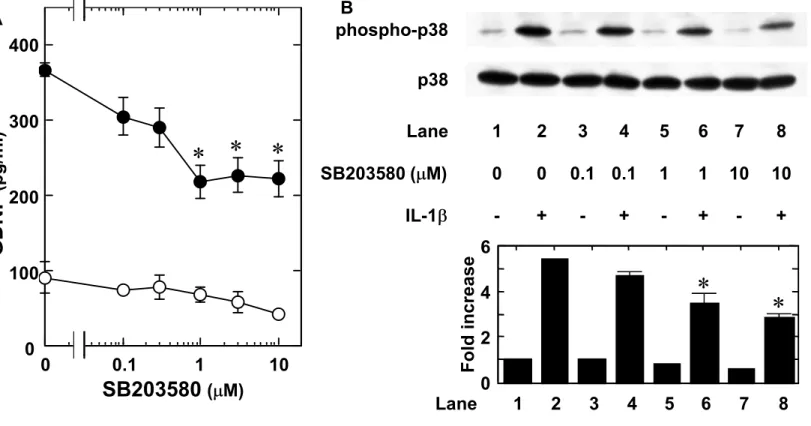
To clarify which three MAP kinases are involved in the IL-1 -induced GDNF release in C6 glioma cells, we examined the effects of each three MAP kinase inhibitor on the IL-1 -induced GDNF release. SB203580, a specific inhibitor of p38 MAP kinase (Cuenda et al., 1995), which by itself slightly decreased the GDNF levels, significantly suppressed the IL-1 -induced GDNF release. The inhibitory effect was concentration-dependent between 0.3 and 10 M (Fig. 4A). SB203580 truly attenuated the IL-1 -induced phosphorylation of p38 MAP kinase (Fig. 4B). Similarly, the GDNF release induced by IL-1 was markedly suppressed by PD98059, a specific
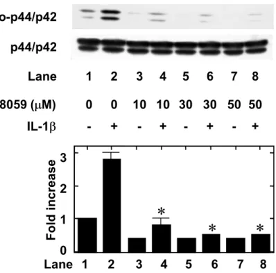
inhibitor of upstream kinase (MAP kinase kinase; MEK1/2) that activates p44/p42 MAP kinase (Alessi et al., 1995). PD98059 alone had little effect on the GDNF levels. The suppressive effect of PD98059 was concentration-dependent between 0.1 and 50
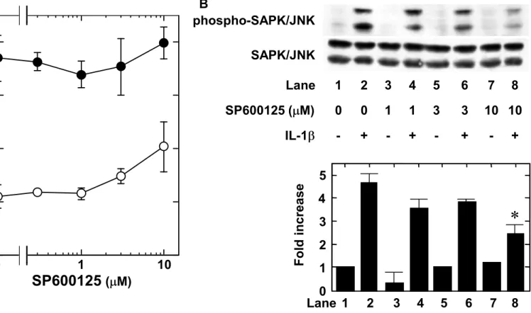
M (Fig. 5A). We found that PD98059 suppressed the IL-1 -induced phosphorylation of p44/p42 MAP kinase (Fig. 5B). However, SP600125, a specific inhibitor of SAPK/JNK (Bennett et al., 2001), failed to reduce IL-1 -induced GDNF release up to 10 M (Fig. 6A). We found that SP600125 (10 M) suppressed the IL-1 -induced phosphorylation of SAPK/JNK (Fig. 6B).
2.5. Effect of JAK inhibitor on the IL-1 -induced GDNF release from C6 cells
The JAK-STAT pathway has an essential role in driving a variety of biological responses to cytokines (Imada and Leonard, 2000). To clarify whether activation of STAT3 is involved in the IL-1 -induced GDNF release in C6 cells, we examined the effect of IL-1 on the phosphorylation of STAT3. IL-1 time-dependently induced the phosphorylation of STAT3 in C6 cells. The phosphorylation was observed at 60 min after the stimulation up to 120 min (Fig. 7). We next examined the effect of JAK inhibitor , an inhibitor of JAK 1, 2 and 3 (Thompson et al., 2002), on the GDNF release. JAK inhibitor , which by itself had little effect on the GDNF levels, significantly suppressed the IL-1 -induced GDNF release. The effect of JAK inhibitor was concentration-dependent between 0.2 and 3 M (Fig. 8A). In addition, JAK inhibitor (1 M) truly suppressed the IL-1 -induced phosphorylation of STAT3 (Fig. 8B).
2.6. Effects of the simultaneous treatment with wedelolactone, SB203580, PD98059 and JAK inhibitor on the IL-1 -induced GDNF release from C6 cells
We examine the combined effect of each inhibitor on the GDNF release. Combination of wedelolactone, SB203580, PD98059, and JAK inhibitor almost completely suppressed the IL-1 -induced GDNF release (Table 2).
3. Discussion
It is well known that the I B-NF B pathway is activated in response to several cytokines such as IL-1 (Rothwell and Luheshi, 2000; Boutin et al., 2003; Viviani et al., 2004). I B is phosphorylated and degradated after stimulation. After stimulation, the activated IKK catalyzes the phosphorylation of I B and subsequent degradation which in turn releases NF B, facilitating its nuclear translocation and gene transcription (Rothwell and Luheshi, 2000; Hayden and Ghosh, 2004). In the present study, we showed that the IL-1 -induced GDNF release was significantly suppressed by wedelolactone, an inhibitor of IKK (Kobori et al., 2004). Wedelolactone inhibits both phosphorylation and degradation of I B (Kobori et al., 2004). We found that wedelolactone truly reduced the IL-1 -induced I B phosphorylation and degradation also in C6 cells. Therefore, our findings suggest that IL-1 induces the GDNF release through the I B-NF B pathway in C6 glioma cells.
The MAP kinase superfamily plays a pivotal role in a variety of cellular functions including proliferation, differentiation and apoptosis (Johnson and Lapadat, 2002). Three major MAP kinases, p38 MAP kinase, p44/p42 MAP kinase and SAPK/JNK, are known to mediate intracellular signaling of extracellular agonists (Johnson and Lapadat, 2002). It is well recognized that MAP kinases are activated by phosphorylation of threonine and tyrosine residues by dual specificity MEK (Johnson and Lapadat, 2002). It has been reported that IL-1 stimulates the activation of p38 MAP kinase and p44/p42 MAP kinase in murine astrocytes (Molina-Holgado et al., 2000). Then, we examined the effects of IL-1 on the MAP kinase superfamily in C6 glioma cells. IL-1 induced the phosphorylation of p38 MAP kinase, p44/p42 MAP kinase and SAPK/JNK. Therefore, our findings suggest that IL-1 activates all three MAP kinases in rat C6 glioma cells. Next, we showed that the IL-1 -induced GDNF release was suppressed by SB203580, a specific inhibitor of p38 MAP kinase (Cuenda et al.,
1995) or PD98059, a specific inhibitor of MEK1/2 (Alessi et al., 1995), but not suppressed by SP600125, a specific SAPK/JNK inhibitor (Bennett et al., 2001). In addition, we confirmed that SB203580, PD98059 or SP600125 truly reduced the IL-1 -induced p38 MAP kinase, p44/p42 MAP kinase or SAPK/JNK phosphorylation respectively. Based on our results, it is probable that IL-1 stimulates GDNF release via p38 MAP kinase and p44/p42 MAP kinase, but not SAPK/JNK in C6 cells. In the present study, it seems that the phosphorylation of p38 MAP kinase in response to IL-1 was more sustained over time than the phosphorylation of p44/p42 MAP kinase or SAPK/JNK. However, it is not clear whether this prolonged phosphorylation of p38 MAP kinase is involved in the IL-1 -induced GDNF release. Further investigation is necessary to clarify the different kinetics of MAP kinases.
It is recognized that JAKs are constitutively associated with many cytokine receptors (Imada and Leonard, 2000). The binding of cytokines to its receptor associated with JAKs, leading to the tyrosine phosphorylation of the receptor, which generates docking site for STATs. (Imada and Leonard, 2000). Then, the STATs are phosphorylated and translocate to the nucleus where they may activates the transcription of several genes (Imada and Leonard, 2000). In the present study, we demonstrated that IL-1 significantly induced the phosphorylation of STAT3 in C6 cells, and that JAK inhibitor , an inhibitor of JAK 1, 2 and 3 (Thompson et al., 2002), suppressed the IL-1 -induced GDNF release. In addition, we found that JAK inhibitor suppressed the IL-1 -induced STAT3 phosphorylation. Therefore, these results suggest that IL-1 induces the GDNF release through the JAK-STAT3 pathway in C6 glioma cells. The IL-1 -induced phosphorylation of STAT3 was observed later than the phosphorylation of MAP kinases. This delayed phosphorylation is consistent with a previous report, showing that IL-1 phosphorylates STAT3 at 60 min after the stimulation in rat myocytes (Ng et al., 2001). It is possible that the IL-1 -stimulated STAT3 phosphorylation is through indirect mechanisms such as the synthesis and release of
other molecules. Each inhibitor used in the present study is specific and selective. However, since they may have the other effects, further investigations are necessary to clarify the details of the IL-1 intracellular signaling.
Many stimuli such as ischemia, some cytokines, serotonin and some drugs, increase GDNF synthesis from neurons, astrocytes and microglia (Verity et al., 1998; Hisaoka et al., 2001, 2008; Airaksinen and Saarma, 2002; Saavedra et al., 2008; Tsuchioka et al., 2008). GDNF has neuroprotective effects through the suppression of apoptosis, excitotoxicity and free radicals-induced injury (Airaksinen and Saarma, 2002; Saavedra et al., 2008). Cytokines are involve in the modulation of several neurological functions and dysfunctions, such as in the communication to the brain of systemic injury, infection and inflammation, in the modulation of the response to peripheral nerve injury, in the control of behaviour and in the progression or inhibition of neurodegeneration (Merrill and Benveniste, 1996; Rothwell and Luheshi, 2000; Viviani et al., 2004). In the CNS, proinflammatory cytokines increase after a damage resulting from exposure to neurotoxicants or from the onset of diseases such as cerebral ischemia, neurodegenerative disease and brain injury (Rothwell and Luheshi, 2000; Viviani et al., 2004). Concerning GDNF, it has been reported that IL-1 increases GDNF gene expression or release from mouse astrocytes or rat C6 glioma cells (Appel et al., 1997; Verity et al., 1998). The dopaminergic neurons damaged by H2O2 induce
IL-1 release from astrocytes (Saavedra et al., 2007). IL-1 , which is released by autocrine or paracrine from asrocytes, binds to IL-1 receptors in astocytes and triggers GDNF upregulation. IL-1 receptor antagonist reduces this GDNF upregulation and the viability of dopaminergic neuron at the same time. Therefore, it is suggested that IL-1 has neuroprotective effect through GDNF upregulation (Saavedra et al., 2007).
A variety of intracellular signaling pathways (MAP kinases, protein kinase C, protein kinase A and intracellular Ca2+ et al.) are reportedly involved in the GDNF release (Saavedra et al., 2008). Regarding the mechanisms of GDNF synthesis and
release from C6 glioma cells, it has been shown as follows; amitriptyline induces GDNF synthesis and release through the activation of p44/p42 MAP kinase but not p38 MAP kinase (Hisaoka et al., 2001). In addition, serotonin induces GDNF synthesis and release through the activation of p44/p42 MAP kinase (Tsuchioka et al., 2008). In the present study, we showed that in addition to p44/p42 MAP kinase pathway, the pathways of I B-NF B, p38 MAP kinase and JAK-STAT3 are involved in the IL-1 -stimulated GDNF release from C6 cells. Additionally, we demonstrated that the IL-1 -induced GDNF release was markedly reduced by cycloheximide. Based on our findings, it is probable that the increased GDNF levels by IL-1 from C6 cells is due to protein synthesis. Further investigation is necessary to clarify the exact mechanisms of GDNF synthesis and release in astrocytes.
In conclusion, our present results strongly suggest that IL-1 stimulates GDNF release through the pathways of I B-NF B, p38 MAP kinase, p44/p42 MAP kinase and JAK-STAT3, but not through the SAPK/JNK pathway in glioma cells.
4. Experimental procedures
4.1. Materials
GDNF enzyme-linked immunosolvent assay (ELISA) kit was purchase from Promega Co. (Madison, WI). IL-1 was obtained from R&D System (Minneapolis, MN). Wedelolactone, SB203580, PD98059, SP600125 and JAK inhibitor were obtained from Calbiochem-Novabiochem Co. (La Jolla, CA). Cycloheximide was obtained from Sigma Chemical Co. (St. Louis, MO). Phospho-specific I B, I B, phospho-specific p38 MAP kinase, p38 MAP kinase, phospho-specific p44/p42 MAP kinase, p44/p42 MAP kinase, phospho-specific SAPK/JNK, SAPK/JNK, phospho-specific STAT3 and STAT3 antibodies were purchased from Cell Signaling (Beverly, MA). An enhanced chemiluminescence Western blotting detection system was obtained from GE Healthcare UK. Ltd. (Buckinghamshire, England). Other materials and chemicals were obtained from commercial sources. Wedelolactone, SB203580, PD98059, SP600125 and JAK inhibitor were dissolved in dimethyl sulfoxide. Cycloheximide was dissolved in methanol. The maximum concentration of dimethyl sulfoxide or methanol was 0.1%, which did not affect the assay for GDNF or Western blot analysis.
4.2. Cell culture
Rat C6 glioma cells, obtained from the American Type Culture Collection (Rockville, MD), were seeded into 35-mm (5 x 10⁴ cells) or 90-mm (2 x 10⁵ cells) diameter dishes and maintained in Dulbecco’s modified Eagle’s medium (DMEM) containing 10% fetal bovine serum at 37℃ in a humidified atmosphere of 5% CO2/95%
air. After 6 days, the medium was exchanged for serum-free DMEM. The cells were then used for experiments after 24 h. When indicated, the cells were pretreated with cycloheximide, wedelolactone, SB203580, PD98059, SP600125 or JAK inhibitor for
60 min, and then stimulated by IL-1 in the presence of the inhibitors.
4.3. Assay for GDNF
The cultured cells (35-mm diameter dishes) were stimulated by IL-1 in serum-free DMEM for the indicated periods. The conditioned medium was collected at the end of the incubation, and the GDNF concentration was measured using an ELISA kit. The absorbance of each sample at 450 nm was measured with Multiscan JX ELISA reader (Thermo Labsystems, Helsinki, Finland). Absorbance was corrected with concentration by means of a standard curve.
4.4. Western blot analysis
The cultured cells (90-mm diameter dishes) were stimulated by 10 ng/ ml IL-1 in serum-free DMEM for the indicated periods. The cells were washed twice with phosphate-buffered saline and then lysed and sonicated during 10 sec on ice in a lysis buffer containing 62.5 mM Tris/HCl (pH 6.8), 2% sodium dodecyl sulfate (SDS), 50 mM dithiothreitol, and 10% glycerol. The sample was used for the analysis by Western blotting as described previously (Tanabe et al., 2008). SDS-polyacrylamide gel electrophoresis (PAGE) was performed by the method of Laemmli (Laemmli, 1970) in 10% polyacrylamide gels. The Western blot analysis was performed using phospho-specific I B antibodies, I B antibodies, phospho-specific p38 MAP kinase antibodies, p38 MAP kinase antibodies, phospho-specific p44/p42 MAP kinase antibodies, p44/p42 MAP kinase antibodies, phospho-specific SAPK/JNK antibodies, SAPK/JNK antibodies, phospho-specific STAT3 antibodies or STAT3 antibodies, with peroxidase-labeled antibodies raised in goat against rabbit immunoglobulin G being used as second antibodies. All antibodies were diluted into 1:1,000. The peroxidase activity on polyvinylidene difluoride membrane was visualized on X-ray film by means of an enhanced chemiluminescence Western blotting detection system. A
densitometric analysis was performed using the Molecular Analyst/Macintosh software program (Bio-Rad, Hercules, CA).
4.5. Statistical analysis
The data were analyzed by ANOVA followed by Bonferroni’s method for multiple comparisons between pairs. P < 0.05 was considered to be significant. Each value represents mean SD of triplicate independent determinations of a representative experiment carried out three times. Each experiment was repeated three times with similar results.
Acknowledgements
We are very grateful to Yoko Kawamura for her skillful technical assistance. This work was supported in part by a Grant-in-Aid for Scientific Research (19209050 and 20591798) from the Ministry of Education, Science, Sports and Culture of Japan.
References
Airaksinen, M.S., Saarma, M., 2002. The GDNF family: signalling, biological functions and therapeutic value. Nat. Rev. Neurosci. 3, 383-394.
Alessi, D.R., Cuenda, A., Cohen, P., Dudley, D.T., Saltiel, A.R., 1995. PD98059 is a specific inhibitor of the activation of mitogen-activated protein kinase kinase in vitro and in vivo. J. Biol. Chem. 270, 27489-27494.
Appel, E., Kolman, O., Kazimirsky, G., Blumberg, P.M., Brodie, C., 1997. Regulation of GDNF expression in cultured astrocytes by inflammatory stimuli. Neuroreport 8, 3309-3312.
Bennett, B.L., Sasaki, D.T., Murray, B.W., O’Leary, E.C., Sakata, S.T., Xu, W., Leisten, J.C., Motiwala, A., Pierce, S., Satoh, Y., Bhagwat, S.S., Manning, A.M., Anderson, D.W., 2001. SP600125, an anthrapyrazolone inhibitor of Jun N-terminal kinase. Proc. Natl. Acad. Sci. U.S.A. 98, 13681-13686.
Boutin, H., Kimber, I., Rothwell, N.J., Pinteaux, E., 2003. The expanding interleukin-1 family and its receptors. Mol. Neurobiol. 27, 239-248.
Cuenda, A., Rouse, J., Doza, Y.N., Meier, R., Cohen, P., Gallagher, T.F., Young, P.R., Lee, J.C., 1995. SB203580 is a specific inhibitor of a MAP kinase homologue which is stimulated by cellular stresses and interleukin-1. FEBS Lett. 364, 229-233.
Hayden, M.S., Ghosh, S., Signaling to NF- B. 2004. Gene Dev. 18, 2195-2224.
Hisaoka, K., Nishida, A., Koda, T., Miyata, M., Zensho, H., Morinobu, S., Ohta, M., Yamawaki, S., 2001. Antidepressant drug treatments induce glial cell line-derived neurotrophic factor (GDNF) synthesis and release in rat C6 glioblastoma cells. J. Neurochem. 79, 25-34.
Hisaoka, K., Nishida, A., Takebayashi M., Koda T., Yamawaki S., Nakata Y., 2004. Serotonin increases glial cell line-derived neurotrophic factor release in rat C6
glioblastoma cells. Brain Res. 1002, 167-170.
Hisaoka, K., Maeda, N., Tsuchioka, M., Takebayashi, M., 2008. Antidepressants induce acute CREB phosphorylation and CRE-mediated gene expression in glial cells: a possible contribution to GDNF production. Brain Res. 1196, 53-58.
Imada, K., Leonard, W.J., 2000. The Jak-STAT pathway. Mol. Immunol. 37, 1-11. Johnson, G.L., Lapadat, R., 2002. Mitogen-activated protein kinase pathways mediated
by ERK, JNK, and p38 protein kinases. Science 298, 1911-1912.
Kobori, M., Yang, Z., Gong, D., Heissmeyer, V., Zhu, H., Jung, Y.K., Gakidis, M.A.M., Rao, A., Sekine, T., Ikegami, F., Yuan, C., Yuan, J., 2004. Wedelolactone suppresses LPS-induced caspase-11 expression by directly inhibiting the IKK complex. Cell Death Differ. 11, 123-130.
Kuno, R., Yoshida, Y., Nitta, A., Nabeshima, T., Wang, J., Sonobe, Y., Kawanokuchi, J., Takeuchi, H., Mizuno, T., Suzumura, A., 2006. The role of TNF-alpha and its receptors in the production of NGF and GDNF by astrocytes. Brain Res. 1116, 12-18.
Laemmli, U.K., 1970. Cleavage of structural proteins during the assembly of the head of bacteriophage T4. Nature 227, 680-685.
Lin, L.-F.H., Doherty, D.H., Lile, J.D., Bektesh, S., Collins, F., 1993. GDNF: a glial cell line-derived neurotrophic factor for midbrain dopaminergic neurons. Sience 260, 1130-1132.
Merrill, J.E., Benveniste, E.N., 1996. Cytokines in inflammatory brain lesions: helpful and harmful. Trends Neurosci. 19, 331-338.
Molina-Holgado, E., Ortiz, S., Molina-Holgado, F., Guaza, C., 2000. Induction of COX-2 and PGE2 biosynthesis by IL-1 is mediated by PKC and mitogen-activated
protein kinases in murine astrocytes. Br. J. Pharmacol. 131, 142-159.
Ng, D.C.H., Long, C.S., Bogoyevitch, M.A., 2001. A role for the extracellular signal-regulated kinase and p38 mitogen-activated protein kinases in
interleukin-1 -stimulated delayed signal tranducer and activator of transcription 3 activation, atrial natriuretic factor expression, and cardiac myocyte morphology. J Biol. Chem. 276, 29490-29498.
Obrig, T.G., Culp, W.J., McKeehan, W.L., Hardesty B., 1971. The mechanism by which cycloheximide and related glutarimide antibiotics inhibit peptide synthesis on reticulocyte ribosomes. J Biol. Chem. 246, 174-181.
Rothwell, N.J., Luheshi, G.N., 2000. Interleukin 1 in the brain: biology, pathology and therapeutic target. Trends Neurosci. 23, 618-625.
Saavedra, A., Baltazar, G., Duarte, E.P., 2007. Interleukin-1 mediates GDNF up-regulation upon dopaminergic injury in ventral midbrain cell cultures. Neurobiol. Dis. 25, 92-104.
Saavedra, A., Baltazar G., Duarte E.P., 2008. Driving GDNF expression: the green and the red traffic lights. Prog. Neurobiol. 86, 186-215.
Tanabe, K., Takai, S., Matsushima-Nishiwaki, R., Kato, K., Dohi, S., Kozawa, O., 2008.
2 Adrenoreceptor agonist regulates protein kinase C-induced heat shock protein 27
phosphorylation in C6 glioma cells. J. Neurochem. 106, 519-528.
Tanaka, T., Oh-hashi, K., Shitara, H., Hirata, Y., Kiuchi, K., 2008. NF- B independent signaling pathway is responsible for LPS-induced GDNF gene expression in primary rat glial cultures. Neurosci. Lett. 431, 262-267.
Thompson, J.E., Cubbon, R.M., Cummings, R.T., Wicker, L.S., Frankshun, R., Cunningham, B.R., Cameron, P.M., Meinke, P.T., Liverton, N., Weng, Y., DeMartino, J.A., 2002. Photochemical preparation of a pyridone containing tetracycle: a Jak protein kinase inhibitor. Bioorg. Med. Chem. Lett. 12, 1219-1223.
Tsuchioka, M., Takebayashi, M., Hisaoka, K., Maeda, N., Nakata, Y., 2008. Serotonin (5-HT) induces glial cell line-derived neurotrophic factor (GDNF) mRNA expression via the transactivation of fibroblast growth factor receptor 2 (FGFR2) in rat C6 glioma cells. J. Neurochem. 106, 244-257.
Uehara, T., Matsuno, J., Kaneko, M., Nishiya, T., Fujimuro, M., Yokosawa, H., Nomura, Y., 1999. Transient nuclear factor B (NF- B) activation stimulated by interleukin-1 may be partly dependent on proteasome activity, but not phosphorylation and ubiquitination of the I B molecule, in C6 glioma cells. J. Biol. Chem. 274, 15875-15882.
Verity, A.N., Wyatt, T.L., Hajos, B., Eglan, R.M., Baecker, P.A., Johnson, R.M., 1998. Regulation of glial cell line-derived neurotrophic factor release from rat C6 glioblastoma cells. J. Neurochem. 70, 531-539.
Villegas, S.N., Poletta, F.A., Carri, N.G., 2003. Glia: a reassessment based in novel data in the developing and mature central nervous system. Cell Biol. Int. 27, 599-609. Viviani, B., Bartesaghi, S., Corsini, E., Galli, C.L., Marinovich, M., 2004. Cytokines
role in neurodegenerative events. Toxicol. Lett. 149, 85-89.
Yi, J-H., Park, S-W., Kapadia R., Vemuganti, R., 2007. Role of transcription factors in mediating post-ischemic cerebral inflammation and brain damage. Neurochem. Int. 50, 1014-1027.
Figure legends
Fig. 1. Effect of IL-1 on GDNF release from C6 cells. (A) The cultured cells were stimulated by 10 ng/ml IL-1 (closed circle) or vehicle (open circle) for the indicated period. (B) The cultured cells were stimulated by the indicated concentrations of IL-1 for 24 h. Each value represents mean SD of triplicate independent determinations of a representative experiment carried out three times. Similar results were obtained with two additional and different cell preparations. *P<0.05 compared to the value of unstimulated cells.
Fig. 2. Effect of wedelolactone on the IL-1 -induced GDNF release from C6 cells. (A) The cultured cells were pretreated with various concentrations of wedelolactone for 60 min, and then stimulated by 10 ng/ml IL-1 (closed circle) or vehicle (open circle) for 24 h. (B) The cultured cells were pretreated with various concentrations of wedelolactone or vehicle for 60 min, and then stimulated by 10 ng/ml IL-1 or vehicle for 30 min. The extracts of cells were subjected to SDS-PAGE with subsequent Western blotting analysis with antibodies against phospho-specific I B, I B or GAPDH. The lower histogram shows quantitative representations of the levels of IL-1 -induced phosphorylation of I B or I B obtained from laser densitometric analysis of three independent experiments, respectively. Each value represents mean SD of triplicate independent determinations of a representative experiment carried out three times. Similar results were obtained with two additional and different cell preparations. *P <0.05 compared to the value of IL-1 alone.
Fig. 3. Effects of IL-1 on the phosphorylation of p38 MAP kinase, p44/p42 MAP kinase or SAPK/JNK in C6 cells. The cultured cells were stimulated by 10 ng/ml IL-1 for the indicated period. The extracts of cells were subjected to SDS-PAGE
with subsequent Western blotting analysis with antibodies against phospho-specific p38 MAP kinase, p38 MAP kinase, phospho-specific p44/p42 MAP kinase, p44/p42 MAP kinase, phospho-specific SAPK/JNK or SAPK/JNK. The lower histogram shows quantitative representations of the levels of IL-1 -induced phosphorylation of p38 MAP kinase, p44/p42 MAP kinase or SAPK/JNK obtained from laser densitometric analysis of three independent experiments, respectively. Each value represents mean SD of triplicate independent determinations of a representative experiment carried out three times. Similar results were obtained with two additional and different cell preparations. *P<0.05 compared to the value of unstimulated cells.
Fig. 4. Effect of SB203580 on the IL-1 -induced GDNF release from C6 cells. (A) The cultured cells were pretreated with various concentrations of SB203580 for 60 min, and then stimulated by 10 ng/ml IL-1 (closed circle) or vehicle (open circle) for 24 h. (B) The cultured cells were pretreated with various concentrations of SB203580 or vehicle for 60 min, and then stimulated by 10 ng/ml IL-1 or vehicle for 20 min. The extracts of cells were subjected to SDS-PAGE with subsequent Western blotting analysis with antibodies against phospho-specific p38 MAP kinase or p38 MAP kinase. The lower histogram shows quantitative representations of the levels of IL-1 -induced phosphorylation of p38 MAP kinase obtained from laser densitometric analysis of three independent experiments, respectively. Each value represents mean SD of triplicate independent determinations of a representative experiment carried out three times. Similar results were obtained with two additional and different cell preparations. *P <0.05 compared to the value of IL-1 alone.
Fig. 5. Effect of PD98059 on the IL-1 -induced GDNF release from C6 cells. (A) The cultured cells were pretreated with various concentrations of PD98059 for 60 min, and then stimulated by 10 ng/ml IL-1 (closed circle) or vehicle (open circle) for 24 h. (B)
The cultured cells were pretreated with various concentrations of PD98059 or vehicle for 60 min, and then stimulated by 10 ng/ml IL-1 or vehicle for 10 min. The extracts of cells were subjected to SDS-PAGE with subsequent Western blotting analysis with antibodies against phospho-specific p44/p42 MAP kinase or p44/p42 MAP kinase. The lower histogram shows quantitative representations of the levels of IL-1 -induced phosphorylation of p44/p42 MAP kinase obtained from laser densitometric analysis of three independent experiments, respectively. Each value represents mean SD of triplicate independent determinations of a representative experiment carried out three times. Similar results were obtained with two additional and different cell preparations. *P <0.05 compared to the value of IL-1 alone.
Fig. 6. Effect of SP600125 on the IL-1 -induced GDNF release from C6 cells. (A) The cultured cells were pretreated with various concentrations of SP600125 for 60 min, and then stimulated by 10 ng/ml IL-1 (closed circle) or vehicle (open circle) for 24 h. (B) The cultured cells were pretreated with various concentrations of SP600125 or vehicle for 60 min, and then stimulated by 10 ng/ml IL-1 or vehicle for 20 min. The extracts of cells were subjected to SDS-PAGE with subsequent Western blotting analysis with antibodies against phospho-specific SAPK/JNK or SAPK/JNK. The lower histogram shows quantitative representations of the levels of IL-1 -induced phosphorylation of SAPK/JNK obtained from laser densitometric analysis of three independent experiments, respectively. Each value represents mean SD of triplicate independent determinations of a representative experiment carried out three times. Similar results were obtained with two additional and different cell preparations. *P <0.05 compared to the value of IL-1 alone.
Fig. 7. Effect of IL-1 on the phosphorylation of STAT3 in C6 cells. The cultured cells were stimulated by 10 ng/ml IL-1 for the indicated periods. The extracts of
cells were subjected to SDS-PAGE with subsequent Western blotting analysis with antibodies against phospho-specific STAT3 or STAT3. Similar results were obtained with two additional and different cell preparations.
Fig. 8. Effect of JAK inhibitor on the IL-1 -induced GDNF release from C6 cells. (A) The cultured cells were pretreated with various concentrations of JAK inhibitor for 60 min, and then stimulated by 10 ng/ml IL-1 (closed circle) or vehicle (open circle) for 24 h. (B) The cultured cells were pretreated with 1 M of JAK inhibitor or vehicle for 60 min, and then stimulated by 10 ng/ml IL-1 or vehicle for 60 min. The extracts of cells were subjected to SDS-PAGE with subsequent Western blotting analysis with antibodies against phospho-specific STAT3 or STAT3. The lower histogram shows quantitative representations of the levels of IL-1 -induced phosphorylation of STAT3 obtained from laser densitometric analysis of three independent experiments, respectively. Each value represents mean SD of triplicate independent determinations of a representative experiment carried out three times. Similar results were obtained with two additional and different cell preparations. *P <0.05 compared to the value of IL-1 alone.
0 0.1 1 10
IL-1
(ng/ml)G
D
N
F
(p g /m l)Figure 1
G
D
N
F
(p g /m l) 0 100 200 300 0 6 18 2 4 12Time
(h)*
*
*
*
0 100 200 300 400 1001 0 2
Figure 2
1 2 3 4 5 6 Lane 0 0 10 10 50 50 Wedelolactone ( M) GAPDH IL-1 - + - + - + Lane 1 2 3 4 5 6 F o ld i n c re a s e 1 0 phospho-I B I B*
*
G
D
N
F
(p g /m l) 0 100 200 300Wedelolactone
( M)*
*
*
0 0.1 1 10 100Figure 3
phospho-p44/p42
p44/p42
phospho-SAPK/JNK
SAPK/JNK
Lane
1 2 3 4 5 6 7 8
Time (min)
0 3 5 10 20 30 60 90
IL-1
- + + + + + + +1 2 3 4 5 6 7 8
* *
*
*
F o ld in cr ea se 0 2 6 4*
*
phospho-p44/p42
phospho-SAPK/JNK
0 2 4 F o ld in cr ea se F o ld in 0 2Figure 4
0 0.1 1 10SB203580
( M)G
D
N
F
(p g /m l) 0 100 200 300 Lane 1 2 3 4 5 6 7 8 SB203580 ( M) 0 0 0.1 0.1 1 1 10 10 IL-1 - + - + - + - + p38 Lane 1 2 3 4 5 6 7 8 F o ld i n c re a s e*
0 2 6 4*
*
*
*
Figure 5
0 0.1 1 10PD98059
( M)G
D
N
F
(p g /m l) 0 100 200 300 Lane 1 2 3 4 5 6 7 8 PD98059 ( M) 0 0 10 10 30 30 50 50 IL-1 - + - + - + - + Lane 1 2 3 4 5 6 7 8 1 0 3 2 F o ld i n c re a s e*
*
*
*
*
**
100Figure 6
0 1 10SP600125
( M)G
D
N
F
(p g /m l) 0 100 200 300 Lane 1 2 3 4 5 6 7 8 SP600125 ( M) 0 0 1 1 3 3 10 10 IL-1 - + - + - + - + SAPK/JNK Lane 1 2 3 4 5 6 7 8 F o ld i n c re a s e 1 0 3 2 4 5*
Figure 7
Lane
1
2
3
4
5
6
7
Time (min)
0
10 20 30 60 90 120
JAK inhibitor Ⅰ
( M) 0 1G
D
N
F
(p g /m l) 0 100 200 300Figure 8
Lane 1 2 3 4 JAK inhibitor Ⅰ - - + + IL-1 - + - + STAT3 Lane 1 2 3 4 1 0 3 2 F o ld in cr ea se*
*
*
*
*
cycloheximide IL1- GDNF (pg/ml)
90 5
399 5*
92 8
227 17**
The cultured cells were pretreated with 30 M cycloheximide or vehicle for 60 min, and then stimulated by 10 ng/ml IL-1 for 24 h. Each value represents mean SD of triplicate independent determinations of a representative experiment carried out three times. Similar results were obtained with two additional and different cell preparations. *P<0.05 compared to the value of unstimulated cells. **P<0.05 compared to the value of IL1- alone.
inhibitors IL1- GDNF (pg/ml)
57 24
380 24*
52 6
48 4**
The cultured cells were pretreated with 10 M wedelolactone, 1 M SB203580, 10 M PD98059 and 1 M JAK inhibitor or vehicle for 60 min, and then stimulated by 10 ng/ml IL-1 for 24 h. Each value represents mean SD of triplicate independent determinations of a representative experiment carried out three times. Similar results were obtained with two additional and different cell preparations. *P<0.05 compared to the value of unstimulated cells. **P<0.05 compared to the value of IL1- alone.