Chitosan (CS) is a plentiful natural polysaccharide that is non-toxic, biocompatible and biodegradable.1—3)It has been
investigated as a vehicle for the sustained release of drugs,4)
and as an absorption enhancer of drugs by mucoadhesion to the gastric cavity and the nasal cavity.5—7)The preparation of CS microspheres has also been studied.8,9)We found that CS
gel spheres formed at around pH 9 in amino acid solution, despite usually forming a gel in solutions with a pH .12.10) Drug release from the gel beads prepared at pH 9 was inhib-ited compared with that from the pH .12 gel beads, because of reduced CS degradation to low molecular weight. Drug re-lease was further slowed by forming a complex between chondroitin sulfate (Cho) and CS.11)Furthermore, the CS gel beads could be implanted into subcutaneous air pouches (AP) prepared on the dorsal surface of mice, with the in vivo release of the drug reflected in the in vitro release.12)
We investigated the preparation of a suitable vehicle, as an example, for intra-articular injection in rheumatoid arthritis to allow sustained drug delivery. Intra-articular injection of steroids is commonly used in the treatment of this condition. Under such conditions, it is difficult to achieve a sustained intra-articular drug level because of the solubility of the drug. It is therefore necessary to use a vehicle that controls drug release. The materials that are implanted in the body must be non-toxic, biocompatible and biodegradable if they are to avoid the host’s defense system during their long-term contact with living structures. Lu et al.13) reported that CS
could act on the epiphyseal cartilage and augment wound healing of articular cartilage. Cho, which is a mucopolysac-charide similar to CS, is biocompatible and biodegradable. It is also a component of cartilage and is used in the clinical treatment of wound healing of articular cartilage. Therefore, CS gel beads modified by Cho (CS-Cho gel beads) might be useful for healing wounds in articular cartilage after fulfilling a role as a vehicle for drug delivery. In this study, we investi-gated in vivo biodegradation and drug release profiles of CS-Cho gel beads by implanting the gel beads into subcutaneous AP prepared on the dorsal surface of mice.
MATERIALS AND METHODS
Materials CS with various degrees of deacetylation
(70% (7B), 80% (8B), 90% (9B), 100% (10B)) was pur-chased from Katokichi Co., Ltd., Japan. The molecular weights of the CS were 2210000 (CS7B), 2140000 (CS8B), 900000 (CS9B) and 950000 (CS10B).1) Cho (chondroitin
sulfate C sodium salt) was purchased from Wako Pure Chemical Industries, Japan. Prednisolone (PS) was pur-chased from Nacalai Tesque Inc., Japan. The other reagents were obtained from either Wako Pure Chemical Industries or Nacalai Tesque Inc.
Preparation of CS Gel Beads CS gel beads were
pre-pared as follows. CS (1—2% w/w) was dissolved in 0.1M ac-etate buffer (pH 4.5) and 1% PS was then added to the CS solution. One gram of this suspension, theoretically contain-ing 10 mg of PS, was dropped slowly into 20 ml of 10% glycine solution (pH 9.0) using a pipette and left at room temperature for 25 min. Hydrogel beads formed sponta-neously and were dried at 37 °C for 24 h in a dish before holding them under vacuum in a desiccator in the presence of P2O5.
Preparation of CS-Cho Gel Beads CS hydrogel beads
were soaked in 20 ml of 1% Cho aqueous solution (1—48 h). The gel beads were removed from the solution and dried at 37 °C for 24 h in a dish, before holding them under vacuum in a desiccator in the presence of P2O5.
Preparation of Gel Beads for Implantation Before
im-plantation the dried beads were washed with distilled/dem-ineralized water and returned to the hydrogel to remove glycine because of the effect of glycine on living tissue.
Dissolution Test The release of PS from the various gel beads into 0.1M phosphate buffer (pH 7.2) was determined. Dried gel beads, corresponding to 1 g of hydrogel, were added to 500 ml of dissolution medium in a JP XIII dissolu-tion test apparatus (paddle method, 100 rpm, 37 °C). A 4-ml aliquot of the solution was removed periodically for analysis and replaced with 4 ml of the dissolution medium (pre-warmed to 37 °C) to maintain a constant volume. The ab-sorbance of each sample was determined with a Hitachi model 200-20 spectrophotometer at 246 nm. All the
dissolu-∗To whom correspondence should be addressed. e-mail: k-kofuji@hokuriku-u.ac.jp © 2002 Pharmaceutical Society of Japan
Effect of Chondroitin Sulfate on the Biodegradation and Drug Release of
Chitosan Gel Beads in Subcutaneous Air Pouches of Mice
Kyoko KOFUJI,* Tomohiro ITO, Yoshifumi MURATA, and Susumu KAWASHIMA
Faculty of Pharmaceutical Sciences, Hokuriku University, 3 Ho, Kanagawa-machi, Kanazawa 920–1181, Japan.
Received July 16, 2001; accepted October 5, 2001
Chitosan (CS) gel beads were prepared in 10% amino acid solution (pH 9) and modified by forming an elec-trostatic complex between the amino group of CS and the carboxyl group of chondroitin sulfate (Cho). Modifica-tion of the CS gel matrix by Cho inhibited the in vitro release of prednisolone (PS) from the gel beads. CS gel beads modified by Cho (CS-Cho) were implanted into air pouches (AP) prepared subcutaneously on the dorsal surfaces of mice. No inflammatory response was observed. The in vivo release of PS from CS-Cho gel beads and their biodegradation in the AP was slower than beads without Cho treatment. After 28 days of implantation, CS-Cho gel beads (deacetylation of CS: 90%) were still detectable, although they had become softer and smaller. Modification of the CS gel matrix by Cho controls the biodegradation of the beads and the release of the drug. This effect makes these beads a promising biocompatible and biodegradable vehicle for sustained drug delivery.
Key words chitosan; chondroitin sulfate; biodegradation; sustained release; gel; air pouch
tion tests were performed in triplicate.
In Vivo Biodegradation and Drug Release of Gel Beads
The biodegradation of gel beads implanted into mice and the in vivo release of PS were investigated as follows. Air (3 ml) was injected subcutaneously into the dorsal surface of mice (ddy, male, 5—6-weeks-old) to form air pouches. An oval AP was formed after an additional 1 ml of air was injected at days 1 and 4. A grain of beads was implanted into the AP under anesthesia with ether 7 d after the first injection of air. The beads were retrieved from the AP under anesthesia with pentobarbital sodium and contents of the AP were collected by washing with 1 ml of physiological saline three times. The abdominal skin was then opened and blood was collected from the vena cava caudalis. The PS concentrations in the plasma and the amount of PS in the gel beads and AP were determined.
Determination of Plasma PS Concentration Blood
samples were centrifuged at 3500 rpm for 5 min. The super-natant (100ml) was then added to 500 ml of 20% trichlo-roacetic acid aqueous solution to remove protein, 200ml of 4mg/ml methyl p-hydroxybenzoate aqueous solution as internal standard, and 200ml of distilled/demineralized water. The mixture was centrifuged at 3500 rpm for 5 min and the supernatant was then loaded into a sample extrac-tion product (OASIS HLB, Waters). The extracted sample was subjected to high-performance liquid chromatography (HPLC). A 20-ml aliquot of the sample was loaded onto a column (YMC-Pack Pro C18; 150 mm33.0 mm) and elut-ed with 25% tetrahydrofuran (THF) aqueous solution as the mobile phase at a flow rate of 0.3 ml/min (Shimadzu LC-10AS). PS in the effluent from the column was detected at 246 nm with a UV spectrophotometer (Shimadzu SPD-10AVVP).
Determination of PS in the Gel Beads CS gel beads
were dissolved in 2 ml of 0.1Macetate buffer (pH 4.5). These samples were added to 50ml of 200 mg/ml internal standard made up in 25% THF aqueous solution, and made up to 5 ml with 25% THF aqueous solution. The mixture was cen-trifuged at 3500 rpm for 5 min, and the supernatant was fil-tered (Cosmonice Filter S, 0.5mm, Millipore) and subjected to HPLC as described above.
Determination of PS in the AP The contents of the AP
were added to 50ml of 200 mg/ml internal standard made up in 25% THF aqueous solution, and made up to 5 ml with physiological saline. One milliliter of the mixture was added to 1 ml of 50% MeOH and centrifuged at 3500 rpm for 5 min. The supernatant was filtered (Cosmonice Filter S, 0.5mm, Millipore) and subjected to HPLC as described above.
Evaluation of Biocompatibility Biocompatibility was
evaluated on the inflammatory response, characterized in terms of the amount of protein in the AP, and compared with injection of 30 mg carrageenin with 1 ml of physiological saline which is known to induce inflammation.14)The protein
content of the AP was determined by the method of Lowry et al.15)using 350ml of the mixture prepared for determination of PS in the AP as described above.
RESULTS AND DISCUSSION
Characteristics of CS Gel Beads The preparation of the
CS gel beads was affected by the properties of the CS. The optimum concentrations for the formation of gel spheres were 1% for both CS7B and CS8B, and 1.5 and 2% for CS9B. Lower concentrations of CS did not form gel spheres and higher concentrations were difficult to drop by pipette because of their high viscosity. CS10B did not form gel spheres even at higher concentrations. The dried CS gel beads retained more than 95% of the theoretical total amount of PS, a glycine content of about 51% of their dry weight and had a diameter of about 3.5 mm. Glycine was detected by a modification of the method of Watanabe and Imai.16)
Characteristics of CS-Cho Gel Beads The amount of
PS retained in the CS-Cho gel beads decreased with rise in soaking time in 1% Cho solution; 76, 70, 39 and 27% of the theoretical total amount of PS was retained in the dried 1% CS7B-Cho gel beads after soaking for 1, 6, 24 and 48 h, re-spectively. The dried 1% CS8B-Cho, 1.5% CS9B-Cho and 2% CS9B-Cho gel beads, soaked in 1% Cho solution for 6 h, retained about 62, 82 and 91% of the theoretical total amount of PS, respectively.
Characteristics of Beads for Implantation The
hydro-gel beads for implantation retained 370—700mg of PS per grain, had a diameter of 4.3—5.4 mm and a glycine content of ,1.56 mg (the limit of detection).
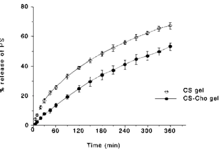
In Vitro Release of PS from CS-Cho Gel Beads The release of PS from 1% CS7B gel beads was inhibited by treatment with Cho (Fig. 1). The sustained release of PS from CS-Cho gel beads resulted from the formation of an electrostatic complex between the carboxyl group of Cho and the amino group of CS.11)However, the profile of PS release
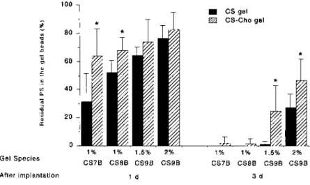
changed very little with soaking time (1—48 h), indicating that the complex between CS and Cho forms on the surface of the CS gel beads. Beads soaked for 6 h in 1% Cho solution were used in subsequent studies, because the amount of PS retained decreased as the soaking time in 1% Cho solution increased. The amount of PS retained in CS-Cho gel beads also differed with CS species. However, the release pattern was not changed by changing the amount of PS retained in the beads. Figure 2 shows the percentage of PS released after 6 h from CS-Cho gel beads that had been soaked in 1% Cho solution for 6 h. The effect of modification of the CS gel by Cho on the release of PS from the beads was observed
re-February 2002 269
Fig. 1. Effect of Cho Treatment of 1% CS7B Gel Beads on the Release of PS
gardless of CS species. The release of PS from 2% CS9B gel beads was inhibited compared with that from the others. This is assumed to be because the matrix of 2% CS9B gel beads is denser than that of the others, because the diameter of 2% CS9B gel beads was the same as others despite higher vis-cosity.
Evaluation of Biocompatibility The amount of protein
in the AP 3 d after implantation of CS-Cho gel beads without PS was determined to evaluate the inflammatory response (Fig. 3). No increase in protein was observed compared to mice injected with 30 mg carrageenin in 1 ml of physiologi-cal saline, which is known to induce inflammation. This re-sult was similar to CS gel beads without Cho treatment,12)
in-dicating the biocompatibility of CS-Cho gel beads.
In Vivo Biodegradation of Gel Beads Gel beads with various degrees of deacetylation, 1% CS7B, 1% CS8B, 1.5% and 2% CS9B, with and without Cho, were implanted in the AP of five mice per treatment. The number of beads that re-mained and maintained their shape over time is shown in Table 1. Biodegradation of the CS gel beads was accelerated as their degree of deacetylation decreased. Both CS7B and CS8B beads degraded in less than 3 d after implantation. However, some of the 1.5% and 2% CS9B gel beads re-mained even after 14 d. Mucopolysaccharides are hydrolyz-ed by enzymes such as lysozymes, it was reporthydrolyz-ed that biodegradation of CS was increased as their degree of deacetylation decreased.1,3,10) In a previous study CS gel beads were degraded in 0.1Mphosphate buffer (pH 7.2) con-taining lysozyme, although no degradation of the beads was observed without lysozyme.11)Degradation of the gel beads
in AP is therefore not erosion but biodegradation. On the other hand, biodegradation of all the CS-Cho gel beads was inhibited compared with beads without Cho treatment. These results indicated that modification of the CS gel matrix by Cho could control biodegradation of these beads. This as-sumes that the matrix is a dense complex between CS and Cho and that erosion by the enzymes is inhibited. However, 1% CS7B-Cho and 1% CS8B-Cho gel were degraded before 5 d, and 1.5 and 2% CS9B-Cho gel beads after 28 d became softer and smaller than before implantation. Therefore, even CS gel beads modified by Cho are susceptible to biodegrada-tion.
In Vivo Release of PS from Gel Beads The in vivo re-lease of PS from gel beads was evaluated by measuring the percentage of residual PS in the beads and AP compared with the amount administered. In a previous study,12)1% PS
suspended in physiological saline was injected into the AP. PS immediately started disappearing from the site: 19.7, 2.4 and 0.2% of administered PS remained in the AP after 1, 5 and 24 h, respectively. The plasma level changed gradually according to the amount of PS in the AP as it passed rapidly into the blood-stream. A similar result was obtained in the case of PS suspended in 1% CS7B acetate buffer solution (pH 4.5). These results showed that when PS powder was dissolved in the exudate in the AP, it did not remain there for long even with viscous CS.
To reduce side-effects and prolong the duration of the ac-tivity of a drug it is necessary to use a vehicle to deliver the drug. As shown in Fig. 4, PS remained in all the gel beads 1 d after implantation. In the case of 2% CS9B gel beads
270 Vol. 25, No. 2
Fig. 2. Effect of CS Species on the Release of PS after Dissolution Test for 6 h
Data represent the mean6S.D. (n53). ∗p,0.05, ∗∗p,0.01: significantly different
from CS gel without Cho. (Student’s t-test).
Fig. 3. Amount of Protein in the AP 3 d after Implantation of CS-Cho Gel Beads
Control: only incision (no implantation). Data represent the mean6S.D. (n53—8).
Table 1. Effect of Gel Species on the Number of Gel Beads Remaining after Implantation
After 1% CS7B 1% CS8B 1.5% CS9 2% CS9B
implantation (d)
CS CS-Cho CS CS-Cho CS CS-Cho CS CS-Cho
1 3 5 5 5 5 5 5 5 3 0 1 0 4 5 5 5 5 5 — 0 — 0 4 5 5 5 7 — — — — 4 5 4 4 14 — — — — 2 5 1 5 28 — — — — 0 5 0 5
after 3 d, about 30% of the PS remained, about 0.05% re-mained in the AP and PS was not determined in plasma. This result indicates that PS was released into the AP more gradu-ally from the CS gel beads. Furthermore, the percentage of residual PS in the beads was increased by modification of the CS gel matrix by Cho. In the case of 2% CS9B-Cho, about 50% of the PS remained in the beads after 3 d. The percent-age of residual PS in the beads increased about 20% com-pared with beads without Cho treatment. However, all of the PS had been released from the beads before 3 d (1% CS7B and CS8B) or 5 d (1% CS7B-Cho and CS8B-Cho) as degra-dation accelerated. On the other hand, PS was detected in the gel beads after 5 d (some 1.5% CS9B) and 7 d (some 2% CS9B, 1.5% CS9B-Cho and 2% CS9B-Cho) as there was no degradation. Changing the type of CS or modifying the CS would control the in vivo release of PS from the beads.
These results show that PS release in AP of mice is gov-erned by both diffusion and degradation of the gel matrix. The control of biodegradation and drug release from a vehi-cle will make possible the supply of the minimum require-ment of a dose by local application, and may result in a pro-longation of drug activity and a reduction in side effects. Control of biodegradation and drug release of CS gel beads in vivo was achieved by forming an electrostatic complex be-tween Cho and CS. Thus, these beads appear promising as a biodegradable vehicle for sustained drug delivery.
Acknowledgment This work was supported by the
spe-cial research fund of Hokuriku University (2000). REFERENCES AND NOTES
1) Yomota C., Komuro T., Kimura T., Yakugaku Zasshi, 110, 442—448 (1990).
2) Onishi H., Machida Y., Biomaterials, 20, 175—182 (1999). 3) Tomihata K., Ikada Y., Biomaterials, 18, 567—575 (1997).
4) Oungbho K., Müller B. W., Int. J. Pharmaceut., 156, 229—237 (1997). 5) Lim S. T., Martin G. P., Berry D. J., Brown M. B., J. Control. Release,
66, 281—292 (2000).
6) Tengamnuay P., Sahamethapat A., Sailasuta A., Mitra A. K., Int. J.
Pharmaceut., 197, 53—67 (2000).
7) Remunan-Lopez C., Portero A., Lemos M., Vila-Jato J. L., Nunez M. J., Riveiro P., Lopez J. M., Piso M., Alonso M. J., S.T.P. Pharm. Sci.,
10, 69—76 (2000).
8) Jameela S. R., Jayakrishnan A., Biomaterials, 16, 769—775 (1995). 9) Tokumitsu H., Ichikawa H., Fukumori Y., Block L. H., Chem. Pharm.
Bull., 47, 838—842 (1999).
10) Kofuji K., Shibata K., Murata Y., Miyamoto E., Kawashima S., Chem.
Pharm. Bull., 47, 1494—1496 (1999).
11) Kofuji K., Ito T., Murata Y., Kawashima S., Chem. Pharm. Bull., 48, 579—581 (2000).
12) Kofuji K., Ito T., Murata Y., Kawashima S., Biol. Pharm. Bull., 24, 205—208 (2001).
13) Lu J. X., Prudhommeaux F., Meunier A., Sedel L., Guillemin G.,
Bio-materials, 20, 1937—1944 (1999).
14) Hambleton P., Miller P., Br. J. Exp. Path., 70, 425—433 (1989). 15) Lowry O. H., Rosebrough N. J., Farr A. L., Randall R. J., J. Biol.
Chem., 193, 265—275 (1951).
16) Watanabe Y., Imai K., J. Chromatogr., 239, 723—732 (1982).
February 2002 271
Fig. 4. Effect of Gel Species on the Percentage of Residual PS in the Gel Beads