The Incidence and Factors Associated With the Occurrence of Pulmonary Vein Narrowing After Cryoballoon Ablation
Ryohsuke Narui, MD1; Michifumi Tokuda, MD, PhD1; Masato Matsushima, MD, PhD, MPH2; Ryota Isogai, MD1; Kenichi Tokutake, MD1; Kenichi Yokoyama, MD1; Mika Hioki, MD1; Keiichi Ito, MD1; Shin-ichi Tanigawa, MD1; Seigo Yamashita, MD, PhD1; Keiichi Inada, MD, PhD1; Kenri Shibayama, MD1; Seiichiro Matsuo, MD, PhD1; Satoru Miyanaga, MD, PhD1; Kenichi Sugimoto, MD, PhD1; Michihiro Yoshimura, MD, PhD1 and Teiichi Yamane, MD, PhD, FHRS1
1. Department of Cardiology, The Jikei University School of Medicine, Tokyo, Japan
2. Division of Clinical Epidemiology, The Jikei University School of Medicine, Tokyo, Japan Short title: Pulmonary vein stenosis after cryoballoon ablation
Correspondence to: Teiichi Yamane, MD, PhD, FHRS
Department of Cardiology, The Jikei University School of Medicine, 3-25-8 Nishi-shinbashi, Minato-ku, Tokyo 105-8461, Japan
Tel: +81-3-3433-1111 Fax: +81-3-3459-6043 E-mail:[email protected]
Word count: 5746 words Number of figures: 5 figures Number of tables: 3 tables The Journal Subject Code: [22] Ablation/ICD/surgery
東京慈恵会 医科大学 電子署名者 : 東京慈恵会医科大学 DN : cn=東京慈恵会医科大学, o, ou, [email protected], c=JP 日付 : 2019.08.02 12:24:14 +09'00'
Abstract Background
In contrast with traditional radiofrequency ablation, little is known about the influence of cryoballoon ablation on the morphology of pulmonary veins (PVs). We evaluated the influence of cryoballoon ablation on the PV dimension (PVD) and investigated the factors
associated with a reduction of the PVD.
Methods and Results
Seventy-four patients who underwent cryoballoon ablation for paroxysmal atrial fibrillation
(PAF) were included in the present study. All subjects underwent contrast-enhanced
computed tomography both before and at 3 months after the procedure. The PVD (cross-
sectional area) was measured using a 3-dimensional electroanatomical mapping system. Each
PV was evaluated according to the PVD reduction rate (ΔPVD), which was calculated as
follows: (1–post-PVD/pre-PVD)×100 (%).
Ninety-two percent of the PVs (271/296) were successfully isolated only by cryoballoon ablation; the remaining 8% of the PVs required touch-up ablation and were excluded from the analysis. Mild (25%-50%), moderate (50%-75%), and severe (≥75%) ΔPVD values were observed in 87, 14 and 3 PVs, respectively, including one case with severe left superior PV stenosis (ΔPVD: 94%) in a patient who required PV angioplasty. In multivariable analysis, a larger PV ostium and lower minimum freezing temperature during cryoballoon ablation were
independently associated with PV narrowing (odds ratio, 1.773: P=0.01; and odds ratio, 1.137:
P<0.001, respectively).
Conclusion
A reduction of the PVD was often observed after cryoballoon ablation for atrial fibrillation.
A larger PV ostium and lower minimum freezing temperature during cryoballoon ablation were associated with an increased risk of PVD reduction.
Key Words: atrial fibrillation, cryoballoon ablation, pulmonary vein narrowing, pulmonary vein isolation
Background
Atrial fibrillation (AF) is the most common cardiac tachyarrhythmia and is associated with an increased risk of stroke, heart failure and mortality.1 The pulmonary veins (PVs) play an important role in the pathogenesis and maintenance of AF.2,3 Although PV isolation (PVI) has been established as an effective treatment for AF, it is associated with the development of PV stenosis, which represents a serious complication. In previous studies that were
performed during the initial AF ablation era, the prevalence of PV stenosis was reported to be 3% to 42%.4-8 Since then, the prevalence has decreased to 0.3% as the ablation site has shifted toward the left atrium (LA),9 and with the optimization of radiofrequency (RF) energy and technical improvements that have been achieved by confirming the catheter position with a 3- dimensional electroanatomical mapping system. Although most cases of PV stenosis are asymptomatic, severe PV stenosis can progress to complete occlusion with potentially life
threatening symptoms of segmental pulmonary hypertension or venous infarction of the corresponding lobe.7,10
PVI using a cryoballoon (CB) has emerged as a safe and effective ablation strategy for patients with paroxysmal AF.11 Immediate and midterm follow-up studies have revealed that its efficacy is similar to that of radiofrequency ablation.12-16 Although some cases of PV stenosis after CB ablation have been reported,17,18 the influence of CB ablation on the morphology of the PV is unknown. The purpose of this study was to evaluate the influence
of CB ablation on the morphology of the PV and to investigate the factors associated with PV
narrowing.
Methods
The study population
The present study included 74 patients with paroxysmal AF without a PV anomaly (such as a left common PV or a right middle PV) whose first catheter ablation procedure was performed using a second-generation cryoballoon at our institution. In the present study, paroxysmal AF was defined as AF of <7 days in duration. All patients received effective anticoagulation therapy for more than 3 months before the procedure. All antiarrhythmic drugs were discontinued at least 5 half-lives before the procedure. All patients provided their written informed consent prior to undergoing the procedure. The study protocol was approved by the ethics committee of the Jikei University School of Medicine.
Electrophysiological study
A steerable 16-polar 2-site (6-polar for the right atrium and 10-polar for the coronary sinus) mapping catheter (InquiryTM Luma-CathTM, St. Jude Medical, St. Paul, MN, USA) was positioned within the coronary sinus at 1–2 o’clock along the mitral annulus in the left anterior oblique projection via the right subclavian vein. The LA and PVs were explored through
either a patent foramen ovale or via transseptal catheterization with two long sheaths. Direct visualization of all 4 PVs was performed using LA angiography and displayed during the procedure to show the venous anatomy and the location of the LA-PV junction. After obtaining venous access, a single bolus of 100 IU/kg body weight of heparin was administered.
During mapping and ablation, the heparin bolus and drips were administered intravenously with a target activated coagulation time of 300–350 seconds; measurements were performed every 20 minutes.
AF ablation
The transseptal sheath was exchanged over a guidewire for a 15Fr steerable sheath (FlexCath AdvanceTM, Medtronic, Minneapolis, MN, USA). An inner lumen mapping catheter (AchieveTM, Medtronic) was sequentially positioned in each PV to obtain the baseline PV potential. A 28-mm cryoballoon (Arctic Front AdvanceTM, Medtronic) was advanced over the inner lumen mapping catheter up to the LA, inflated and positioned in the PV ostium of each vein. We did not use 23-mm cryoballoon in the present study. In order to avoid esophageal injury, a nasogastric thermometer (Sensi Therm™, St. Jude Medical, St. Paul, MN, USA) was inserted to identify the course of the esophagus and to measure the esophageal temperature during CB ablation. After verifying the complete occlusion of the PV ostium, the cryoballoon was applied for 180 seconds. If the PV potential disappeared, a bonus
application was performed for 120 seconds at the same position according to the previously recommended protocol.19,20 In the cases with either a minimum temperature of <-60 °C, an esophageal temperature of <20 °C or those in which the PV potential remained after a 100- second application of cryoenergy, CB ablation was discontinued. If the PV potential did not disappear after the 3rd application at the same PV, touch up ablation with an 8-mm-tip non- irrigated ablation catheter or an 8-mm-tip conventional cryocatheter (Freezor MAXTM, Medtronic) was added. In order to avoid phrenic nerve injury, the cryoballoon applications for right PVs were performed with the monitoring of the diaphragmatic compound motor action potentials (C-MAP) during phrenic nerve pacing.21 After the CB ablation of all 4 PVs, a bi- directional block at the PV antrum was confirmed using a variable circular mapping catheter (OPTIMATM, St. Jude Medical). Dormant PV conduction was induced by the administration of adenosine triphosphate (20-40 mg) under isoproterenol infusion and was subsequently eliminated with touch-up ablation.
The evaluation of the pulmonary vein morphology
Contrast-enhanced multidetector computed tomography (MDCT) (Somatom DefinitionTM, Siemens Medical Solutions, Forchheim, Germany) was performed before and 3 months after the procedure. Three-dimensional images of the LA and PVs were reconstructed from multidetector computed tomographic images using a 3-demensional electroanatomical
mapping system (EnSite NavXTM, St. Jude Medical) and the PV dimension (PVD) was
measured by 2 independent experts who were unaware of the procedural results. The PVD was measured on the 3-dimensional electroanatomical mapping system by tracing the area within the PV plane at 5-mm intervals from the PV ostium in a distal direction until 15 mm or bifurcation in each PV (Figure 1). The PVD reduction rate was calculated at each segment using the following formula: (1-post-PVD/pre-PVD)×100 (%). The ΔPVD was defined as the maximum PVD reduction rate in each PV. Severe PV narrowing was defined as a reduction of 75% in the cross-sectional area, just as in the previous large randomized trial.15 In the present study, “PV narrowing” was defined as a ΔPVD value of ≥25%. PV narrowing was classified into 3 groups according to ΔPVD value: mild narrowing (25–50%), moderate narrowing (50–75%) and severe narrowing (>75%), respectively. In the present study, the change in the PVD was evaluated in PVs treated only with the CB, in order to examine the influence of CB ablation. We excluded PVs with additional touch-up ablation from the analysis.
Statistical analysis
The presence of a normal distribution was assessed by the Shapiro-Wilk test.
Continuous variables are expressed as the mean ± standard deviation. Data were analyzed by a one-way analysis of variance (ANOVA) or Kruskal–Wallis test, with Tukey’s or Steel- Dwass’s post-hoc test, as appropriate. Correlations were made with the Spearman’s rank
correlation test, and probabilities were calculated for each correlation coefficient. The categorical variables, expressed as numbers or percentages, were analyzed using the chi- squared test unless the expected values in any cells were less than 5, in which case Fisher’s exact test was used. The receiver operator characteristic curve was determined in order to evaluate the performance of the predictor of PV narrowing after CB ablation. The optimal cut-off point was chosen as the combination with the highest sensitivity and specificity. All of the tests were 2-tailed. P values of <0.05 were considered to indicate statistical significance.
To check the multi-collinearity of explanatory variables, we calculated variance inflation factor among explanatory variables. The mixed-effect random-intercept logistic regression was employed to consider the clustering of the data coming from the same patients in the analysis.
The statistical analyses were performed using the SPSS software program (version 21.0.0; SPSS, Chicago, IL, USA) and Stata 14 software (StataCorp. 2015. Stata Statistical Software: Release 14. College Station, TX: StataCorp LP).
Results
Patient Characteristics
The baseline patient characteristics are shown in Table 1. The mean age was 58±10 years, and 91% of the patients were male. The mean LA diameter was 36.1±4.5 mm, and the
mean left ventricular ejection fraction was 65.1±4.5%. Hypertension was observed in 18 (24%) patients and 2(2.7%) patients had evidence of structural heart disease.
The results of ablation
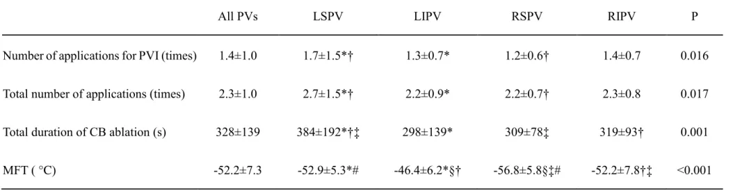
In total, 296 PVs were isolated from the LA during the procedure. Among these, 271 PVs (92%) were successfully isolated by CB ablation alone; the remaining 25 PVs (8%) required additional touch-up ablation and were excluded from the analysis. In the 271 PVs that were isolated by CB ablation alone, the total number and duration of CB ablations in each PV were 2.3±1.0 and 328±139 seconds, respectively. The minimum freezing temperature (MFT) during CB ablation was -52.2±7.3°C (Table 2). When we compared the CB ablation parameters among the 4 PVs, the number of CB ablations for PVI in the left superior PV (LSPV) was significantly greater than that in the right superior PV (RSPV) (1.7±1.5 vs. 1.2±0.6, P=0.013). The total number of applications in the LSPV was significantly greater than that in the left inferior PV (LIPV) and RSPV (2.7±1.5 vs. 2.2±0.9, P=0.037, 2.7±1.5 vs. 2.2±0.7, P=0.026) and the total CB ablation time in the LSPV was significantly longer than that in the other three PVs (LSPV vs. LIPV, 384±192 vs. 298±139 s, P=0.001; LSPV vs. RSPV, 384±192 vs. 309±78 s, P=0.006; and LSPV vs. right inferior PV [RIPV], 384±192 vs. 319±93 s P=0.041) (Table 2). The MFT in the RSPV was significantly lower than that in the other 3 PVs, while that in the LSPV was significantly lower than that in the LIPV (RSPV vs. LSPV, -56.8±5.8 vs.
-52.9±5.3 °C, P=0.001; RSPV vs. LIPV -56.8±5.8 vs. -46.4±6.2 °C, P<0.001; RSPV vs. RIPV, -56.8±5.8 vs. -52.2±7.8 °C, P<0.001; and LSPV vs. LIPV, -52.9±5.3 vs. -46.4±6.2 °C, P=0.001).
Major complications occurred in 4 (5.4%) patients, 3 (4.1%) of whom suffered from transient right-side phrenic nerve palsy during CB ablation of the RSPV. One case of arteriovenous fistula requiring vascular surgery occurred on the seventh postoperative day.
There were no cases of stroke or transient ischemic attack, cardiac tamponade, or any other major complications.
The dimensions of the pulmonary veins
Figure 2 shows a comparison of the PVDs before and after CB ablation. The
baseline PVDs of the LS, LI, RS and RIPV were 2.8±0.9, 1.6±0.5, 3.2±0.9 and 2.3±0.7cm2, respectively. The PVDs of the LS, LI, RS and RIPV after CB ablation were 2.3±0.9, 1.4±0.5, 2.5±0.9 and 2.0±0.7cm2, respectively. In each of the PVs, the PVD was significantly smaller after CB ablation than it was at baseline. The mean ΔPVD value in all PVs was 18±22%, while PV narrowing (ΔPVD ≥25%) were observed in 36.9% (100/271) of the PVs. Mild (25- 50%), moderate (50-75%), and severe (≥75%) narrowing were observed in 83 (30.6%), 14 (5.2%), and 3 (1.1%) PVs, respectively. The ΔPVD value was found to be significantly correlated with the baseline PV ostium dimension (r=0.318, P<0.0001) (Figure 3A) and the
MFT (r=0.489, P<0.0001) (Figure 3D). The results of a detailed analysis of the factors associated with the different degrees of PV narrowing are shown in Table 3A. The baseline PV ostial dimension in the PVs with mild narrowing was significantly larger than that in those without narrowing (2.8±1.1 vs. 2.4±0.9 cm2, P<0.001). Furthermore, the MFT during CB ablation in PVs with mild narrowing and moderate to severe narrowing was significantly lower than that in those without narrowing (-53.8±7.4 vs. -51.0±7.0 °C, P=0.018 and -56.0±7.1 vs. - 51.0±7.0 °C, P=0.011). When we compare PVs with and without PV narrowing (ΔPVD
≥25% vs. ΔPVD <25%), the PV ostial dimension before the procedure was significantly larger and the MFT during CB ablation was significantly lower in PVs with narrowing than in those without (2.9±1.1 vs. 2.4±0.9cm2, P<0.001 and -54.2±7.4 vs. -51.0±7.0 °C, P<0.001).
The variance inflation factors of number of applications for PVI and total number of applications were more than 10. Of these 2 variables, the number of applications for PVI was excluded from the mixed-effect logistic regression by considering a clinical importance.
After excluding number of applications for PVI, all variance inflation factors were less than 10.
According to a multivariate analysis, the PV ostial dimension before the procedure and the MFT during CB ablation were found to be independent predictors of PV narrowing (P=0.01 and P<0.001, respectively) (Table 3B). Residual plots of each explanatory variable vs.
Pearson residuals identified one point which was thought to be an influential point. However, the model without this point did not almost change compared with the model with this point.
Therefore, we employed the model with this point. A likelihood ratio test revealed that the mixed effect model was fitted better than the ordinary logistic model. Due to the 7 missing values of explanatory variables, 264 observations were used in the calculation of variance inflation factors and Pearson residuals, receiver operator characteristic curve analysis and also the mixed-effect logistic model.
The receiver operator characteristic curve for MFT as a predictor of PV narrowing (ΔPVD ≥25%) after CB ablation showed an area under the curve of 0.751 (95% confidence interval: 0.687 to 0.814, p<0.0001) (Figure 4). A cut-off point of -53.5 °C for the MFT had a specificity of 76.5% and a sensitivity of 66.0% in predicting PV narrowing after CB ablation.
In the present study, severe PV narrowing (ΔPVD ≥75%) was observed in 3 LSPVs in 3 (4.1%) cases (Figure 5). The PV ostial dimension before CB ablation in these 3 cases was 4.2, 3.4 and 2.1 cm2, respectively. Additional CB ablation was performed in all cases, and the MFT in cases 1, 2 and 3 was -59 °C, -63 °C and -48 °C, respectively. Although the patient in case 3 remains free from any symptoms due to PV stenosis, Tc-99m macroaggregated albumin lung perfusion scintigraphy revealed diminished blood flow to the left upper lobe in the LSPV. PV angioplasty was performed 9 months after the procedure.
Discussion Main findings
In the present observational study, we evaluated the incidence of PV narrowing after CB ablation by comparing computed tomographic images obtained before and 3 months after CB ablation. In total, 36.8% of the PVs demonstrated PV narrowing; the mean reduction of the PVD (PVD) after CB ablation was 18%. The PV narrowing was classified as mild, moderate and severe in 30.6%, 5.2% and 1.1% of the cases, respectively. There was no significant difference in the mean PVD value among the 4 PVs. However, the baseline PV
ostial dimension was larger and the MFT was lower in PVs with narrowing after CB ablation than in those without. To the best of our knowledge, this is the first report to quantitatively evaluate the impact of CB ablation on the PV morphology after PVI using multidetector computed tomographic imaging.
PV stenosis after PVI
PV stenosis is a major complication that is known to result from the use of radiofrequency energy in PVI. As the methods and technology of PVI have advanced, the region of PVI has shifted to the more proximal side of the PV antrum. As a result there has been a decrease in the incidence of PV stenosis requiring intervention (0.74% to 0.29%, in worldwide surveys).9,22 Now, with the extensive use of the wide PVI strategy with the
application of radiofrequency energy, the issue of PV stenosis during AF ablation has almost been overcome. On the other hand, the incidence and mechanisms of PV narrowing/stenosis after CB ablation remain unknown. In an early animal study, Feld et al. reported that no PV stenosis occurred after CB ablation inside the canine PVs.23 As for the influence of CB ablation on PV morphology, Sarabanda et al. reported that no significant PV narrowing was seen in the long-term follow-up after PVI using CB ablation.24 Although it was initially suggested that cryoablation was associated with little or no risk of PV stenosis in humans,11,25,26 a few reports have described cases of PV stenosis after CB ablation (28 mm diameter).17,18 A previous study reported a 3.1% incidence of severe PV stenosis (>75%);15 however, their balloon size for the stenosed PV was smaller than that in our study (23 mm vs. 28 mm).
Predictors of PV stenosis
The predictors of PV narrowing/stenosis have been proposed previously. Traullé et al. reported that the supplementation of cryoballoon ablation of the PV with focal, irrigated ostial RF ablation may be associated with a higher risk of PV stenosis.27 We excluded PVs in which touch-up ablation was performed from the analysis and showed that a lower MFT during CB ablation was associated with a higher PV reduction rate.
Arentz et al. previously reported that distal ablation inside smaller PVs was associated with a higher risk of stenosis after radiofrequency catheter ablation.28 In contrast, a larger
baseline PV ostial dimension was related with a higher incidence of PV narrowing in the present study. This discrepancy is likely due to the size of the cryoballoon that was used in this study. Cryoenergy application sites may be located in a more distal portion of PVs with a larger ostial dimension in comparison to those with a smaller ostial dimension because the maximum size of the cryoballoon is currently 28 mm. It is possible that cryoballoon application at a more distal portion in larger PVs may cause a stronger seal and a lower MFT, resulting in the narrowing of the PVDs. Additionally, the ability of the second-generation cryoballoon to cool the distal hemisphere as well, and not merely the equatorial belt of the balloon's surface, may have affected the results.
The mechanism of PV stenosis after CB ablation
The mechanism of cellular damage due to freezing by cryoenergy has been shown to be a complex process with 3 primary factors: direct cellular damage, vascular failure and immunological effects.29 It has been reported that cryolesions are distinguished from hyperthermic injury by the preservation of the basic underlying tissue architecture with preserved endocardial contours, minimal cartilage formation and the absence of signs of chronic inflammation or evidence of viable myocytes within the lesions, which can result in minimal tissue shrinkage and PV contraction in comparison to radiofrequency lesions.29-31 Although the true mechanism of the PV narrowing that was observed in the present study
remains to be elucidated, we found that the baseline PV size and the MFT were significantly associated with the occurrence of PV narrowing in the present system of CB ablation. At the very least, need to keep in mind that CB ablation can cause PV narrowing/stenosis in human PVI procedures.
Limitations
There present study is associated with several limitations. First, we regarded a PVD reduction of ≥25% as the definition of PV narrowing. Since mild narrowing of the PVD can occur due to reverse remodeling after AF ablation, the results might reflect both PV narrowing and PV reverse remodeling. In a future analysis of a larger study population, PV narrowing should be defined by a ≥50% reduction, as this would provide more accurate results.
Secondly, we used the twice-freezing method as a standard in this study, since it was the recommended strategy at the time of this study. The newer strategy of the single-shot freezing method, which has been widely accepted, might yield different results.
Conclusion
A reduction in the PVD is not a rare occurrence after CB ablation for PVI in AF patients. A larger PV ostium and a lower MFT during CB ablation were associated with a higher risk of PVD reduction. Larger-sized PVs are not optimal candidates for CB ablation
under the current system in which balloon catheters with a maximum diameter of 28 mm are used.
Acknowledgments
We are grateful to Dr. Brian Quinn (Editor-in-Chief, Japan Medical Communication) for providing linguistic comments on the article. We thank Mr. Takehiro Tojo for providing technical support.
Funding Sources: None Disclosures: None
Figure Legends Figure 1.
A typical example of the pulmonary vein dimension (PVD) measurement and the method of calculating the pulmonary vein dimension rate (ΔPVD) value. The PVD was measured before and after cryoballoon (CB) ablation at 5mm intervals from the PV ostium in a distal direction until 15 mm. The PVD reduction rate was calculated at each point of the PV using the following formula: (1-post-PVD/pre-PVD)×100 (%). The ΔPVD was defined as the maximum PV dimension reduction rate among the 0 mm (1), 5 mm (2), 10 mm (3) and 15 mm points (4) from the ostium. The PVD at the 0 mm point (1) indicates pulmonary vein ostial
dimension. CB indicates cryoballoon; PV, pulmonary vein; and PVD, pulmonary vein dimension.
Figure 2. A-D,
The PVD at baseline and after CB ablation in each PV. The PVD was significantly smaller at baseline than after CB ablation in each of the PVs. LSPV indicates left superior pulmonary vein; LIPV, left inferior pulmonary vein; RSPV, right superior pulmonary vein; and RIPV, right inferior pulmonary vein. The other abbreviations are the same as those in the previous figure.
Figure 3.
The ΔPVD is plotted against the PV ostial dimension before ablation (A), total number of applications (B), total duration of CB ablation (C) and MFT (D). A regression line is shown for each graph, and the correlation coefficient is also shown. MFT indicates minimum freezing temperature. The other abbreviations are the same as those in the previous figures.
Figure 4.
The receiver operator characteristic curve analysis of the MFT according to PV narrowing after cryoballoon ablation procedures. AUC indicates are under the curve; and CI, confidence
interval. The other abbreviations are the same as those in the previous figures.
Figure 5.
Three cases of severe narrowing. Severe PV narrowing occurred in the LSPV in all of the cases. The PV with a ΔPVD value of 94% (Case 3) required PV angioplasty at 9 months after the procedure. PVI indicates pulmonary vein isolation. The other abbreviations are the same as those in the previous figures.
References
1. Fuster V, Rydén LE, Cannom DS, Crijns HJ, Curtis AB, Ellenbogen KA, Halperin JL, Le Heuzey JY, Kay GN, Lowe JE, Olsson SB, Prystowsky EN, Tamargo JL, Wann S, Smith SC Jr, Jacobs AK, Adams CD, Anderson JL, Antman EM, Hunt SA, Nishimura R, Ornato JP, Page RL, Riegel B, Priori SG, Blanc JJ, Budaj A, Camm AJ, Dean V, Deckers JW, Despres C, Dickstein K, Lekakis J, McGregor K, Metra M, Morais J, Osterspey A, Zamorano JL. ACC/AHA/ESC 2006 guidelines for the management of patients with atrial fibrillation—a report of the American College of Cardiology/American Heart Association Task Force on Practice Guidelines and the European Society of Cardiology Committee for Practice Guidelines (Writing Committee to Revise the 2001 Guidelines for the Management of Patients With Atrial Fibrillation). J Am Coll Cardiol. 2006;48:854-906.
2. Haïssaguerre M, Jaïs P, Shah DC, Takahashi A, Hocini M, Quiniou G, Garrigue S, Le Mouroux A, Le Métayer P, Clémenty J. Spontaneous initiation of atrial fibrillation by ectopic beats originating in the pulmonary veins. N Engl J Med. 1998;3:659-666.
3. Jaïs P, Hocini M, Macle L, Choi KJ, Deisenhofer I, Weerasooriya R, Shah DC, Garrigue S, Raybaud F, Scavee C, Le Metayer P, Clémenty J, Haïssaguerre M. Distinctive electrophysiological properties of pulmonary veins in patients with atrial fibrillation.
Circulation. 2002;106:2479-2485.
4. Yu WC, Hsu TL, Tai CT, Tsai CF, Hsieh MH, Lin WS, Lin YK, Tsao HM, Ding YA, Chang MS, Chen SA. Acquired pulmonary vein stenosis after radiofrequency catheter ablation of paroxysmal atrial fibrillation. J Cardiovasc Electrophysiol. 2001;12:887-892.
5. Robbins IM, Colvin EV, Doyle TP, Kemp WE, Loyd JE, McMahon WS, Kay GN.
Pulmonary vein stenosis after catheter ablation of atrial fibrillation. Circulation.
1998;98:1769-1775.
6. Scanavacca MI, Kajita LJ, Vieira M, Sosa EA. Pulmonary vein stenosis complicating catheter ablation of focal atrial fibrillation. J Cardiovasc Electrophysiol. 2000;11:677-681.
7. Saad EB, Marrouche NF, Saad CP, Ha E, Bash D, White RD, Rhodes J, Prieto L, Martin DO, Saliba WI, Schweikert RA, Natale A. Pulmonary vein stenosis after catheter ablation of atrial fibrillation: emergence of a new clinical syndrome. Ann Intern Med.
2003;138:634–638
8. Saad EB, Rossillo A, Saad CP, Ha E, Bash D, White RD, Rhodes J, Prieto L, Martin DO, Saliba WI, Schweikert RA, Natale A. Pulmonary vein stenosis after radiofrequency ablation of atrial fibrillation: functional characterization, evolution, and influence of the ablation strategy. Circulation. 2003;108:3102-3107.
9. Cappato R, Calkins H, Chen SA, Davies W, Iesaka Y, Kalman J, Kim YH, Klein G, Natale A, Packer D, Skanes A, Ambrogi F, Biganzoli E. Updated worldwide survey on the methods, efficacy, and safety of catheter ablation for human atrial fibrillation. Circ Arrhythm Electrophysiol. 2010;3:32-38.
10. Ravenel JG, McAdams HP. Pulmonary venous infarction after radiofrequency ablation for atrial fibrillation. Am J Roentgenol. 2002;178:664–666
11. Kuck KH, Brugada J, Fürnkranz A, Metzner A, Ouyang F, Chun KR, Elvan A, Arentz T, Bestehorn K, Pocock SJ, Albenque JP, Tondo C; FIRE AND ICE Investigators.
Cryoballoon or Radiofrequency Ablation for Paroxysmal Atrial Fibrillation. N Engl J Med.
2016;374:2235-2245.
12. Luik A, Radzewitz A, Kieser M, Walter M, Bramlage P, Hörmann P, Schmidt K, Horn N, Brinkmeier-Theofanopoulou M, Kunzmann K, Riexinger T, Schymik G, Merkel M, Schmitt C. Cryoballoon Versus Open Irrigated Radiofrequency Ablation in Patients With Paroxysmal Atrial Fibrillation: The Prospective, Randomized, Controlled, Noninferiority FreezeAF Study. Circulation. 2015;132:1311-1319.
13. Aytemir K, Oto A, Canpolat U, Sunman H, Yorgun H, Şahiner L, Kaya EB. Immediate and medium-term outcomes of cryoballoon-based pulmonary vein isolation in patients with paroxysmal and persistent atrial fibrillation: single-centre experience. J Interv Card Electrophysiol. 2013;38:187–195.
14. Kuck K-H, Furnkkranz A. Cryoballoon Ablation of Atrial Fibrillation. J Cardiovasc Electrophysiol. 2010;21:1427–1431.
15. Packer DL, Kowal RC, Wheelan KR, Irwin JM, Champagne J, Guerra PG, Dubuc M, Reddy V, Nelson L, Holcomb RG, Lehmann JW, Ruskin JN. Cryoballoon Ablation of Pulmonary Veins for Paroxysmal Atrial Fibrillation. J Am Coll Cardiol. 2013;61:1713–
1723.
16. Defaye P, Kane A, Chaib A, Jacon P. Efficacy and safety of pulmonary veins isolation by cryoablation for the treatment of paroxysmal and persistent atrial fibrillation. Europace.
2011;13:789–795.
17. Thomas D, Katus HA, Voss F. Asymptomatic pulmonary vein stenosis after cryoballoon catheter ablation of paroxysmal atrial fibrillation. J Electrocardiol. 2011;44:473-476 18. Miyazaki S, Ichihara N, IesakaY. Pulmonary vein stenosis after cryoablation using 28-mm
second-generation balloon. J Cardiovasc Electrophysiol. 2015;26:570-571
19. Klein G, Oswald H, Gardiwal A, Lüsebrink U, Lissel C, Yu H, Drexler H. Efficacy of pulmonary vein isolation by cryoballoon ablation in patients with paroxysmal atrial fibrillation. Heart Rhythm. 2008;5:802-806
20. Metzner A, Reissmann B, Rausch P, Mathew S, Wohlmuth P, Tilz R, Rillig A, Lemes C, Deiss S, Heeger C, Kamioka M, Lin T, Ouyang F, Kuck KH, Wissner E. One-year clinical outcome after pulmonary vein isolation using the second-generation 28mm cryoballoon.
Circ Arrhythm Electrophysiol. 2014;7:288-292
21. Mondésert B, Andrade JG, Khairy P, Guerra PG, Dyrda K, Macle L, Rivard L, Thibault B, Talajic M, Roy D, Dubuc M. Clinical experience with a novel electromyographic approach to preventing phrenic nerve injury during cryoballoon ablation in atrial fibrillation. Circ Arrhythm Electrophysiol. 2014;7:605–611.
22. Cappato R, Calkins H, Chen SA, Davies W, Iesaka Y, Kalman J, Kim YH, Klein G, Packer D, Skanes A. Worldwide survey on the methods, efficacy, and safety of catheter ablation for human atrial fibrillation. Circulation. 2005;111:1100-1105.
23. Feld GK, Yao B, Reu G, Kudaravalli R. Acute and chronic effects of cryoablation of the pulmonary veins in the dogs as a potential treatment for focal atrial fibrillation. J Interv Card Electrophysiol. 2003;8:135-140.
24. Sarabanda AV, Bunch TJ, Johnson SB, Mahapatra S, Milton MA, Leite LR, Bruce GK, Packer DL. Efficacy and safety of circumferential pulmonary vein isolation using a novel balloon ablation system cryothermal. J Am Coll Cardiol. 2005;46:1902–1912.
25. Tse HF, Reek S, Timmermans C, Lee KL, Geller JC, Rodriguez LM, Ghaye B, Ayers GM, Crijns HJ, Klein HU, Lau CP. Pulmonary vein isolation using transvenous catheter cryoablation for treatment of atrial fibrillation without risk of pulmonary vein stenosis. J Am Coll Cardiol. 2003;42:752– 758.
26. Mugnai G, de Asmundis C, Ciconte G, Irfan G, Saitoh Y, Velagic V, Ströker E, Wauters K, Hünük B, Brugada P, Chierchia GB. Incidence and characteristics of complications in the setting of second-generation cryoballoon ablation: a large single-center study of 500 consecutive patients. Heart Rhythm. 2015;12:1476-1482.
27. Traullé S, Miyazaki S, Mouquet V, Kubala M, Hermida JS. The anatomic impact of sequential, additional, ostial radiofrequency ablation following pulmonary vein cryo- isolation. Pacing Clin Electrophysiol. 2012;35:1420-1427.
28. Arentz T, Jander N, von Rosenthal J, Blum T, Fürmaier R, Görnandt L, Josef Neumann F, Kalusche D. Incidence of pulmonary vein stenosis 2 years after radiofrequency catheter ablation of refractory atrial fibrillation. Eur Heart J. 2003;24:963–969.
29. Avitall B and Kalinski A. Cryotherapy of cardiac arrhythmia: from basic science to the bedside. Heart Rhythm. 2015;12:2195-2203.
30. Avitall B, Urboniene D, Rozmus G, Lafontaine D, Helms R, Urbonas A. New cryotechnology for electrical isolation of the pulmonary veins. J Cardiovasc Electrophysiol.
2003;14:281-286.
31. Thibault B, Villemaire C, Talajic M. Catheter cryoablation in a more effective and potentially safer method to create atrial conduction block: comparison with radiofrequency ablation. Semin Interv Cardiol. 1997;2:251-265.
Table 1. The characteristics of the study population (n=74)
N=74
Age (years) 58±10
Sex (male)- n (%) 67 (91)
BMI (kg/m²) 23.5±2.8
AF history (years) 3.7±4.2
LAD (mm) 36.1±4.5
LVEF (%) 65.1±4.5
BNP (pg/ml) 55.8±95.1
Hypertension - n (%) 18 (24)
Structural Heart Disease - n (%) 2 (2.7)
BMI indicates body mass index; AF, atrial fibrillation; LAD, left atrial diameter; LVEF, left ventricular ejection fraction; and BNP, brain natriuretic peptide. Age, BMI, AF history, LAD, LVEF, and BNP are expressed as the mean ± SD.
Table 2. The results of ablation in PVs isolated by CB ablation alone
All PVs LSPV LIPV RSPV RIPV P
Number of applications for PVI (times) 1.4±1.0 1.7±1.5*† 1.3±0.7* 1.2±0.6† 1.4±0.7 0.016 Total number of applications (times) 2.3±1.0 2.7±1.5*† 2.2±0.9* 2.2±0.7† 2.3±0.8 0.017 Total duration of CB ablation (s) 328±139 384±192*†‡ 298±139* 309±78‡ 319±93† 0.001
MFT ( °C) -52.2±7.3 -52.9±5.3*# -46.4±6.2*§† -56.8±5.8§‡# -52.2±7.8†‡ <0.001
CB indicates cryoballoon; PV, pulmonary vein; LSPV, left superior pulmonary vein; LIPV, left inferior pulmonary vein; RSPV, right superior pulmonary vein; and RIPV, right inferior pulmonary vein. The number of applications for PV isolation, total number of applications, total duration of CB ablation, and minimum freezing temperature are expressed as the mean ± SD.
*, †, ‡, §, # indicates a pair with a significant difference (P<0.05) in a post hoc test.
Table 3.
(A) Characteristics of cryoballoon application among the degrees of PV narrowing
Narrowing (-) (n=172PVs)
Mild narrowing (n=83PVs)
Moderate to severe narrowing (n=17PVs)
P
PV ostial dimensionbefore ablation (cm2) 2.4±0.9* 2.8±1.1* 2.9±1.1 <0.001
Number of applications for PV isolation 1.4±1.0 1.5±1.1 1.2±0.4 0.56
Total number of applications 2.3±0.9 2.4±1.1 2.0±0.6 0.39
Total duration of CB ablation (sec) 331±137 331±155 271±65 0.21
MFT (°C) -51.0±7.0†‡ -53.8±7.4† -56.0±7.1‡ 0.001
*, †, ‡ indicates a pair of values with a significant difference (P<0.05) in a post hoc test.
(B) The factors of the PVs with / without narrowing (≥25%) after cryoballoon ablation
Univariate analysis Multivariate analysis
ΔPVD ≥25%
(n=100PVs)
ΔPVD <25%
(n=171PVs) P Odds ratio Standard error 95% CI P PV ostial dimensionbefore ablation (cm2) 2.9±1.1 2.4±0.9 <0.001 1.773 0.401 1.137-2.765 0.01
Number of applications for PV isolation 1.4±1.0 1.4±1.0 0.98 - - - -
Total number of applications 2.3±1.1 2.3±1.1 0.97 1.201 0.442 0.583-2.473 0.620
Total duration of CB ablation (s) 321±145 333±135 0.60 0.999 0.003 0.994-1.004 0.707
MFT (°C) -54.2±7.4 -51.0±7.0 <0.001 1.137 0.0002 1.066-1.212 <0.001
Constant - - - 0.0001 0.0002 3.86*10-6-0.038 <0.001
PV indicates pulmonary vein; CB, cryoballoon; ΔPVD, maximum pulmonary vein dimension reduction rate; and MFT, minimum freezing
temperature. The PV ostium dimension before CB ablation, the number of applications for PV isolation, the total number of applications, the total duration of CB ablation, and MFT are expressed as the mean ± SD.
The estimate of variance of the level 2 random term was 3.139 with standard error 1.403.
cm2
PVD 4/4’ 3/3’ 2/2’ ΔPVD (1/1’)
(1-post PVD/pre PVD)㽢100(%) 12% 33% 45% 65%
3 2 4
5mm
1 4’3’2’
1’
2 1 3
4 4’ 3’ 2’ 1’
Baseline Post ablation
2.0 2.0 2.1
1.8 1.6 1.4 1.1 0.7
(cm2) (cm2)
Figure 1.
Baseline Post-ablation RIPV
䠆
Baseline Post-ablation RSPV
䠆
PV dimension(cm2 )
Baseline Post-ablation LSPV
䠆
Baseline Post-ablation LIPV
䠆
(A) (B)
(C) (D)
P<0.0001
䠆
PV dimension(cm2 )PV dimension(cm2 ) PV dimension(cm2 )
Figure 2.
Figure 3.
1 - specificity
Sensitivity
AUC=0.751 P<0.0001
95% CI: 0.687-0.814 Figure 4.
Baseline
Post-ablation
Case 1 Case 2 Case 3
Baseline Baseline
C 2
Post-ablation
C 3
Post-ablation
Case 1 Case 2 Case 3
PV ostium dimension (cm2) 4.2cm2 3.4cm2 2.1cm2
Number of CB ablation until PVI 1 1 2
Total number of CB ablations 2 2 3
Total duration of CB ablation (s)(sec) 300 287 400
MFT (ºC) -59℃ -63℃ -48℃
Figure 5.