Introduction
Since we experienced our first living donor liver transplantation(LDLT)at Fukuoka University Hospital on May 14th 2005, six LDLT had been per- formed by the end of 2007. The procedure of im- plantation of partial liver graft has been steadily improving and becoming more consistent. In this article, we discuss our procedure for LDLT focus- ing on the surgical techniques of a implantation of partial liver graft.
Subjects and Methods
Six LDLT operations performed from May 2005 to December 2007 were analyzed. Data were ob- tained from the operative record in the charts.
Surgical technique Preparation
A total hepatectomy was carried out for the preparation of LDLT. The hepatic artery and the
Implantation of a Partial Liver Graft from a Live Donor
―Surgical Technique of Living Donor Liver Transplantation―
Tomoaki N
ORITOMI1), Yasushi Y
AMAUCHI1), Koji M
IKAMI2),
Takafumi M
AEKAWA2), Naotaka N
ODA1), Ryosuke Y
AMAGUCHI1), Nobuharu Y
AMAMOTO1), Noritsugu M
ORISHIGE3), Tadashi T
ASHIRO3),
Masaaki K
OSAKA4), Hiroyuki O
JIMI4), Yasuaki T
AKEYAMA5), Takashi T
ANAKA5), Shotaro S
AKISAKA5)and Yuichi Y
AMASHITA1)1)
Department of Gastroenterological Surgery, Faculty of Medicine, Fukuoka University
2)
Department of Surgery, Chikushi Hospital, Fukuoka University
3)
Department of Cardiovascular Surgery, Faculty of Medicine, Fukuoka University
4)
Department of Plastic Surgery, Faculty of Medicine, Fukuoka University
5)
Department of Gastroenterology and Medicine, Faculty of Medicine, Fukuoka University
Abstract:We have experienced six living donor liver transplantations(LDLT)so far. In this article, we discuss our procedure of LDLT focusing on the surgical techniques of implantation for a partial liver graft. Six LDLT operations performed from May 2005 to December 2007 were analyzed. Data were obtained from the operative record in the charts. A total hepatectomy was carried out for the preparation of LDLT. The inferior vena cava(IVC)was preserved, and the hepatic veins were divided at their orifices. Among our 6 cases, 5 were left liver grafts and one was right lateral sector graft. A single large hepatic vein anastomosis orifice was reconstructed. The portal vein anastomosis was made in an end to end fashion. From case number 4, microsurgery was applied for hepatic artery anastomosis. Duct to duct anastomosis was utilized for biliary reconstruction. There was no anastomosis related postoperative compli- cation in vascular anastomosis. Two out of our 6 cases developed delayed stricture at biliary an- astomosis 6 month after surgery. The procedure of LDLT is meticulous. The further establishment of the surgical technique and the accumulation of patients for transplant team are thus mandatory.
Key words:Livingdonor liver transplantation(LDLT) , Partial liver graft
LDLT:Living Donor Liver Transplantation MDCT:multidetector computed tomography IVC:Inferior Vena Ca- va MHV:Middle Hepatic Vein LHV:Left Hepatic Vein PVT:Portal Vein Thrombosis HAT:Hepatic Artery Throm- bosis PTBD:Percutaneous Transhepatic Biliary Drainage
Correspondence to:Tomoaki N
ORITOMI, MD
Department of Gastroenterological Surgery, Faculty of Medicine, Fukuoka University 7 45 1 Nanakuma, Jonan ku Fukuoka 814 0180, Japan
Tel:092 801 1011, Ext. 3425, 3426, 3435, (PHS)6295 Fax:092 861 8271 E mail:[email protected] u.ac.jp
portal vein were divided at the distal end of their first order branch. The biliary tree was dissected from the liver parenchyma including connective tis- sue and peribiliary vessels of the hilar plate. The bilateral hepatic ducts were also divided at the dis- tal end of their first order branch. The liver was dissected from the inferior vena cava. The infe- rior vena cava(IVC)was preserved, and the hepatic veins were divided at their orifice.
Harvesting of the graft
The choice of a partial liver graft was deter- mined by three dimensional volumetry based on multi detector computed tomography(MDCT). A partial liver graft was chosen for LDLT when the calculated volume of the graft was greater than 35% of standard liver volume of the recipient. This is our policy that the volume of partial liver graft should be 35% or greater of the standard liver vol- ume of the recipient to avoid the postoperative he- patic failure of the recipient. The remnant liver volume of the donor has to be 35% or greater to avoid the postoperative hepatic failure, too.
Harvesting the partial liver graft from the live donor was essentially the same as for an ordinary hepatectomy. However, the blood vessels such as hepatic artery, portal vein and hepatic vein had to be preserved until the hepatic transaction was completed. Following the dissection of the liver, the bile duct was divided. Next, the hepatic ar- tery, the portal vein and the hepatic vein were clamped and divided.
Back table surgery
The partial liver graft was set on the back table immediately after the removal from the donor.
The blood in the graft was flushed out with a lac- tate Ringer solution followed by replacement with the University of Wisconsin solution. Thereafter, the preparation of the blood vessels and the bile duct were attempted.
Hepatic vein plasty was important to prevent ve- nous outflow block that caused postoperative insuf- ficient graft function.
1)Making a single large orifice of the hepatic vein of the graft is essential in LDLT. We perform venous patch plasty for he- patic vein reconstruction. The venous patch graft obtained from the right or left portal vein, hepatic
vein, the greater saphenes vein of the recipient was utilized. In our first case the cryopreserved graft obtained from the cadaver donor was available.
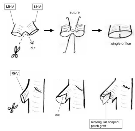
2)In the left liver graft, a vertical cut was made on the spur between the left and the middle hepatic vein and reconstructed to the single orifice with a non absorbable monofilament suture. When the orifice of the left and the middle hepatic vein were separated, their medial walls are then sutured to make a single orifice. Then venous patch grafts were attached to the anterior and the posterior wall of the reconstructed hepatic vein(Fig. 1 a). In case number 4, a hepatic vein tributary was present on the distal side of the cut surface of the right posterior segment(right lateral sector)
graft. In that case, a side to end anastomosis be- tween the distal hepatic vein tributary of the graft and the right hepatic vein graft obtained from the recipient was done. Then the proximal side of this right hepatic vein graft was sutured to the right hepatic vein of the graft and the single big orifice was reconstructed. The same technique was ap- plied for the anastomosis of the short hepatic vein of the left caudate lobe(Spiegel portion)in case number 5(Fig. 1 b).
The portal vein of the graft did not need recon- struction on the back table in all cases, because they were properly dissected during a hepatectomy in the donor. In one of our 6 cases, the hepatic ar- tery reconstruction was needed using free arterial graft obtained from the right gastroepiploic ar- tery of the recipient.
There was one case that needed bile duct recon- struction on the back table. In that case, the bile duct of the graft as separated two orifices. They were reconstructed into one orifice by suturing their medial wall.
Implantation of the partial liver graft
The implantation of the partial liver graft was completed starting with hepatic vein anastomosis followed by portal vein and hepatic artery anastomosis. The biliary anastomosis was done after the blood perfusion of the graft had been restored.
Hepatic vein anastomosis
The hepatic vein of the graft was reconstructed
to make a single large orifice using a vein patch ex situ. In case of left liver graft, a big orifice was previously reconstructed with the left and the mid- dle hepatic vein of the recipient (Fig. 2 a.). When the right lateral sector graft was implanted(case number 4), the right hepatic vein of the recipient was used for hepatic vein anastomosis. The ante- rior wall of the right hepatic vein was cut longitu- dinally and a rectangular shaped vein patch was attached to enlarge the orifice(Fig. 2 b.). Hepatic vein anastomosis was done in an end to end manner.
Portal vein anastomosis
The portal vein anastomosis was made between the first order branch of the recipient portal vein and the portal vein of the partial liver graft. The portal vein anastomosis was completed in an end
to end continuous suture with 6 0 non absorbable monofilament suture.
Hepatic artery anastomosis
End to end hepatic artery anastomosis was done between the left or right hepatic artery of the re- cipient and the hepatic artery of the partial liver graft with 8 0 non absorbable monofilament suture. Either end to end anastomosis or branch patch reconstruction was chosen for each case. Mi- crosurgery was applied for hepatic artery anasto- mosis in last three cases (cases 4 to 6).
Biliary anastomosis
The biliary reconstruction was the final phase of partial liver graft implantation. Among our cas- es, we apply duct to duct anastomosis between the left or right hepatic duct of the recipient and the
Fig. 1. Venoplasty of the hepatic vein orifice in the partial liver graft.In case of left liver graft. The venous patch grafts are attached to the anterior and the posterior wall of the reconstructed middle
(MHV)and left hepatic vein(LHV). The technique in case num- ber 4 and 5. A hepatic vein tributary is anastomosed to the right hepatic vein graft from the recipient. Then single large hepatic vein orifice is reconstructed in the proximal side of the graft.
hepatic duct of the partial liver graft. A single layer, interrupted suture was carried out using 4 0 or 5 0 absorbable suture for bile duct anastomosis.
The biliary drainage tube was placed into the he- patic duct of the partial live graft through the tributary of the hepatic duct on the other side of
the recipient(Fig. 3.).
Results
A total of 6 LDLTs were performed. The patient’s age ranged 30 to 60. Four were male and
Fig. 2. Venoplasty of the recipient’s hepatic vein. The confluence of the
middle(MHV)and the left hepatic vein (LHV) is cut and recon- structed to the single large orifice. The ante4rior wall of the right hepatic vein is cut longitudinally. A rectangular shaped patch graft is attached and the orifice is enlarged.
Fig. 3. Biliary reconstruction. A duct to duct anastomosis between the
left hepatic duct of the recipient and the hepatic duct of the partial liver graft. A single layer interrupted suture is applied using 4
0or 5
0 absorbable suture. The biliary drainage tube is placed intothe hepatic duct of the partial liver graft through the orifice of the right hepatic duct of the recipient.
2 were female. The diagnosis was 2 hepatitis C re- lated cirrhosis with hepatocellular carcinoma, 2 al- coholic cirrhosis, 1 fulminant liver failure and 1 Wilson’s disease.
Among our 6 cases, 5 were left liver grafts and one was a right lateral sector graft(case 4). Usu- ally, the left liver graft was accompanied by the left caudate lobe(Spiegel portion). In our case 4, the right lateral sector graft was selected because the volume of the left liver of the donor was less than 35% of the standard liver volume of the recipi- ent, and the volume of the right lateral sector of the donor was greater than 40% of the standard liver volume of the recipient. The clinical charac- teristics of partial liver grafts are shown in Table 1. There were 2 mortality cases in our series. One was a left liver graft case who died due to postop- erative hepatic failure. The other patient used a right lateral sector graft, and who died due to bili- ary and pulmonary infection.
There has not been any problem, such as venous outflow block or postoperative bleeding in hepatic venous anastomosis.
There has also not been any problem regarding portal vein anastomosis. However, we have experi- enceda case of partial portal vein thrombosis that developed portal vein thrombus in the peripheral portal branch of segment 7 due to inflammation of the adjacent bile duct(case 4). Anticoagulation therapy was not effective in this case because the
cholangitis of the adjacent bile duct could not be resolved.
We have not experienced any anastomosis re- lated complication in the hepatic artery among our 6 cases so far.
The two out of our 6 cases developed delayed stricture at the biliary anastomosis 6 month after surgery. One had a tube stent, and the other one has been undergoing percutaneous transhepatic bil- iary drainage(PTBD).
Discussion
A graft size mismatch is a problem that some- times arises in adult to adult LDLT. Because only a part of the liver is implanted in LDLT, the graft mass is not enough for the metabolic demand of the recipient. To prevent this problem, the number of right liver graft implantations which are usually larger than the left liver graft has thus been increasing. According to the annual report of the liver transplantation society of Japan, 60.3% of adult to adult LDLT used right liver graft.
3)However, the major complications of the donor occur more often in right liver graft harvest- ing than left liver graft. Hwang et al. reported that the incidence of major donor complication was 1.4% for left liver graft cases, whereas it was 4.9%
for right liver graft cases.
4)Moreover, the mortal- ity cases of right liver donor have been reported in
Table 1. Characters of LDLT casesOutcome,/cause of death Bile Duct
Anastomosis Hepatic
Artery Anas- tomosis Portal
Vein Anastomosis Hepatic
Vein Reconstruction Graft
Diagnosis Sex
Case Age
#
Survive 31months DucttoDuct
Endtoend EndtoEnd
Cryopreserved Graft Left Liver
CLC, HCC Male
60 1
Survive 21 months DucttoDuct
Endtoend EndtoEend
Venous Patch Left Liver
Fulminant hepatic failure Female
30 2
Survive 19 months DucttoDuct
Branch patch EndtoEnd
Venous Patch Left Liver
CLC, HCC Male
59 3
Died 4 months after LDLT/Biliary and Pulmo- nary infection (aspergillosis) DucttoDuct,
Bile Duct Plasty Endtoend,
Microsurgery EndtoEnd
Right He- patic Vein
Graft Right
Lateral Sector Alcoholic
cirrhosis Male
41 4
Survive 2 months DucttoDuct
Endtoend, Microsurgery EndtoEnd
Right He- patic Vein
Graft Left Liver
Wilson’s Disease Female
36 5
Died 3 days after LDLT/Hepatic failure DucttoDuct
Endtoend, Microsurgery EndtoEnd
Venous Patch Left Liver
Alcoholic cirrhosis Male
53 6
C LC:type C hepatitis related cirrhosis, HCC:hepatocellular carcinoma
western country and Japan.
5)From the point of donor safety, we use the left liver graft when the graft volume is 35% or greater of the standard liver volume of the recipient that is considered to meet the recipient’s metabolic demand. The right lateral sector graft is chosen when the volume of the left liver of the donor was less than 35% of the standard liver volume of the recipient, and the vol- ume of the right lateral sector of the donor was greater than 35% of the standard liver volume of the recipient. In our series, the use of these two kind of grafts did not show any difference in the outcome. There was one mortality case each for both types of graft, and the causes of death were hepatic failure and infection.
Venous outflow block which causes the conges- tion of the liver and the liver dysfunction is one of the major concerns in LDLT. Because venous out- flow block is caused by a kink or flexion of the he- patic vein anastomosis that related to regeneration and enlargement of the partial liver graft. Making a large anastomosis orifice of the hepatic vein is thus essential to prevent the kink of hepatic vein anastomosis. For this purpose various venous re- construction have been contrived using venous graft either autograft or cryopreserved graft.
1)2)6)8)In our series, 1 case out of 6 cases used cryopreserved venous graft, others used autograft.
The portal vein thrombosis(PVT)and the hepatic artery thrombosis(HAT)are common postopera- tive complications in liver transplantation. It is said that the incidence of postoperative PVT is 1 3%, while HAT is 3.7%. In our series, there was 1 case of partial portal vein thrombosis, whereas no HAT was experienced. That portal vein thrombo- sis was developed in the peripheral portal branch of segment 7 due to cholangitis of the adjacent bile duct. The application of microsurgery in hepatic artery anastomosis lowers the risk for HAT.
9)10)In our last three cases(cases 4 to 6), microsurgery has been applied for hepatic artery anastomosis.
Biliary complications remain another serious problem in liver transplantation. The bibliographi- cal incidence of biliary complications is 4.7% for bile leakage, and 26.6% for biliary stricture in duct to duct anastomosis.
11)The treatment are biliary
decompression, drainage or balloon dilation.
11)12)We performed duct to duct anastomosis all of our
6 cases, and experienced 2 cases of biliary stricture.
In conclusion, we have had success in performing LDLT at Fukuoka University Hospital so far.
However, the procedure of LDLT is meticulous.
Further establishment of surgical technique and the accumulation of appropriately indicated pa- tients for transplant team are mandatory.
References
1)Sano K, Makuuchi M, Miki K, Maema A, Sugawara Y, Imamura H, Matsunami H, Takayama T:Evalua- tion of hepatic venous congestion:Proposed indica- tion criteria for hepatic vein reconstruction. Ann Surg 236:241 247, 2002.
2)Sugawara Y, Makuuchi M, AkamatsuN, Kishi Y, Niiya T, Kaneko J, Imamura H, Kokudo N.:Refine- ment of venous reconstruction using cryopreserved veins in right liver grafts. Liver Transplant 10:541 547, 2004.
3)The Japanese liver transplantation society. Liver Transplantation in Japan. ― Registry by the Japa- nese Liver transplantation society:Ishoku 41:599 608, 2006.(in Japanese)
4)Shin Hwang, Sung Gyu Lee, Kyu Bo Sung, Kwang Min Park, Ki Hun Kim, ChulSoo Ahn, Deok Bog Moon, Gyu Sam Hwang, Kyung Mo Kim, et al. Les- sons learned from 1,000 living donor liver transplan- tations in a single center:How to make living donations safe:Liver Transplant 12:920927, 2006.
5)Kiyosawa K, Ichida T, Umeshita K, Kawasaki S, Mi- zokami M, Mochida S, Yanaga K, Yonemoto S, Nakanuma Y. Verification of lethal prognosis of liv- ing donor and proposal for prevention of recidivation.
Ishoku 39:47 55, 2004.(in Japanese)
6)Hwang S, Lee SG, Park KM, Kim KH, Ahn CS, Moon DB, Ha TY.:Quilt venoplasty using recipient saphe- nous vein graft for reconstruction of multiple short hepatic veins in right liver grafts. Liver Transplant 11:104 107, 2005.
7)Hashimoto T, Sugawara Y, Kishi Y, Akamatsu N, Matsui Y, Kokudo N, Makuuchi M.:Reconstruction of the middle hepatic vein tributary in a right lateral sector graft. Liver Transplant 11:309 313, 2005.
8)Takemura M, Sugawara Y, Hashimoto T, Akamatsu N, Kishi Y, Tamura S, Makuuchi M.:New hepatic vein reconstruction in left liver graft. Liver Trans- plant 11:356 360, 2005.
9)Jia YP, Lu Q, Gong H, Ma BY, Wen XR, Peng YL, Lin L, Chen HY, Qiu L, Luo Y.:Postoperative com- plications in patients with portal vein thrombosis af- ter liver transplantation:Evaluation withDoppler ultrasonography. World J Gastroenterol 13:4636 4640, 2007.