Yuichi Hirose
A Thesis
for the Degree of Doctorate of Philosophy Department of Chemistry
Tokyo Metropolitan University 2011
In this thesis, we proposed a model for lipid bilayers, and discussed the mechanism of the finite-sized domain formation together with the effects of coupling of the two monolayers.
A lipid monolayer consisting of saturated lipid, hybrid lipid and cholesterol was con- sidered. We proposed a phenomenological Ginzburg-Landau model in which the coupling of the lipid composition and chain orientational vector field is considered. This coupling arises from the liquid crystalline nature of the hybrid lipid which adjusts its orientational order in the tails to reduce the line tension. As a result, finite-sized domains can be formed. The minimization of the free energy with respect to the vector field yields an effective free energy which is analogous to that of 3D microemulsions (above the transition temperature) and modulated structures (below the transition temperature).
Then we considered the model for lipid bilayers comprised of two modulated mono- layers which are coupled via interleaflet interactions. The structure and the dynamics of the coupled modulated bilayer are investigated theoretically.
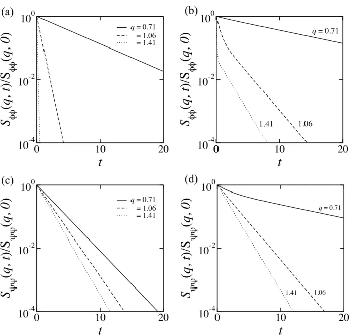
We first studied concentration fluctuations in bilayers occurring above the transition temperature. We have calculated the static structure factors, and obtained the bilayer phase diagrams close to the critical temperature. In both monolayers, fluctuations are induced due to the coupling, and the spectrum of the induced fluctuations is determined by the cross correlation of the structure factor. When the two monolayers having differ- ent preferred wavenumbers are coupled, the peak height at smaller wavenumber becomes larger, whereas that at a larger wavenumber remains the same. We studied the dynam-
equations, and calculated the intermediate structure factors of the bilayer. In general, concentration fluctuations exhibit a double-exponential decay. Due to the coupling, the time for the cross over of the two concentration fluctuations appears when the character- istic length scale of each monolayer is different.
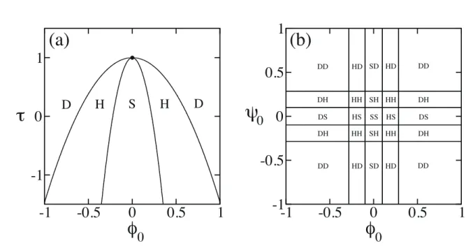
Then we discussed the phase separation of the coupled modulated bilayers. Within the mean field approximation, we constructed phase diagrams of the bilayer when the two monolayers have the same wavenumber of the modulations. The phase behavior of the bilayers is described by the combinations of the stripe and the hexagonal morphologies.
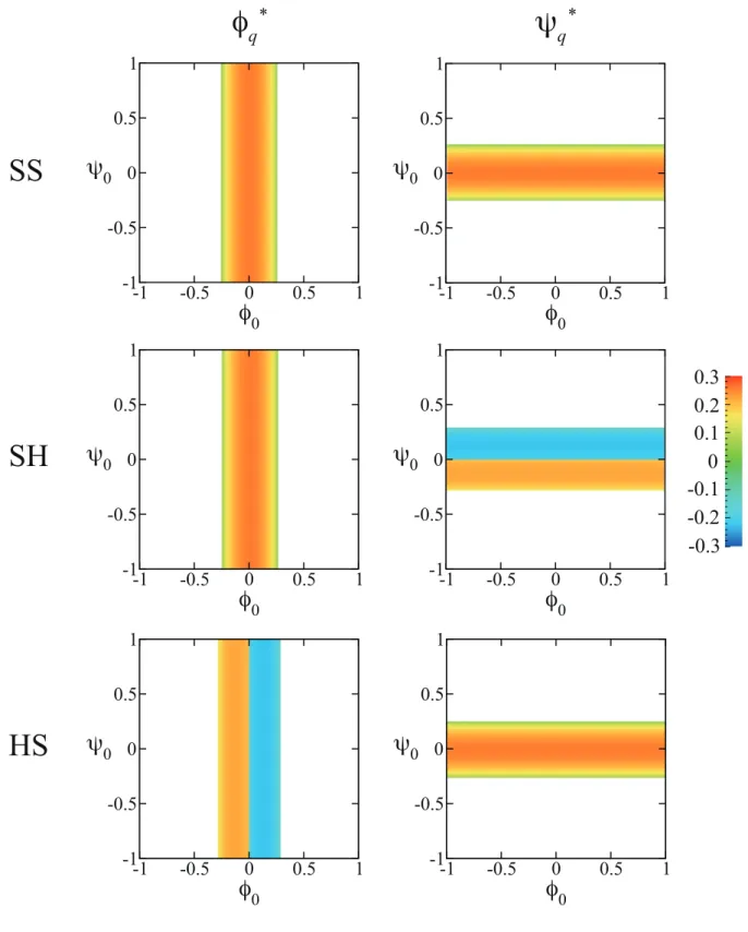
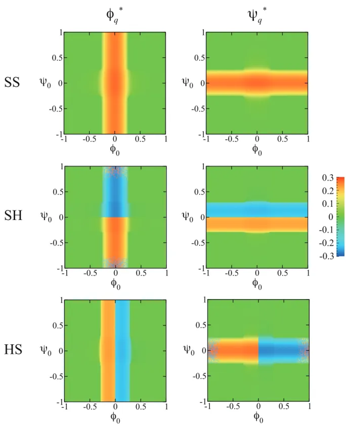
Due to the coupling effect, one of the monolayers induces micro-phase separation in the other monolayer. As the coupling strength increases, the asymmetric phases tend to disappear. By performing numerical simulations, we obtained various phase separated patterns when the two monolayers have different modulations. The obtained patterns are approximately classified into “independent”, “intermediate” and “coincident” cases.
The degree of the overlap between the two monolayers is characterized by the inter-leaflet correlation of the local compositions. We showed that the initial growth rates of the most unstable mode are essentially identical to the decay rates of the concentration fluctuations.
We showed that the formation of the intermediate structures is related to the stability of the concentration fluctuations occurring above the transition temperature.
Using the mechanism of finite-sized domain formation proposed above, we shall also discuss its relation to the biologically relevant context of lipid rafts.
Contents
1 Introduction 1
2 Model 13
2.1 Lipid monolayer . . . . 13
2.2 Lipid bilayer . . . . 19
3 Concentration Fluctuations 23 3.1 Statics . . . . 23
3.1.1 Decoupled case (Λ = 0) . . . . 25
3.1.2 Coupled case (Λ6= 0) . . . . 29
3.2 Dynamics . . . . 43
4 Phase Separations 51 4.1 Bilayers with the same q∗ . . . . 51
4.1.1 Decoupled case (Λ = 0) . . . . 51
4.1.2 Coupled case (Λ6= 0) . . . . 57
4.1.3 Dynamics . . . . 73
4.2 Bilayers with different q∗ . . . . 78
4.2.1 Phase separated patterns . . . . 78
4.2.2 Dynamics . . . . 96
5 Discussions 101
5.1 Emergence of the characteristic length scale . . . 101
5.2 Concentration fluctuations of 2D micelles . . . 103
5.3 Coupling of different modulations . . . 106
5.4 Non-equilibrium nature of asymmetrical bilayers . . . 107
6 Conclusion 109
Appendix A 111
Appendix B 114
Appendix C 117
Appendix D 119
References 145
List of Publications 151
Acknowledgments 153
Chapter 1 Introduction
An amphiphilic molecule possesses both hydrophilic and hydrophobic moieties in a sin- gle molecule. The typical examples are surfactants, lipids and block copolymers. Due to hydrophobic interactions between the hydrocarbon chain and water molecules, these molecules self-assemble to form supermolecular structures in aqueous solutions as shown in Fig. 1.1. As the surfactant concentration is increased, micelles, bilayer vesicles, hexag- onally packed cylinders, lamellae (which consist of stack of bilayers) and more complex cubic structures emerge due to the competition between entropy and intermolecular in- teractions [1].
The characteristic length scale of these mesoscopic structures is much larger than the molecular size and is typically 10∼1000 nm. The mesoscopic structures that are formed can be easily deformed upon the application of an external force. This is why amphiphilic systems are considered to be good examples of soft matter. The entropic modulus of a soft material is typically given byG∼kBT /d3, where kB is the Boltzmann constant,T is the temperature and d is the characteristic length scale of the internal structure. Living matter is essentially soft matter in terms of its softness because the typical cell size is on the order of micron. Many researchers have been trying to clarify the border between living matter and soft matter. For example, the mechanics of the cell and its relations with the biochemical properties has attracted attention [2].
hydrophilic
hydrophobic amphiphile
lamellae vesicle hexagonal cylinders cubic
micelle i l vesicle cubic
Figure 1.1: A schematic representation of an amphiphilic molecule and their organized structures in water solvent.
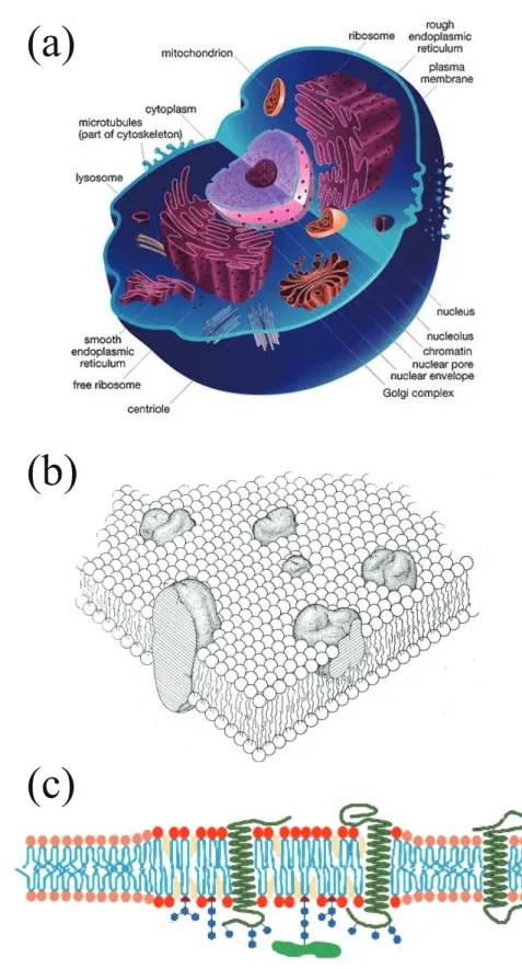
proteins. As presented in Fig. 1.2(a), they separate inner and outer environment of the organelles in the cell. Membrane proteins are known to play essential roles for the biological functions. For example, they transduce external stimulation into chemical sub- stances which are transmitted to internal environment of the cell. Such a process is called as signal transduction. Singer and Nicolson proposed the “fluid mosaic” model for biomembranes [3]. In this picture, membrane proteins are floating on the sea of lipids as schematically depicted in Fig. 1.2(b). Later, Simons and Ikonen proposed the “lipid raft” hypothesis in which some of the lipid components and/or proteins are incorporated into finite sized domains as shown in Fig. 1.2(c) [4]. Raft domains are expected to play important roles for the signal transduction processes by providing a field for the specific proteins to interact each other and increasing their efficiency.
Numerous studies have been conducted to identify the raft domains in biological mem- branes. For instance, detergent extraction of the membrane components from the cells was performed [5]. Although the detergent resistant membrane (DRM) fractions are con- sidered as raft components, the extracts are not the representatives of the rafts because of the perturbation by the detergents and the low temperature (4 ◦C) of the process.
Electron microscopic (EM) images of cell membranes were captured by labeling target lipids with gold nano particles of 10 nm size. A typical lipid cluster size was found to be about 10–100 nm [6, 7]. According to the fluorescence resonance energy transfer (FRET) study, the cluster size of GPI-anchored proteins was reported to be 4–5 nm [8]. Although the size and the existence of lipid rafts are still controversial, the former is expected to be in the range of 10–200 nm [9]. The interactions between lipid molecules and proteins are thought to be the driving force for the raft formation.
Among many studies, dynamics of a single lipid molecule in living cell membranes have been tracked using fluorescent microscopy, and the effect of cytoskeleton on the diffusion
(a)
(b)
(c)
Figure 1.2: (a) A schematic representation of a single cell. There are many biomembranes inside the cell. Each internal organ has its own lipid membrane. (b) Fluid mosaic model of a biomembrane taken form the original paper [3]. The membrane proteins are embedded in the phospholipid bilayers. (c) A biomembrane with raft domains which are rich in certain components of the lipids and proteins.
lipid dynamics in living cells is achieved using stimulated emission depletion (STED) fluorescent microscopy [11]. They reported that sphingomyeline and membrane proteins are transiently (10-20 ms) trapped in 20 nm diameter areas. These experimental results suggest that lipid rafts are dynamical assemblies of molecules with finite decay time. In the latter study, the entrapment was not observed when the cholesterol is depleted from the membrane by the addition of cyclodextrin, suggesting the specific interactions with proteins or lipids on biomembranes.
In the area of “lipidomics”, the main concern is to understand the functional speci- ficity of lipids on various metabolic diseases such as obesity or diabetes [12]. The diversity in the lipid species (about one thousand) is expected to have some biological significance.
However, domain formation in artificial membranes requires only three components; (i) saturated lipid having two saturated acyl chains, (ii) unsaturated lipid having one or more double bonds in one or both acyl chains, (iii) cholesterol having four cycloalkanes (see also Fig. 1.3). For both saturated and unsaturated lipids, the acyl chains are frozen in the low temperature (the gel phase denoted asSo phase), and they are melted in the high temperature (the fluid phase denoted as Ld phase). This phase transition is called as the
“main transition”. The transition temperature of unsaturated lipids is generally lower than that of saturated lipids. This is because the double bonds introduce geometrical frustrations and act against the crystallization of the chains. Cholesterol interacts more favorably with saturated lipids than unsaturated lipids since the rigid structure of choles- terol fits well with saturated chains. The phase behavior of the membranes composed of these lipids is complex because of the interactions among them.
Many researches have been conducted to reveal the domain structures on artificial lipid membrane vesicles consisting of saturated lipid, unsaturated lipid and cholesterol (Fig. 1.3) [13, 14, 15, 16, 17]. When the membranes are below the transition temperature,
micron sized domains can be observed using fluorescent microscopy. Domain formation is a consequence of the phase separation between the liquid ordered (Lo) phase and the liquid disordered (Ld) phase. The acyl chains are more ordered in the Lo-phase than in the Ld-phase, while the lateral mobility is much higher in the both phases compared to the So-phase. Such a demixing of lipids is caused by the difference in the packing nature of hydrophobic chains of saturated and unsaturated lipids. Without cholesterol, the phase separation between the So-phase and the Ld-phase is observed. The preferential affinity of cholesterol to the saturated lipids introduces more fluidity in theLo phase. The effects of cholesterol on the phase behavior of lipid mixtures have been investigated theoretically such as using a phenomenological model [18], a microscopic model [19], or coarse grained simulations [20]. Typical size of the phase separated domains on artificial membranes is micron, and the domain growth continues until it reaches the size of the vesicle. In contrast to this, phase separated domains with distinct size (micron order) have been observed in artificial membranes [21, 22, 23, 24]. Although the characteristic size of the phase separated domains is different from that of the biomembranes, it is worthwhile to consider the formation mechanism of the finite sized domains on biomembranes in relation to the artificial membranes.
Another interesting research direction is to probe concentration fluctuations above the transition temperature in ternary lipid mixtures, as shown in Fig. 1.4 [25]. Analyzing fluorescent microscope images, the correlation length of critical concentration fluctuations in model membranes was measured [26, 27]. The obtained critical exponent characterizing the critical behavior of the correlation length suggests that these lipid mixtures belong to the universality class of 2D Ising model. Critical concentration fluctuations were also observed in membranes extracted from living cells [28]. It is rather surprising that plasma membranes may be naturally at their critical compositions considering the fact that they consist of thousands of different species of lipids and proteins.
Ld Lo d
Figure 1.3: The phase diagram of the model membrane consisting of POPC (palmitoy- loleoylphosphatidylcholine, unsaturated lipid), PSM (palmitoylsphingomyeline, saturated lipid) and cholesterol. The chemical structures of these molecules are illustrated. Phase separa- tions occur in the light gray regions of the phase diagram with a typical microscope im- age of the phase separated structure. (The microscope picture is taken from the bilayer of DOPC/PSM/cholesterol.) Dark and bright domains in the micrograph are the liquid ordered (Lo) phase and the liquid disordered (Ld) phase, respectively. Cholesterol is preferentially partitioned in the Lo-phase than the Ld-phase. Figures are taken from Refs. [14, 38, 56].
Figure 1.4: Fluorescent microscope images of the surface of a single giant unilamellar vesicle for different times [25]. The transition temperature isTc= 31.9◦C. Concentration fluctuations can be observed when the temperature is higher thanTc. The vesicle is composed of 25:20:55 mol% of diphytanoylphosphatidylcholine (DiPhyPC), dipalmitoylphosphatidylcholine (DPPC) and cholesterol. The scale bar is 20µm.
mains (clusters) rather than macroscopically phase separated domains. This question is primarily concerned with the question whether biomembranes are above or below the miscibility transition temperature. Suppose biomembranes are above the transition tem- perature, the correlation length characterizing the concentration fluctuations is the only length scale which diverges at the critical point. If biomembranes are below the transi- tion temperature, there should be a mechanism to suppress the coarsening of domains. In this case, lipid membranes may undergo micro-phase separation similar to block copoly- mers. This naturally explains the typical length scale below the transition temperature in equilibrium.
One possibility to account for the finite sized domains in lipid membranes both above and below the transition temperature is the existence of “hybrid lipids” which have one saturated chain and one unsaturated chain [29, 30, 31]. Hybrid lipids are one type of unsaturated lipids, and are abundant in the biological membranes [32, 33]. Such lipids tend to localize at the 2D domain boundaries in order to reduce the frustration of the chain mismatch and act as line active molecules similar to the surfactant. As a result, hybrid lipids can drive the line tension to become negative. Hence a ternary mixture of saturated lipid (having two saturated chains), unsaturated lipid (having two unsaturated chains), and hybrid lipid has a close resemblance to a microemulsion consisting of oil, water, and surfactant [25, 29, 34]. It is known that three-dimensional (3D) microemulsions are thermodynamically stable solutions in which oil and water are isotropically mixed on the scale of 50–100 nm. Although the physical mechanisms for the stabilization of interfaces are different between surfactants and hybrid lipids, the ternary lipid mixtures can be regarded as 2D microemulsions. It was proposed that the hybrid lipid is a more efficient linactant in the mixture of saturated lipid, hybrid lipid, and cholesterol [31]. When the two chains of the hybrid lipid are oriented towards the domain interface, the line tension
can become negative. The lipid domains generated in the membrane can then be termed as “2D micelles”.
The important feature of biological membranes is that the lipid composition in each monolayer is notably different. For example, it was reported that the distribution of lipid species composing inner and outer leaflets in human red blood cells is asymmet- ric [35]. More importantly, these asymmetric monolayers are coupled and do not behave independently. The coupling of the domains in monolayers might be important because interleaflet interactions between proteins of outer and inner leaflets may connect the outer environments and inner signaling pathway of the cell. Such a coupling between the two leaflets was experimentally verified by looking at the phase behavior of combined monolay- ers which have different lipid compositions [36]. It was shown that one of the monolayers either induce or suppress phase separation in the other monolayer. One of the possibilities to account for this inter-monolayer coupling is the lipid chain interdigitation occurring at the mid-plane of the bilayer [37]. The phase behavior of the coupled lipid bilayer was theoretically studied using either the regular solution theory [38] or the Landau the- ory [39]. These works have been successful in describing the experimentally observed phase behaviors of coupled bilayers below the transition temperature. Later, concentra- tion fluctuations of coupled bilayers above the transition temperature was investigated in Ref. [40].
In this thesis, the statics and dynamics of inhomogeneous structures in lipid bilay- ers are studied. We propose a model describing the formation of finite sized domains in membranes containing hybrid lipids. Within the Ginzburg-Landau theory, we consider a coupling between the chain orientation and the composition field in lipid monolayers. For both above and below the transition temperature, the characteristic length of the com- positional modulations emerges due to the coupling. Such a characteristic length scale might possibly regulate the raft size in biomembranes. Especially, we discuss the concen-
decay time of the concentration fluctuations can be interpreted as a life time of dynamical domains. Then the effects of coupling of two modulated monolayers are considered by introducing interleaflet interactions as argued before [36, 38, 39]. We discuss both static and dynamic properties of concentration fluctuations in coupled modulated bilayers. We obtain the static and dynamical structure factors of coupled bilayers above the transi- tion temperature. We construct the phase diagrams of the coupled modulated bilayers, and determine the regions of the ordered phase, the structured-disorder phase and the disorder phase in each monolayer. Due to the coupling between the two leaflets, concen- tration fluctuations are induced in both the monolayers. We especially focus on the case when the fluctuations in the two monolayers have different characteristic wavelengths. We also discuss the phase separation in the coupled modulated bilayers below the transition temperature. Various kinds of intermediate phase separated structures arise due to the two competing structures of different wavelengths. We show that the formation of the intermediate structures can be explained by the stability of the concentration fluctuations occurring above the transition temperature. Finally we discuss the biological relevance of the suggested model.
This thesis is organized as follows. In Chap. II, the model for modulated monolayers and coupled modulated bilayers are presented. The static and dynamical structure factors of the bilayers are computed in Chap. III. In Chap. IV, the phase behavior of the bilayer below the transition temperature is discussed. Some discussions are given in Chap. V.
Finally, we conclude in Chap.VI.
Chapter 2 Model
In this chapter, we present models for a lipid monolayer and a bilayer. Firstly, a mecha- nism for the finite sized domain formation in lipid monolayers is discussed. The coupling between the concentration field of the lipids and the orientational vector field of lipid chains generates finite sized domain (modulated) structures in monolayers. Then we show a model for coupled bilayers which consist of two modulated monolayers. The physical origin of the inter-leaflet coupling is also discussed.
2.1 Lipid monolayer
As a model for the lipid monolayer, we consider a 2D lattice composed of saturated and hybrid lipids, and these lipids constitute the liquid ordered phase (Lo) and the liquid disordered phase (Ld), respectively (Fig. 2.1). Hybrid lipids possess both saturated chain
lipid S lipid H
unsaturated saturated
Figure 2.1: 2-dimensional lattice compose of saturated lipids and hybrid lipids. Hybrid lipids possess both saturated acyl chain and unsaturated acyl chain in a single molecule.
and unsaturated chain in a single molecule. We define the number of the lipid species as NS and NH. The Flory-Huggins free energy of mixing for the binary lipids per unit area is expressed as
f0 kBT = 1
d2[φSlogφS+φHlogφH+χφSφH], (2.1) whereφS=NS/(NS+NH) andφH=NH/(NS+NH) are the area fraction of each species, χis the Flory-Huggins parameter, andd is the typical size of the lipid, such as the size of the head group. By defining φ = (φS−φH)/2 and using the incompressibility condition φS+φH= 1, Eq. (2.1) is expanded to give
f0−fc
kBT = τ¯
2φ2+u
4φ4, (2.2)
with
¯
τ = 2(2−χ)
d2 , (2.3)
and
u= 16
3d2. (2.4)
Then the free energy is given by Fc[φ(˜r)] =
∫ d˜r
[σ
2( ˜∇φ)2+ ¯τ
2φ2+ u
4φ4−µφ¯ ]
. (2.5)
Where ˜∇ = (∂/∂x, ∂/∂˜ y) and d˜˜ r = d˜xd˜y, characters with tildes have the dimension of length. The term with σ(>0) generates the energy penalty for spatially inhomogeneous distribution ofφ, and is related to the line tension between two adjacent domains. ¯µis the chemical potential which regulates the average composition in the membranes. This free energy describe the phase separation. When ¯τ < 0,fchas two minima atφ1,2 =±√
−τ /u (when spatially averaged compositionφ0 is zero), corresponding to the composition ofLo and Ld phases, respectively. The dynamics of the phase separation is described by the time evolution equations of the compositions
∂φ
∂t =Lφ∇˜2δFc
δφ , (2.6)
the above equation, one can show that phase separated domains merge to grow in time driven by the penalty σ. The growth continues until the size of the domain reaches the
“macroscopic” size (i.e., comparable to the system size). Thus there is no characteristic size of the domain for the phase separation phenomena.
We define the orientation of a hybrid lipid molecule as a vector from the unsatu- rated tail towards the saturated tail by b = (bx, by) as shown in Fig. 2.2. The vector m(˜r) = (mx(˜r), my(˜r)) is defined as the spatial average of the b vectors over areas large as compared with molecular size but still small enough as compared with macroscopic scales. The energy of these orientational vector is expressed by
Fo[m(˜r)] =
∫ d˜r
[a
2m2+K1
2 ( ˜∇ ·m)2+ K2
2 ( ˜∇ ×m)2 ]
. (2.7)
The term ofa(>0) is related to the orientational entropy, andK1, K2(>0) are the elastic constants for the orientation. Without the gradient terms, the above energy is minimized when the orientational field is m= 0.
When the two lipid species are spatially inhomogeneous both above Tc (concentration fluctuations) and below Tc (phase separations), the coupling of the concentration and the orientational vector fields emerges. Here the coupling term is given by
Fcoup[φ(˜r),m(˜r)] = −g
∫
d˜r m·( ˜∇φ), (2.8) where g >0 is the coupling constant. Notice that this term is negative when m and ˜∇φ point to the same direction. Hybrid lipids are responsible for the emergence of the cou- pling, because the saturated chain and the unsaturated chain of the lipid have preferences to theLo-phase and theLd-phase, respectively. Especially at the boundary between theLo and the Ld-domains, the hybrid lipids are expected to reduce the chain mismatch by ori- enting their saturated/unsaturated chains toward the Lo and Ld-phases. The mechanism of chain direction alignment at the boundary was proposed by Yamamotoet.al. [31], and
φ
m
lipid H
unsaturated saturated
b
Lo
Ld
lipid S
Figure 2.2: A lipid monolayer consisting of saturated lipids and hybrid lipids. The 2D vector of a lipid molecule from the less packing chain toward the packed chain is defined as b.
Spatially averaged orientational vector is defined as m.
the mechanism we introduce here is a coarse grained and intuitive picture of this effect.
The Ginzburg-Landau free energy functional for the lipid monolayers based on two order parameters φ(˜r) and m(˜r) is given by the sum of these contributions (Fc+Fo +Fcoup),
Fm[φ(˜r),m(˜r)] =
∫ d˜r
[σ
2( ˜∇φ)2+ τ¯
2φ2+u
4φ4−µφ¯ +a
2m2+K1
2 ( ˜∇ ·m)2+K2
2 ( ˜∇ ×m)2−gm·( ˜∇φ) ]
. (2.9) In the Fourier space, Fm[φ,m] is expressed as
Fm[φ(˜q),m(˜q)] =
∫ d˜q
[1 2
(
σq˜2+ ¯τ )
φ(˜q)φ(−q)˜ (2.10)
+1 2
(
a−K1q˜x2−K2q˜y2 )
mx(˜q)mx(−q)˜ +1
2 (
a−K1q˜y2−K2q˜x2 )
my(˜q)my(−q)˜
−(K1+K2)˜qxq˜ymx(˜q)my(−q)˜
+ig[˜qxmx(˜q)φ(−˜q) + ˜qymy(˜q)φ(−q)]˜ ]
−µφ(0)¯ +
∫ d˜q
∫ d˜q0
∫ d˜q00
[u
4φ(˜q)φ(˜q0)φ(˜q00)φ(−q˜−q˜0−q˜00) ]
,
where i is the imaginary unit, ˜q = (˜qx,q˜y) is the wavevector in the monolayer, and
˜
q = |q˜| is the wavenumber. Minimizing Fm[φ(˜q),m(˜q)] with respect to m(˜q) yields the
m∗(˜q) = igq˜
a+K1q˜2φ(˜q). (2.11)
Note that the constant K2 vanishes in this minimization process. By putting Eq. (2.11), the free energy depends only on the composition,
Fm[φ(˜q)] =
∫ d˜q
(σ
2q˜2+ τ¯
2 − g2q˜2 2(a+K1q˜2)
)
φ(˜q)φ(−q)˜ −µφ(0)¯ +
∫ d˜q
∫ d˜q0
∫ d˜q00
[u
4φ(˜q)φ(˜q0)φ(˜q00)φ(−q˜−q˜0 −q˜00) ]
. (2.12)
Again, the constant K2 does not appear in the minimized energy. For small q, the coefficients of the coupling term can be expanded, and the free energy is given by
Fm[φ(˜q)] =
∫ d˜q
(
2Bq˜4−2A˜q2+τ¯ 2
)
φ(˜q)φ(−˜q)−µφ(0)¯ +
∫ d˜q
∫ d˜q0
∫ d˜q00
[u
4φ(˜q)φ(˜q0)φ(˜q00)φ(−q˜−q˜0 −q˜00) ]
, (2.13)
with
B˜ = K1g2
4a2 , (2.14)
and
A= 1 4
(g2 a −σ
)
. (2.15)
Notice thatAis dimensionless quantity. The wavenumber ˜q-dependence of the free energy Fm[φ(˜q)] is plotted in Fig. 2.3. When the coupling is weak (g2/a−σ < 0), the line tension has a positive value (A <0) and the minimum is at ˜q∗ = 0, indicating the macro- phase separation (below Tc). If the coupling is strong (g2/a−σ >0), the line tension can become negative (A >0) and the energy has a minimum at ˜q∗ 6= 0. The negative gradient squared term favors spatial modulations, whereas the positive coefficients of the Laplacian squared term ( ˜B > 0) suppresses modulations. As a result of these contributions, a stable modulation of finite wavenumber emerges. The characteristic wavenumber of this
0 0.5 1
q
-0.5 0 0.5 1
f(q)
q4 - q2 q4 + q2
Figure 2.3: Wavenumber q dependence of the free energy.
modulated structure is obtained by minimizing Eq. (2.13) with respect to ˜q
˜ q∗ =
√ A 2 ˜B =
√
a(1−aσ/g2)
2K1 . (2.16)
Here the length scale of the modulation is determined by a, the line tension σ, the elastic coefficientK1, and the coupling constantg. These coefficients are related to the properties of the lipid mixture.
In real space, Eq. (2.13) is expressed as Fm[φ(r)] =
∫ dr
[
2B(∇2φ)2−2A(∇φ)2+ τ
2φ2+ 1
4φ4−µφ ]
, (2.17)
Here r = u˜r and ∇ = ˜∇/√
u are the dimensionless area and Laplacian, respectively.
B = uB,˜ τ = ¯τ /u, and µ = ¯µ/u are the dimensionless quantities. Equation (2.17) has been used successfully in the past to describe a variety of modulated systems: magnetic garnet films [41], Langmuir films [42], lipid membranes [43] and diblock copolymers [44, 45]. This free energy functional also describes the structure of 3D microemulsions [46, 47]. It should be noted that the free energy functional of Eq. (2.9) was proposed in order to explain the phase behavior of the 3D microemulsion systems [48]. In magnetic
energy (favoring large domain) and the demagnetizing energy (dipolar interactions which favoring small domain) energy. In the case of Langumuir films, a balance between the line tension (favoring large domain) and the dipolar electrostatic repulsion (favoring small domain) generates modulations. In lipid membranes, the coupling of lipid composition and curvature of the membranes generates the modulated structures [21, 22, 23, 24]. In our model of lipid monolayers, a competition between the negative line tension (favoring small domain) and the elastic energy (favoring large domain) generates modulations.
All of these modulated structures emerge as a balance between short and long range interactions. In the next subsection, we present the model for lipid bilayers in which two modulated monolayers are coupled each other.
2.2 Lipid bilayer
In order to illustrate the coupling effect between two modulated monolayers, we consider a pair of lipid monolayers forming a coupled bilayer. Each of the monolayer have finite sized modulations due to the above mentioned mechanism. As shown in Fig. 2.4, we assume the other monolayer is also a mixture of lipid S and lipid H. Area fractions of lipids in the other monolayer is defined by ψS(r) and ψH(r). Assuming that the monolayer is incompressible ψS(r) +ψH(r) = 1, order parameter of the lower monolayer is defined by ψ(r) = ψS(r) − ψH(r). The coarse-grained free-energy functional for the coupled modulated bilayer is written as:
Fb[φ, ψ] =
∫ dr
[
2B(∇2φ)2−2A(∇φ)2+ τφ
2φ2+1
4φ4−µφφ +2D(∇2ψ)2−2C(∇ψ)2+τψ
2ψ2 +1
4ψ4−µψψ−Λφψ ]
. (2.18) This is a modified dimensionless Ginzburg-Landau free energy expanded in powers of the order parameters φ and ψ and their derivatives. The first five terms depend only
φ ψ
−Λφψ
high temperature
−Λφψ
φ ψ
low temperature
Figure 2.4: Schematic illustration of two coupled modulated monolayers forming a bilayer membrane. Each monolayer is composed of a binary S/H lipid mixture, which can have a spatial modulation. The relative composition of lipids of S and H in the upper and the lower leaflets are defined by φ and ψ, respectively. In general, the average composition in the two monolayers can be different. The lipid tails interact across the bilayer mid-plane. The phenomenological coupling term between these two variables are assumed to be bi-linear of the form−Λφψ in the free energy of Eq. (2.18).
on φ and its derivatives and describe the upper monolayer and its possible modulations.
Similarly, the next five terms that are only functions of ψ and its derivatives describing the lower monolayer. The last term represents the coupling between the two leaflets. The coefficients of the gradient squared and the Laplacian squared terms (C, D) are related to the properties of the lower monolayer. The φ2, φ4, ψ2 and ψ4 terms in Fb are the usual Landau expansion terms withτφandτψ being the scaled temperatures. Finally, the linear term coefficients, µφ and µψ are the scaled chemical potentials.
Here we address the physical origin of the coupling term. We first note that this quadratic term is invariant under the exchange of φ ↔ ψ. When Λ > 0, this term can be obtained from a (φ−ψ)2 term [38, 39], which represents a local energy penalty when the upper and lower monolayers have different compositions. In the case of mixed
lipid chains, and hence would have entropic origin [38]. By estimating the degree of the lipid chain interdigitation, the magnitude of the coupling parameter Λ was estimated by May [37]. In general, the coupling constant Λ can also be negative depending on the specific coupling mechanism [37]. However, it will be explained later that the phase diagram for Λ <0 can easily be obtained from the Λ > 0 one. Hence, it is sufficient to consider only the Λ>0 case without loss of generality. The case when the other coupling terms including non-local coupling between the monolayers is discussed in Appendix A.
Chapter 3
Concentration Fluctuations
In this chapter, we consider concentration fluctuations in modulated bilayers occurring above the critical temperature. We obtain analytical expressions for the static struc- ture factors, the phase diagrams, and the intermediate structure factors of the coupled modulated bilayers.
3.1 Statics
We write the order parameters φ and ψ as
φ=φ0+δφ, ψ =ψ0+δψ, (3.1)
where φ0 =hφi and ψ0 =hψi are the spatially averaged compositions in each monolayer, and δφ and δψ describe the deviations of the composition from their average values. In equilibrium, the average compositions satisfy the following conditions
τφφ0+φ30−µφ−Λψ0 = 0, (3.2)
τψψ0+ψ03−µψ −Λφ0 = 0. (3.3)
Inserting Eq. (3.1) to Eq. (2.18) and leaving only up to quadratic terms in δφandδψ, we obtain the Gaussian free energy
FG[δφ, δψ] =
∫ dr
[
2B(∇2δφ)2−2A(∇δφ)2+ τ˜φ 2 (δφ)2 +2D(∇2δψ)2−2C(∇δψ)2+ τ˜ψ
2 (δψ)2−Λ(δφ)(δψ) ]
, (3.4)
where we have used the notations ˜τφ=τφ+ 3φ20 and ˜τψ =τψ + 3ψ20.
The above free energy can be expressed in Fourier space. For this purpose, we first define the Fourier coefficients of δφand δψ byδφ(q) and δψ(q), respectively, i.e.,
δφ(q) =
∫
drδφ(r) exp(−iq·r), (3.5) δψ(q) =
∫
drδψ(r) exp(−iq·r). (3.6) Then Eq. (3.4) becomes
FG[δφ(q), δψ(q)] = 1 2
∫ dq[
δφ(−q) δψ(−q) ]
S−1(q)
[ δφ(q) δψ(q)
]
, (3.7) where
S−1(q) =
[ 2Γφ(q) −Λ
−Λ 2Γψ(q) ]
, (3.8)
with
Γφ(q) = 2Bq4−2Aq2+τ˜φ 2, Γψ(q) = 2Dq4−2Cq2+ τ˜ψ
2 . (3.9)
Note thatq =|q|.
The inverse matrix of Eq. (3.8) gives the structure factor matrix S(q) S(q) =
[ Sφφ(q) Sφψ(q) Sψφ(q) Sψψ(q)
]
, (3.10)
in which the partial structure factors Sφφ(q), Sφψ(q),Sψφ(q),Sψψ(q) are defined by Sφφ(q) = hδφ(q)δφ(−q)i, (3.11)