Acta med. Nagasaki. 12 : 37-51
Histological Observations of Cysticercus Cyst Walls of the Liver of Rats in Relation to the Precancerous Stage in the Watanabe I Tumor
Fumitomo WATANABE*
Laboratory of Animal Tumor, Nagasaki University, School of Medicine,
Nagasaki, Japan
Received for publication, March 1, 1968
Histological observation of 342 cysticercus cysts of adult rat livers has revealed that disarranged and spindle-shaped liver cells compressed by cysticercus fasciolaris, larvae of Tenia crassicollis were present in the inner layer of some cyst walls observed. Active proliferation of some type of the deformed liver cells in the cysts leads probably to the formation of sarcomatoid hepatoma. Following above findings, the occurrence of the histological picture of the original tumor generally depends largely upon the condition of the precancerous tissue. Histological change of the premalignant tissue cells adapted to their new environment may continue after the onset of outonomous transformation. This suggestion will interprete the origin of Watanabe I tumor, the sarcomatoid hepatoma of free cell type and the formation of sarcomatoid hepatoma and mixed tumor by exfoliated premalignant liver cells of two cysticercus cysts of rats from which the enclosed larvae were removed.
The Watanabe I tumor, a strain of free-celled hepatoma of rats, in ascites form, was established by the author by means of the artificial transformation of a solid liver tumor into an ascites form in 1953.
Originally, it was derived from the cysticercus cyst induced by cysti- cercus fasciolaris, larva of Tenia crassicollis, parasitic to the liver of one of the rats in which repeated subcutaneous injections of hot water were made during a period of about one and a half years.
The tumor was preliminarily reported as a strain of ascites sarcomas by WATANABE and MATSUNAGA22) based on some sarcomatoid figures occurring in the original tumor. A detailed histological re-examination of the primary and subcutaneously transplanted tumors carried out by WATANABE and TONOMURA23I have revealed that though its early growth
showed sarcomatoid appearance, it was a hepatoma originally derived
*渡 辺文友
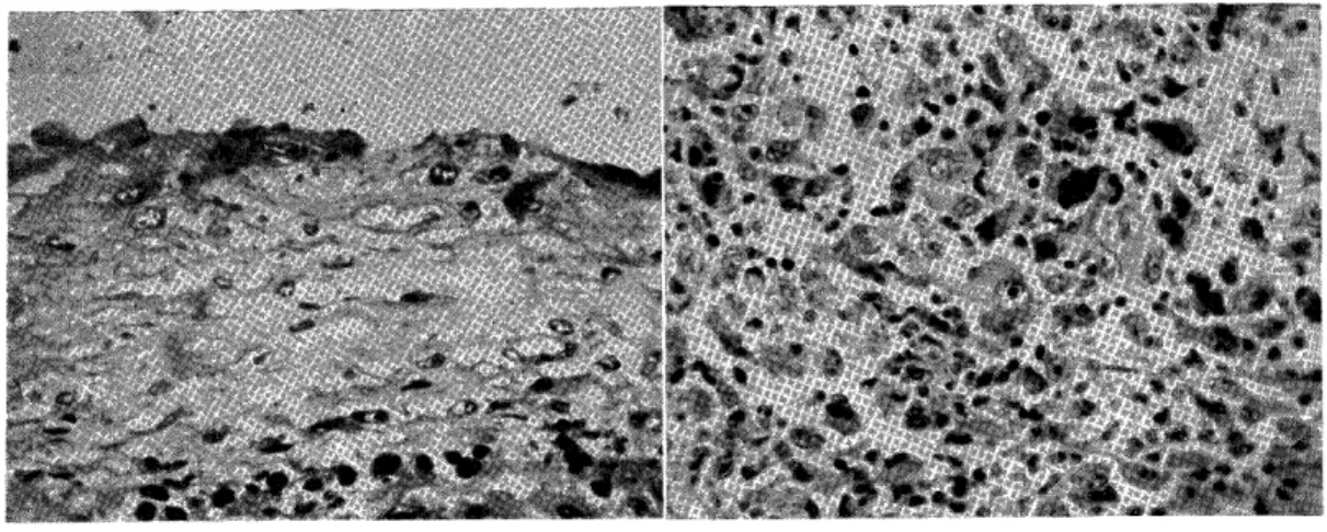
from liver cells in the cysticercus cyst wall (Fig. 1) .
There are many data previously published which showed no or infrequent development of the hepatoma in the cysticercus cysts of the rats liver (BORREL2),HIR`CHFELDI1), HEIM10), BULLOCK and CURTIS3)4)5)6), LIANG13), LARROUSSES12), ARAI1)).
In the present study, a histological observation of the cysticercus cyst spontaneously developed in livers of rats inbred in our laboratory was undertaken with the hope of solving the question as to why the hepatoma did not usually originate in the wall of the cysticercus cyst of liver.
MATERIALS AND METHODS
The cysticercus cysts, 342 in number, which were developed spontaneously in livers of rats of both sexes weighing from 60 to 300 grams were provided as the material for the present study. The cysticercus cysts were found growing in any one or all lobes of the livers and were approximately 0.2 to 1.2 cm in diameter. With the exception of a few cases, all of the cysts contained living Tenia larvae or segmented worms with a small amount of fluid. Cysticercus cysts were taken out with adjacent liver tissues and preserved in 10 percent formalin (Fig. 2.) They were cut into small pieces with or without larval fragments, and imbedded in paraffin. The sections were stained with hematoxyllin and eosin. The argyrophil fibers were stained by
means of modified BIELSHOWSKY' S method .
With the purpose of studying the relation between the larval body and the cysticercus cyst wall during their growth, the following procedures were made in 12 rats; after the removal of the larvae from the incised cysts the intact livers bearing the empty cysts were replaced in the peritoneal cavities and the abdomen were closed.
The treated rats were reared for a period from 12 to 309 days.
Some of them were sacrificed and the cysts having no parasites together with the adjacent parts of the liver were submitted to histological investigations.
OBSERVATIONS
The cys ticercus cysts under study can be divided into following 4
classes according to the degree of proliferation of cellular components
in the course of the developmental stages of cysts; they are 1) cysts
at early stage, 2) cysts at the cell-proliferation stage, 3) thin-walled
cysts, and 4) fibrous and hyaloid cysts. Histological classification of
342 cysticercus cysts is shown in table 1.
1) Cysts at early stage
Two of the cysticercus cysts studied were referred to as cysts at early stage. The incomplete cyst wall consisted of inner, middle and outer layers. In one of the two cysts, the inner layer of the wall
showed thick infiltration of polymorphonucleated and eosinophilic leucocytes together while a few isolated liver cells lying near the surface of the parasite, while in the other slightly compressed liver tissue occured in direct contact with the skin of the parasite. The middle layer of the former cyst wall contained atrophic liver tissue
and scattered polymorphonucleated leucocytes, and the middle layer of the latter was apparently similar to the inner layer in structure. The outer layer of both cysts was made up of some granulation tissue
usually accompanied by dilated capillaries (Fig. 3) . 2) Cysts at the cell-proliferation stage
In this category, a diffuse proliferation of cellular elements of several kinds was observed in the course of the developmental stages of the cyst wall. Following the absorption of the infiltrated leucocytes
in the inner layer of the wall at the beginning of the cell-proliferation stage, liver cells of irregular shape come to touch directly with the surface of parasites. In the inner layer of the cyst wall, stagnated bile pigments were infrequently observed intra- and intercellularly in the compressed liver tissue (Fig.4). As the larvae grow in the cyst.
the liver cells lining the inner layer of the cyst wall may undergo gradual transformation into an elongated or spindle-shaped one, probably due to the direct compression of the larval or worm bodies.
In advanced stages, the entire wall or a localized area of the cyst became thickened with an increase in number of cellular elements through their proliferation. This proliferation may be induced by compression as a result of growth and movement of the parasites and the effects of their chemical agents as suggested by MENDELSOHN, and DUNNING and GURTIS. The outer layer of. the cyst wall showed at this stage an active proliferation of small vessels and capillaries and infiltration of eosinophilic leucocytes, histocytes and lymphocytes.
Although there are some difficulties in the histological classification of the dividing and resting cells of the compressed cyst tissue the following features of proliferation of cells are distinguishable:
(1) Proliferation of liver cells
It is important to find that the disarranged and transformed liver
cells of the inner cyst layer seem to be able to proliferate in some
instances probably through an adequate irritation from the larvae
enclosed in the cyst, but it is difficult to distinguish the transformed
liver cells from connective tissue cells and endothelial cells. Slightly
transformed and disarranged liver cells located in the inner cyst layer
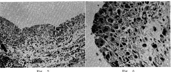
together with endothelial cells of KUPFFER' S type show active proliferation in all the cyst wall in some cases or in particular portions in some other instances (Figs. 5, 6) .
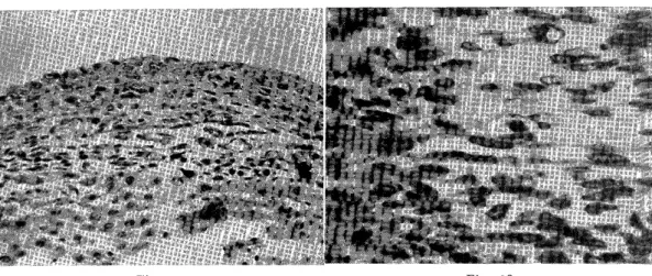
These two types of proliferation were found in the cyst walls at early stage. Disarranged liver cells in inner cyst layer seem to be able to proliferate and to increase their intercellular polymorphs with frequent mitotic figures (Fig. 7) .
Apparently isolated plump liver cells together with a large number of endothelial cells lying in inner and middle layers of some other cyst walls seem to show a greater or less degree of groliferative activity, because there are present dividing cells (Figs-8,9).
It is likely that the changes induced by the proliferation of liver cells as mentioned above seem to represent a sarcomatoid feature.
In some of the cysts containing dead larvae, a number of large polynucleated cells having 5-20 nuclei were observed in same parts of walls surrounding partially organized larval bodies (Fig. 10).
(2) Proliferation of endothelial cells
Usually in the inner cyst layer at relatively later stages of deve- lopment of cysts, there are observable a few areas in which liver cells of spindle shape were found, while a slight proliferation of endothelial cells occurred throughout the walls (Figs.11,12).
Generally, most of the proliferating endothelial cells contained enlarged nuclei of irregular spindle-shape (Fig.13). They possessed a small amount of cytoplasm which was slightly acidophilic and non- granular, and showed intercellular connections through fine fibrillar processes. Argyrophil fibres of alveolar networks found in inner layer of cyst walls corespond to proliferating endothelial cells (Fig.14).
In the outer layer, it is difficult to distinguish endothelial cells from flat fibroblasts.
(3) Hyperplasia of the capillary
In most of the cysts found at relatively advanced stages of develo- pment, there were hyperemic capillaries in the outer layer. Some of the extremely proliferated capillaries in this layer show the general appearance of a hemangioma (Fig.15).
3) Cysts of thin wall
In a total of 342 cysts observed 274 cysts were thin walled showing no progressive proliferation of the cells, 45 showed cell proliferation, 19 were fibrous and hyaloid degeneration and 4 were worm-organized cysts (Table 1).
In almost all the thin walled cysts there were present large and
elongated fusiform liver cells having hyper-chromatic nuclei and rich
cytoplasm lining the inner cyst layer. In the middle part of the cyst
Table 1. Histological Classification of 342 Cysticercus Cysts.
Histologic diagnosis of cysticercus cysts Number of cysts observed
Inflammatory walled cyst at an early stage ... 2
Proliferation of liver cells and Kupffer's cells in certain localized areas of inner layer ... 2
Polymorphous cell sarcoma at an early stage ... 9
Microscopically small, localized polymorphous-cel1-sarcoma at an early stage ... 4
Proliferation of endothelial cells, fibroblasts and isolated liver cells ... 6
Slight proliferation of fibroblasts and endothelial cells ... 10
Rather extensive proliferation of endothelial cells ... 3
Slight proliferation of endothelial cells and polymorphous cells in localized areas ... 1
Proliferation of endothelial cells in localized areas ... 3
Proliferation of spindle shaped liver cells and polymorphous endothelial cells ... 2
Angiomata ... 2
Lipomatous proliferation in localized areas ... 1
Fibroid and hyaloid cyst wall ... 19
Organization of dead larva' ... 4
Thin walled cyst ... 274
Total ... 342
wall, there were oval, stellate or irregulary transformed endothelial cells and fibroblasts. In the outer layer of certain thin walled cysts, there were liver cell-islands or-strands and highly wided capillaries accompanied by a number of large eosinophilic and mononuclear leucocytes (Fig. 16).
4) Fibrous and hyaloid walls
The thin walled cysts consisting mainly of the connective tissue tend at their later stages to shift into the thinner fibrous and partially hyaloid ones. They contain a small amount of cellular elements imbedded in the rich fibrous matrix, and do not show three layers.
There are areas showing hyalinization near the surface of the worms.
In some aged cysts, however, many isolated and intact liver cells are present within the hyaloid wall, while in the fibrous walls there occur groups or strands of liver cells without any sign of proliferation (Fig. 17).
5) Histologic changes of the cyst walls following the removal of larvae.
Except for two cases, in which malignant growth was found, all the treated animals showed no marked cell-proliferation though there occurred edematous and fibrous thickening of the cyst walls.
In the animals in which the parasitic larvae were removed from the
liver, two cases of tumorous growth were found: the animals showed an
accumulation of a moderate amount of tumorous ascites and nodules in their peritoneal cavities. There is a possibility that a malignant change of exfoliated cyst cells, especially lying in the inner layer of the cyst wall, might have already occurred before the operation. General data of the two tumors produced are shown in Table 2.
Table 2. Abdominal Tumors from Cysticercus Cysts.
No. of Larva removed Histological Survivals of
rat rat cyst liver of Abdominal tumor tumor diagnosis in abdomen of the animals treatment after
10 cc of
hemorrhagic
No tumor in tumorous ascites.
8 shrinked adhered to cyst, A number of small Sarcomatoid 45 days
nodules on hepatoma
abdominal wall
diaphragms, mesentery and
omentum
4 cc of hemorrhagic
No tumor in tumorous ascites.
slightly
11 thickened A large number of Mixed tumor 76 days
cyst, adhered nodules on
to omentum omentum,
mesentery and
peritoneal wall
One of the two cysticercus tumors thus formed was recognized as a sarcomatoid solid hepatoma: it is characterized by multinucleated or bizarre shaped giant cells (Fig.18) .
Histological examination of the other tumor revealed that in the sarcoma tissue there were clusters of large epithelial cells, having rich and slightly acidiphilic cytoplasm, which probably originated from disarranged liver cells of the incised cysts. This tumor should be diagnosed as a mixed tumor.
It is premature to conclude based on the above findings that the hepatoma may be produced from the isolated liver cell-islands or-strands lying in the outer layer of the cyst wall following the removal or worms, because of the small number of treated animals and merely two cases of the tumor produced.
DISCUSSION
Detailed observations of the histogenesis of the experimentally
induced cysticercus tumors in rat livers have been carried out by
j3ULLOCK anal CURTIs and CURTIS, DUNNING and ]~ULLOCK.7)8191 Most authors
have reported that the cyst icercus tumors are of mesodermal origin.
According to BULLOCK and CURTIS, all of 1400 cysticercus tumors expe- rimentally induced were mesodermal in origin and malignant except for one benign tumor.
On the other hand YOSHIDA25) reported a case of sarcomatoid cysti- cercus tumor which was histologically regarded as an atypical hepatoma.
BULLOCK and CURTIS5) found two cases of epithelial neoplasms (adenomas) among more than 2100 cysticercus tumors developing in rat livers.
They also described an instance of a spontaneous benign hepatoma in a rat. Further, BULLOCK and CURTIS have described a case of carcino- osteochondro-sarcoma which originated from a cysticercus cyst of the rat liver: the animal bearing this sarcoma had straw-colored fluid in its abdominal cavity. This tumor is very interesting in comparison with two induced cysticercus tumors developed from treated cysts in the present experiment, and with the Watanabe II tumor, an ascites endothelioma of a rat, which was spontaneously transformed from a solid cysticercus tumor in the original host (WATANABE and AZUMA24)).
WATANABE and URANO21I have published a preliminary note on the histology of the cysticercus cyst walls. They indicated that the cysticercus cysts consisted of not only the connective tissue but also transformed liver cells, endothelial cells, liver-cell strands, bile duct epithelium, well-developed capillaries, and matrix fibres of various kinds.
In other words, the cysticercus cyst walls can be designated as extremely compressed liver tissue. Unfortunately BULLOCK and CURTIS have given no detailed descriptions of the deformed and disarranged liver-cells in the inner layer of the cyst. On the basis of their findings, they should regard some tumors as the sarcomatoid hepatoma. They described further a few cases of cysticercus sarcomas which show alveolar structure indicating the general appearance of carcinoma.
There is some question as to whether some of these polymorphous sarcomas originating from the inner layer of cysts of the liver may or may not be atypical large cells of a solid hepatoma as classified recently by OHTA.15)
The frequent occurrence of mitotic figures in disarranged and transformed liver cells lying in the inner cyst layer suggests that the sarcomatoid hepatoma may originate from the transformed liver-cells in the cyst wall, as a result of continuous irritations of larvae enclosed.
A sarcomatoid transformation of hepatomas induced by O-amidoazotoluol during the transplant generations has been reported by SATOMII7) .
There are two possibilities for the recognition of the sarcomatoid
alteration suggested by Watanabe I tumor: the first is that the tumor
tissue showing sarcomatoid features in some parts of the specimen
undergoes a gradual change into small cell soild hepatoma as designated
by OHTA. This was clarified by the re-examination of a large number
of serial sections prepared from the original tumor of the Watanabe
tumor. The second possibility is to be shown by the finding that the histological picture of the typical alveolar hepatoma found in the subcutaneous tumor of the first subcutaneous passage (Fig. 1) . Probably, the histological features of this alveolar structure may be due to the recovery into an epithelial nature of the malignant tumor cells, as well as through the proliferative activity of the malignant liver-cells by means of transplanting them into the subcutaneous tissue.
On the basis of the data from the histological observations of the cysticercus cyst wall and of the solid Watanabe I tumor, it seems very probable that the liver cells may be able to transform their morphology into the seemingly mesenchymal type during their multiplication in order to adapt to new circumstances such as cysticercus walls, and that they may be able to aquire an autonomous character during their continous proliferation, through the change of cellular elements. If the cells may be isolated to proliferate in the body fluid, such as the blood stagnated in the outer layer of the cyst wall, the malignant or premalignant liver-cells tend to lose their own intercellular characteris- tics. This condition was called "ascites tumor bud" by WATANABE20):
it was suggested that the development of the ascites tumor bud might be the result of mutation selected by the body fluid, and that the free-cell population which thus occurred seemed to be the basic condition which may result in the formation of the free celled ascites tumors such as the Watanabe I tumor, the Watanabe II tumor, the ascites endothelioma and the MTK sarcomas of rats reported by MAK1NO, TANAKA14 and others.
Based on the histogenesis of the cysticercus cyst in the present study the occurrence of three layers surrounding the body of the cysti- cercus larvae seems to recall the growth of three layers in infarctor implantnecrosis described by SCHUERMAN 18' and WATANABE19j . Working with the development of the cysticercus cysts of rat livers, BULLOCK and CURTIS3) reported that the liver tissue surrounding the parasites showed necrobiosis and inflammation similar to those in infarct necrosis about a week after the beginning of the growth of the larvae, and two weeks later the cyst wall showed typical cell-proliferation while after three weeks the cell-proliferation was reduced.
Two cases at an initial stage of the cyst development in the present study showed either infiltration of leucocytes or degeneration of the surrounding liver tissue, and the other two cases showed the proliferation. of liver-cells which underwent transformation.
Twenty-seven cases represented a transformation of liver-cells into a malignant condition and proliferation of endothelial cells including fibroblastes, while 13 cases were found to be the polymorphous cell sarcomas at the early stage developing in the middle or inner layers of the cyst wall (Table 2).
It is noteworthy that the cysts in which dead larvae were found,
showed no evidende of progressive proliferation: this fact seems to suggest that the living parasitic larvae may produce an adequate cell- proliferating stimuli which may act as a neoplastic agent upon the cells in the cyst wall in the early cell-proliferation stage.
On account of the supposition that the malignant transformation of both types of cells; i. e., fibroblasts and endothelial cells, may occur
in the cyst wall, the endothelioma and sarcoma are here designated as sarcoma according to BULLOCK and CURTIS31. The sarcomatoid hepatomas might probably be included in the cysticercus sarcomas as po'ymorphous cell sarcoma arising in the cyst wall. As already mentioned, this atypical hepatoma should be separated from the true sarcoma by their histological pictures.
The potentiality of the transformation may be menifested and strengthened by the gradual loss of specificity of their own histological structures during the course of proliferation in order to adapt to new circumstances occurring in the precancerous tissue, such as the cysticercus cyst of the rat liver.
The data from the histological investigations of empty cysts taken at different intervals following the removal of Tenia larvae, indicated that intact` liver cell-islands or -strands lying in the connective tissue of the outer cyst layer provided no evidence of proliferation at all.
This may furnish an important suggestion that the isolation of a cell- group or -groups alone may not be the fundamental factor of acquiring malignancy as emphasized by RIBBERT16' , and that irreversible inter- and intracellular disarrangement and resulting polymorphie, as shown by isolated liver cell clusters found in the mixed tumor which developed from a treated cyst, are expected to be one of causative processes for the development of autonomy.
SUMMARY
1) With the purpose of inquiring into the origin of the Watanabe I tumor, the tat ascites hepatoma, developed from the cysticercus
cyst wall of a rat liver, some histological studies were undertaken
with 342 cysticercus cysts with particular reference to the prolifer- ation and transformation of the cellular elements during the
developmental stages of the wall of the cysts containing Tenia
larvae.
2) In all the cysts under study, the wall consists of different cellular elements: spindle shaped liver-cells are frequently found in the
inner layer of the wall, while endothelial cells, fibroblasts,
capillaries, bile-duct epithelium, liver cell islands are formed supported by the fibre matrix of the wall.
3) Based on the results of the histological study of the premalignant
cysticercus cyst wall, it seems probable that among so-called
cysticercus sarcomas there are some hepatomas originating from the transformed liver-cells in the cysticercus wall.
4) One of two tumors developed in the cyst after the removal of Tenia larvae may be a sarcomatoid hepatoma.
The author wishes to express his thanks to Mrs. AZUMA for her technical assistance.
REFERENCES
1) ARAI, M. . Uber die spontanen Geschwuelste bei weissen Ratten. Gann 43 : 137-142 (1940).
2) BORREL, A. : Tumours cancirenses et helminthes. Bull. de I' Acad. de med.
56 : 141 (1906).
3) BULLOCK, F. D. and CURTIS, M.R. : A study of the reactions of the tissues of the rat's liver to the larvae f Tenia Crassicollis and the histogenesis of cysticercus
sarcoma. J. Cancer Research 8 : 446 (1924).
4) BULLOCK, F.D. and CURTIS, M.R. . Types of Cysticercus Tumors. J. Cancer Research 9 : (1925).
5) BULLOCK, F.D. and CURTIS, M. R. : A cysticercus carcino-osteo-chondrosarcoma
of the rat liver with multiple cysticercus sarcomata. J. Cancer Research. 12 : 32E (1923).
6) BULLOCK, F.D. and CURTIS, M. R. : Spontaneus tumor of the rat. J. Cancer Research. 14 : 1-115 (1930).
7) CURTIS, M. R., DUNNING, W. F. and BULLOCK, F. D. Genetic factors in relation to the ethiology of malignant tumor. Am. J. Cancer. 17 : 894 (1933).
8) CURTIS, M.R., DUNNING, W. F. and BULLOCK, F.D. : Is malignancy due to a process analogous to somatic mutation? Science u. s. 77 : 175 (1933).
9) DUNNING W. F. and CURTIS, M. R. : Attempt to isolate the active agent in cysticercus fasciolaris. Cancer Research. 13 : 838 (1953).
10) HEIM, Fr. : Sarkombildung bei der Ratte durch Cysticercus fasciolaris. Z.
Krebsforsch 26 : 418 (1928).
11) HIRSCHFELD, H. : Cysticercus fasciolaris als Erreger eines Angiosarkoms bei einer Ratte. Z. Krebsforsch. 16 : 95 (1919).
12) L2 RROUSSE, F. : Bemerkungen zur Bildung eines Cysticercussarkoms in der Leber der Ratte. Z. Krebsforsch. (Ref.) 38 : 5 (1933).
13) LIANG, B. : Beitrag zum Lebersarkom bei Ratten durch cysticercus fasciolaris.
Z. Krebsforsch. 33 : 654 (1931).
14) MAKINO,S. and TANAKA, T. . Chromosome feature in the pregenerating rat liver following partial extripation. Texas report on biology and Medicine. 11
588-592 (1953).
15) OHTA, K. and MATUMOTO, S. : Collision of multicentric cancerous foci agatinsi each other in experimental rat liver cancer.Gann 45 : 581-589 (1954).
16) RIBBERT,O. : Uber die Histogenese and das Wachstum des Carcinomas. Archiv fur Pathologischen Anatomie u. Physiology. (1895).
17) SATOMI, M. : Uber die struktur des Leberzellekrebs mit besonderer Beriicksi- chtigung der Stromafrage. Gann 35 : 270 (1941).
18) SCHURMAN, P. : Uber die Entstehung der Infarkt-nekrose. Verh der dent, path.
Gesellschaft. 29 : Tagung (1937).
19) WATAN!?BE, F. : Study on the infarctnecrosis (1 report). Nagasake Medical Journal 23 : No. 1, 16-20 (1948) (Japanese).
20) WATANL BE, F. : Study on the premalignant cells (1 report). 'Proliferative
behaviour of the liver cells in the cysticercus cyst walls. Acta Pathologica.
Japonica. 6: No. 4 (1956),
21) WATAN'XBE,F. and UR'NO,K. : Histological studies on walls of cysticercus cysts of rat livers. Transactiones Societatis Pathologicae Japonicae. 43 : 324 (1954)
(Japanese).
22) WA TANA.BE,F. and MFTSUNAG'k, T. : A hepatoma of rats induced by repeated injection of hot water with its experimental transformation into the ascites tumor.
Gann 45 :. 443-445 (1954) (Japanese).
23) WATANABE, F. and TONOMURA, A.: On the Watanabe ascites hepatoma, a new ascites tumor of rats, produced after the application of hot water. Gahn 47 15-21 (1956).
24) WATAN2.BE,F. and AZUMA,M. A: new ascites tumor showing fragments with histoid structure in the ascites fluid. Gann 48 : 547-549 (1957) (Japanese).
25) YOSHID., T. . Uber die experimentelle Erzeugung der Geschwulst durch subcutane Injection der oliven 011osung des O-Amidoazotoluol. Gann 28 : No. 4,
448 (1934) (Japanese).
EXPLANATION OF PIATES
Fig. 1. Alveo'ar structure presenting the typical hepatoma found in subcutaneous tumor in first passage of Watanabe ascites hepatoma.
Fig. 2. Two cysticercus cysts of a rat liver, through the walls of which larvae can been seen.
Fig. 3. Early cyst wall showing wided capillaries and cell infiltration in granulation tissue as well as almost intact liver tissue touched to the surface of the
larva suggesting presence of liver cells in inner zone at later stages of cyst
formation. Left line presenting larval skin.
Fig. 4. Early cyst showing inter- and intra-cellular staguation of bile pigments in compressed liver tissue of the inner zone of a cyst.
Fig. 5. Diffuse proliferation of slightly transformed liver cells in inner zone of a cyst wall.
Fig. 6. Proliferation of the polymorphously transformed liver cells at localized area in inner zone of the wall.
Fig. 7. Localized proliferation of morphologically changed liver cells in inner zone and isolated liver cells in middle zone of the walls.
Fig. 8. Isolated large plump liver cells scattered with endothelial cells in middle and inner zones showing possible proliferative activities.
Fig. 9. A mitotic figure of a liver cell in inner zone of a cyst wall.
Fig. 10. Polynucleated giant cells in the wall of a cyst containing dead larva.
Fig. 11. Elongated fusiform liver cells lining the inside of the inner zone of thin- walled cyst and endothelial cells in outer zone. Lower part showing liver tissue.
Fig. 12. Endothelial cells of inner zone of a cyst wall shown by high magnification.
In middle part a capillary is found.
Fig. 13. Increased polymorphie of the endothelial cells in inner zone of a wall.
Fig. 14. Argyrophil fibers of inner zone showing alveolar network as well as those of outer zone presenting stratiform corresponding to difference of cellular morphology in each zone of the walls.
Fig. 15. Wided hyperemic capillaries in outer zone of the wall.
Fig. 16. Liver cell-island showing no proliferation found in outer zone of the cyst wall.
Fig. 17. Many evident liver cells scattered in hyaloid matrix found in the old cyst wall.
Fig. 18. Apparent sarcomatoid hepatoma originated in the exfoliated cells from the treated cysts consisting of epithelial cells, multinucleated and bizarre shaped
giant cells mixing with endothelial cells. A tumor (1.2cm x l .Ocm x l .Ocm)
of peritoneal metastatic nodules accompanied with hemorrhagic ascitic fluid
10 cc containing a large number free tumor cells. A tumor-bearing rat died
in 76 days following operation.
Fig. 1 Fig. 2
Fig. 3 Fig. 4
Fig. 5 Fig. 6
Fig. 7 Fig. 8
Fig. 9 Fig. 10
Fig. 11 Fig. 12
Fig. 13 Fig. 14
Fig. 15 Fig. 16
Fig. 17 Fig. 18