Introduction
Preeclampsia is a progressive, multisystem disorder unique to pregnant women and is a leading cause of maternal death and con- tributes significantly to premature deliveries. It is characterized by hypertension and proteinuria, and its incidence is influenced by various factors, such as parity, race, and environmental factors. It is estimated to be about 5~10% in European countries and at 5.8%
in first pregnancies and 0.4% in second pregnancies.1The etiology of preeclampsia is still unknown, but genetic factors have been im- plicated since the syndrome shows a familial tendency.2Published reports of pedigree analysis suggest that development of preeclampsia may be based on a single recessive gene or dominant gene with in- complete penetrance.3 However, more recent studies have sug- gested that the pattern of inheritance is multifactorial and depends on several genetic loci with greater or smaller contributions from environmental factors. In addition, not only maternal gene but also
fetal gene may be implicated, and maternal-fetal interaction could not be ignored.
It is unlikely that a particular genotype is involved in the develop- ment of preeclampsia. Rather, many loci confer genetic liability that predisposes individuals to the disease. During the last decade, a growing number of genetic variants have been implicated in the development of preeclampsia. To date, reported preeclampsia- related genes are classified in following categolies,
Renin-Angiotensin system association genes
(AGT, Angiotensin Ⅱ Type 1 Receptor (AGTR1), AGTR1 Agonistic Antibodies, AngiotensinⅡType 2 Receptor),
Endothelial Nitric Oxide Synthase (eNOS),
Coagulopathy and Vascular injury genes (MTHFR, FactorⅤ Leiden, Prothrombin),
Oxidative Stress Candidate Genes (Lipoprotein Lipase, Apolipoprotein E), and
Immunoregulatory Candidate Genes (HLA, TNF- .
Address correspondence:Atsushi Yoshida, M.D., Department of Obstetrics and Gynecology, Nagasaki University School of Medicine, 1-7-1 Sakamoto Nagasaki 852-8501 JAPAN
TEL: +81-(0)95-819-7363, FAX: +81-(0)95-819-7365, E-mail: [email protected] Received June 13, 2008; Accepted October 14, 2008
MS#AMN 07029
Correlation Between Preeclampsia and Prevalence of Polymorphism of Angiotensinogen, Methyleneteterahydrofolate Reductase and FactorⅤ, Prothrombin Genes Among Japanese Women
Atsushi YOSHIDA, Kiyonori MIURA, Daisuke NAKAYAMA, Hideaki MASUZAKI
Department of Obstetrics and Gynecology, Nagasaki University School of Medicine, Nagasaki, Japan
OBJJECTIVE: To determine the genotypes of four candidate genes in Japanese women with a history of preeclampsia, and in a control group of parous woman.
STUDY DESIGN: Fifty-two pregnant women with a history of preeclampsia in their first pregnancy and 113 normotensive gravid women were studied. All subjects were Japanese women with singleton gestations. Genomic DNA was extracted, and genotypes of angiotensinogen (AGT), methyleneteterahydofolate reductase (MTHFR), factorⅤLeiden, and prothrombin genes were analyzed.
RESULTS: The frequencies of homozygous AGT gene mutation and homozygous MTHFR gene mutation in preeclampsia were significantly higher than that in control. The calculated risk associated with the presence of both mutations did not exceed the risk with polymorphism of each gene. None of the examined cases showed polymorphism of factorⅤLeiden and prothrombin G20210A genes.
CONCLUSION: In Japanese patients with preeclampsia, the angiotensinogen gene and particularly MTHFR gene may play a role in the patho- genesis of preeclampsia
ACTA MEDICA NAGASAKIENSIA 53: 37−41, 2008
Keywords:Preeclampsia; Polymorphism; Angiotensinogen; Methlenetetrahydrofolate; FactorⅤ; Prothrombin
Among them, mutations of angiotensinogen (AGT), methlenetetrahydrofolate reductase (MTHFR), Factor Ⅴ Leiden, and prothrombin genes seem to be the most likely candidate in Japanese although no mu- tation in single gene can correctly predict the disease. The detec- tion rate of AGTand MTHFR gene polymorphisms are high in Japanese, and relationship between preeclampsia and DNA poly- morphism in these two genes were also reported. And, in European countries, strong relationship between preeclampsia and Factor
ⅤLeiden, prothrombin genes polymorphisms were reported.
In the present study, we determined the composite genetic risk for preeclampsia by simultaneous genotyping of these loci.
Material And Methods
Patients participating in this study were recruited from the ob- stetric population followed at Nagasaki University Hospital be- tween December 1997 and March 2000. Informed consent was ob- tained from all patients, and the ethics committee of our university approved the study protocol. Fifty-two pregnant women with preeclampsia in their first pregnancy and 113 normotensive gravid women were recruited for this retrospective study. All subjects were Japanese women with singleton gestations. According to the working group of the National High Blood Pressure Education Program (NHBPEP 2000), preeclampsia is defined by the de novo appearance of hypertension after 20weeks of gestation, systolic blood pressure ≧140 mmHg or diastolic blood pressure ≧90 mmHg, accompanied by new-onset proteinuria, defined as≧300 mg per 24 h. Genomic DNA was extracted from peripheral blood leukocytes by standard procedures (Quiagen DNA extract kit), and the genotype was determined by a PCR restriction fragment length polymorphism method (RFLP) as described below.
Angiotensinogen M235T polymorphism
A 165-base-pair DNA fragment of the angiotensinogen gene was amplified by polymerase chain reaction (PCR) using the sense primer 5' CAG GGT GCT CAC ACT GGA CCC C-3' and antisense primer 5' CCG TTT GTG CAG GGC CTG GCT CTC T- 3', according to the method described by Kamitani et al.4 After endonuculease digestion with Tth111-I, PCR fragments were electrophoresed in 10% polyacrylamide gels and stained with ethidium bromide.
MTHFR C677T polymorphism
A 189-base-pair DNA fragment of theMTHFRgene was ampli- fied by the PCR using the sense primer 5' TGA AGG AGA AGG TGT CTG CGG GA-3' and antisense primer 5' AGG ACG GTG CGG TGA GAG TG-3', according to the method described by Morita et al.5After endonuclease digestion withHinfI, PCR frag- ments were electrophorased in 10% polyacrylamide gels and stained with ethidium bromide.
FactorⅤgene analysis
A 287-base-pair DNA fragment of FactorⅤgene was amplified by the PCR using the sense primer 5' GGA ACA ACA CCA TGA TCA CAG CA-3' and antisense primer 5' TAG CCA GGA GAC CTA ACA TGT TC-3', according to the method described by Zoller et al.6 After endonuclease digestion with MnlI, PCR frag- ments were electrophorased in 8% polyacrylamide gels and stained with ethidium bromide.
Prothrombin G20210A polymorphism
A 640-base-pair DNA fragment of the prothrombin gene was amplified by PCR using the sense primer 5' CGG TGT GTG TGT AGG AAC TCC-3' and antisense primer 5' CAA TGT CAG ATG CTG GGG ACT-3', according to the method described by Poort et al.7 After endonuclease digestion with Alw 44I, PCR fragments were electrophoresed in 1% agarose gels and stained with ethidium bromide.
Statistical analysis
Differences between groups were examined for statistical sig- nificance using the Fisher's exact test or chi-square test. Odds ratio and 95 percent confidence intervals (95%CI) were calculated. Ap value less than 0.05 denoted the presence of a statistically signifi- cant difference.
RESULTS
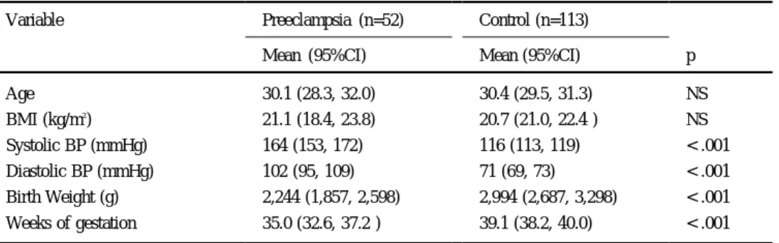
We studied 52 pregnant women with preeclampsia in their first pregnancy and 113 normotensive gravid women (Table 1).
Angiotensinogen
Table 2 shows the M235T genotyping results. M235T homozygosity (genotype TT) was detected in 33 of 52 (63.5%) patients with preeclampsia and 51 of 113 (45.1%) control subjects. The fre- quency of each genotype in the control group was consistent with those reported previously by other investigators.4,8,9 Compared with the other genotypes (TM and MM), the frequency of aa was significantly higher in preeclampsia than in controls (p<0.05). The observed genotype frequencies were not different from those pre- dicted from Hardy-Weinberg equilibrium.
MTHFR
Table 3 shows the results of C677T genotyping.
C677T homozygosity (TT) was identified 18 of 52 (34.6%) in con- trol subjects. The frequency of each genotype in the control was consistent with those reported previously by other investigators.5,10 Compared with the other genotypes (CT and CC), the frequency of
TT was significantly higher in preeclampsia than in control sub- jects (p<0.01). The observed genotype frequencies were not differ- ent from those predicted from Hardy-Weinberg equilibrium.
Analysis of the effect of modification was performed to evaluate the estimates of risk associated withAGTgene mutation (genotype aa) andMTHFRgene mutation (genotype aa), and their combina- tion (Table 4). The frequency of double homozygous (TT/TT) in the Preeclampsia is 13.4% (8/52), and 3.5% (4/113) in control. The calculated risk associated with the presence of both mutations (odds ratio 4.79, 95%CI: 1.66-13.8) did not exceed the risk of MTHFRsingle gene mutation (odds ratio 4.91, 95%CI: 2.11-11.4).
FactorⅤand prothrombin gene
Previous studies have shown thatfactorⅤLeidenmutation and prothrombin G20210A mutation are very rare in Asia.11 In our population sample, no Leiden or prothrombin G20210A mutations were detected in all 165 women.
Discussion
Preeclampsia is one of the most serious complications during pregnancy and a syndrome that affects virtually all-maternal organ systems. The condition is still the leading cause of maternal and in- fant morbidity and mortality.12Several studies have examined the etiology and pathophisiology of preeclampsia, but the exact mecha- nism remains unknown. Furthermore, several studies reported the possible association between genetic factors and preeclampsia.13,14 In the present study, we investigated the genotypes of four can- didate genes, AGT, MTHFR, factor ⅤLeiden, and prothrombin gene in the same patients with history of preeclampsia. Our results demonstrated that pregnagnt woman with both homozygous for AGTgene mutations and MTHFRgene mutations have a higher risk for preeclampsia comparing to the controls with other geno- types.
Jeunemaitre et al.15were the first to report the genetic associa- tion between the AGTgene and hypertension. Ward et al.8 pro- posed that preeclampsia could be viewed as a pregnancy-induced proteinuric hypertension, and thus the condition could be associ- ated with AGTgene mutation. They were able to demonstrate a significant association between preeclampsia and a molecular vari- ant ofAGT, M235T. Based on this finding they suggested that in- creased concentration of angiotensinogen in individuals carrying variants of AGT, such as M235T, might be associated with in- creased production of angiotensinogen Ⅱ. They also argued that chronic stimulation in M235T carriers could increase vascular tone and promote vascular hypertrophy, consequently causing preeclampsia during pregnancy. In Japanese, the frequency of M235T is higher than in Caucasians, but as in Caucasians, several reports confirmed the association between M235T mutation and preeclampsia,8,15but Table 1.Clinical Characteristcs
Variable Preeclampsia (n=52) Control (n=113)
Mean (95%CI) Mean (95%CI) p
Age BMI (kg/m2) Systolic BP (mmHg) Diastolic BP (mmHg) Birth Weight (g) Weeks of gestation
30.1 (28.3, 32.0) 21.1 (18.4, 23.8) 164 (153, 172) 102 (95, 109) 2,244 (1,857, 2,598) 35.0 (32.6, 37.2 )
30.4 (29.5, 31.3) 20.7 (21.0, 22.4 ) 116 (113, 119) 71 (69, 73) 2,994 (2,687, 3,298) 39.1 (38.2, 40.0)
NS NS
< .001
< .001
< .001
< .001 BMI: Body mass index
CI : confidence interval; BP : blood pressure, NS : not significant
Table 2.AGT genotype distribution in patients with pre-eclampsia and control subjects
MM (%) MT (%) TT (%)
PE (n=52) Controls (n=113)
4 (7.7) 11 (9.7)
15 (28.9) 51 (45.1)
33 (63.5) 51 (45.1) PE : preeclampsia
Table 3.MTHFR genotype distribution in patients with pre-eclampsia and control subjects.
CC (%) CT (%) TT (%)
PE (n=52) Controls (n=113)
17 (32.7) 48 (42.4)
17 (32.7) 54 (47.8)
18 (34.6) 11 (9.7) PE : Preeclampsia
Table 4.Odds ratios of preeclampsia in the presence ofAGT, MTHFRand combinedAGT + MTHFRgene polymorphisms.
Odds ratio 95%CI AGT homozygous mutation (TT) (n=33)
MTHFR homozygous mutation (TT) (n=18) Both homozygous mutaiton (TT/TT) (n=8)
2.11 4.91 4.79
1.08-4.15 2.11-11.4 1.66-13.8
others could not confirm these findings.16,17
Hyperhomocysteinemia has been identified as an important risk factor for occlusive vascular disease,18and some reports suggested a relationship between repeated fetal loss or abruptio placentae and hyperhomocysteinemia.19 MTHFRis one of the key enzymes es- sential for normal homocystein metabolism, and abnormalities in this enzyme can lead to hyperhomocysteinemia. Frosst et al.20were the first group to identify a common mutation in MTHFR gene (C677T) in hyperhomocysteinemia, and significantly higher con- centrations of plasma homocystein in individuals homozygous for this mutation. Based on these findings, they suggested that this mu- tation is an important genetic factor in vascular disease. Other groups later supported these results.4Vascular damage consisting of thrombosis of small uterine arteries may be involved in the pathophisiology of preeclampsia.21,22Souda et al.9reported the as- sociation between preeclampsia and MTHFR gene mutation, C677T. However, other investigators could not confirm such rela- tionship.23-27
Our results showed a significant correlation between mutations of these two genes and preeclampsia, and that the presence of MTHFR gene mutation has a strong impact on the incidence of preeclampsia. Our analysis also demonstrated that AGT and MTHFR gene mutations play a role in the pathogenesis of preeclampsia. Our results indicate that combination analysis of ge- netic mutations is potentially clinically useful for genetic screening of patients at risk for the development of preeclampsia. However, in our combination study we could not find additive effects both mutations; i.e., the calculated risk associated with the presence of both mutations was not higher than that ofMTHFRmutations only.
The reason for the lack of the additive effects may be simply due to the small sample size. To examine this issue, we are currently attempting to analyze more patients for mutations of these genes.
It is probable that a group of genes could accurately predict the risk of preeclampsia, while individual genes may have only a limited predictive power. Therefore, combination analysis of only two genes may not establish significant predictive power. However, available data suggest that a very common allele is necessary to explain the observed inheritance of preeclampsia. Thus, we believe in the existence of a few "major loci", and many "minor loci", and postulate thatMTHFRgene may be at the front andAGTgene at the back in such a scheme.
Our results showed the lack of mutations of factor ⅤLeiden and prothrombin genes in our 165 patients with preeclampsia.
These results are different from those of previous studies.28 In Caucasian, several reports suggested the presence of a significant correlation between mutation of factor Ⅴ Leiden gene and preeclampsia.29,30However, the majority of these studies were from European countries and to our knowledge, no such correlation has been previously reported in Asian-based studies. In this regard, Rees et al. reported the world distribution offactorⅤLeidenmu- tation, and demonstrated confinement offactorⅤLeidenmutation to European countries with the exception of two cases.11 These genodemographic findings are in agreement with our results.
In conclusion, based on the results of the present study, we speculate that AGT and MTHFR are likely to be implicated in preeclampsia in Japanese women. We suggest that preeclampsia is a heterogeneous disease. To identify the underlying genetic factors associated with preeclampsia, one has to consider the variability in the incidence of preeclampsia among different countries and races, and that such variability probably reflects different prevalence of mutations of candidate genes.
References
1. Roberts JM, Cooper DW (2001) Pathogenesis and genetics of pre-eclampsia.
Lancet357: 53-56, 2001
2. Chesley L. Annito J. Cosgrove R. The familial factor in toxemia of pregnancy.
Obstet Gynecol32: 303-311, 1968
3. Chesley LC, Cooper DW. Genetics of hypertension in pregnancy: possible single gene control of pre-eclampsia and eclampsia in the descendants of eclamptic women.Br J Obstet Gynaecol93: 898-908, 1986
4. Kamitani A. Rakugi H, Higaki J et al. Association analysis of polymorphism of the angiotensinogen gene with essential hypertension in Japanese.J Hum Hypertens8:
521-524, 1994
5. Morita H, Taguchi J, Kurihara H et al. Genetic polymorphism of 5,10- methylenetetrahydrofolate reductase (MTHFR) as a risk factor for coronary artery disease.Circulation95: 2032-2036, 1997
6. Zoller B, Svensson P, He X, Dahlback B. Identification of the same factor 5 gene mutation in 47 out of 50 thrombosis-prone families with inherited resistance to ac- tivated protein C.J Clinical Invest94: 2521-2524, 1994
7. Poort S, Michiels J, Reitsma P, Bertina R. Homozygosity for a novel missense mu- tation in the prothrombin gene causing a severe bleeding disorder. Thromb Haemost72: 819-824, 1994
8. Ward K, Hata A, Jeunemaitre X, Helin C et al. A molecular variant of angiotensinogen associated with preeclampsia.Nature Genet4: 59-61, 1993 9. Hata A, Namikawa C, Sasaki M, Sato K et al. Angiotensinogen as a risk factor for
essential hypertension in Japan.J Clin Invest93: 1285-1287, 1994
10. Souda S, Arinami T, Hamada H, Yamada N, Hamaguchi H, Kubo T.
Methylenetetrahydrofolate reductase polymorphism and pre-eclampsia. J Med Genet34: 525-526, 1997
11. Rees D, Cox M, Clegg J. World distribution of factorⅤLeiden.Lancet346:
1133-1134, 1995
12. Roberts J, Redman C. Pre-eclampsia: more than pregnancy-induced hypertension.
Lancet341: 1447-1451, 1993
13. Klipatrick D, Liston W, Gibson F, Livinstone J. Association between susceptibility to pre-eclampsia within families and HLA DR4.Lancet2: 1063-1065, 1989 14. Liston W, Klipatrick D. Is genetic susceptibility to pre-eclampsia conferred by
homozygosity for the same single recessive gene in mother and fetus?Br J Obstet Gynecol98: 1079-1086, 1991
15. Jeunemaitre X, Soubrier F, Kotelevtsev Y et al. Morecular basis of human hyper- tension: role of angiotensinogen.Cell71: 7-20, 1992
16. Kobashi G, Hata A, Fujimoto S. Association of pregnancy-induced hypertension with a molecular variant of angiotensinogen gene.Nippon Sanka Fujinka Zassi47:
497-498, 1995
17. Morgan L. Preeclampsia and the angiotensinogen gene.Br J Obstet Gynecol70:
649-657, 1995
18. Clarke R, Daly L, Robinson K et al. Hyperhomocysteinemia: -26An independent risk factor for vascular disease.N England J Med324: 1149-1155, 1991 19. Kupferminc MJ, Baram A, Jaffa AJ, Wolman I, Shenhav M, Leesing JB. Increased
frequency of genetic thrombophilia in women with pre-eclampsia.N Engl J Med 340: 9-13, 1999
20. Frosst P, Blom HJ, Millos R et al. A candidate risk factor for vascular disease: a common mutation in methylenetetrahydrofolate reductase.Nature Genet10: 111- 113, 1995
21. Ballegeer V, Spitz P, De Baene L, Van Assche A, Hidajat M, Criel A. Platelet ac- tivation and vascular damage in gestational hypertension.Am J Obstet Gynecol 166: 629-633, 1992
22. Haller H, Hempel A, Homuth V et al. Endometrial-cell permeability and protein kinese C in pre-eclampsia.Lancet351: 954-959, 1998
23. Kobashi G, Yamada H, Asano T et al. Absence of association between a common mutation in the methyleneteterahydrofolate reductase gene and preeclampsia in Japanese women.Am J Med Genet93(2): 122-125, 2000
24. Stonek F, Hafner E, Philipp K et al. Methylenetetrahydrofolate reductase C677Y polymorphism and pregnancy complications.Obstet Gynecol110: 363-368, 2007 25. Jarvenpaa J, Pakkila M, Savolainen E.-R, Perheentupa A, Jarvela I, Ryynanen M.
Evaluation of FactorⅤLeiden, Prothrombin and Methylenetetrahydrofolate reductase gene mutations in patients with severe pregnancy induced complications in northern Finland.Gynecol Obstet Invest62: 28-32, 2006
26. Prasmusinto D, Skrablin S, Fimmers R, van der Ven K. Ethnic differences in the association of factorⅤLeiden mutation and the C677T methylenetetrahydrofolate reductase gene polymorphism with preeclampsia.Eur J Obstet Gynecol Reprod Biol112: 162-169, 2004
27. Zahed L, Rayes R, Mahfouz R et al. Prevalence of factorⅤLeiden, prothorombin
and methylene tetrahydrofolate reductase mutations in women with adverse out- comes in Levanon.Am J Obstet Gynecol195: 1114-1118, 2006
28. Dizon-Townson D, Nelson L, Easton K, Ward K. The factorⅤLeiden mutation may predispose women to severe preeclampsia.Am J Obstet Gynecol175: 902- 905, 1996
29. Suzan R Kahn. Severe preeclampsia associated with coinheritance of factorⅤ Leiden mutation and protein S deficiency.Obstet Gynecol91: 812-814, 1998 30. Grandone E, Margaglione M, Colarizzo D et al. FactorⅤLeiden, C>T MTHFR
polymorphism and genetic susceptibility to preeclampsia.Thromb Haemost77:
1052-1054, 1997