Acta Med. Nagasaki 26 : 73-81
Studies on the Effects of Growth Hormone on the Retinal Vascular Elements of Streptozotocin
Induced Diabetic Rats
Naoyuki TSUDA and Isao TAKAKU Department of Ophthalmology
Nagasaki University School of. Medicine Nagasaki, Japan
Received for publication, May 1, 1981
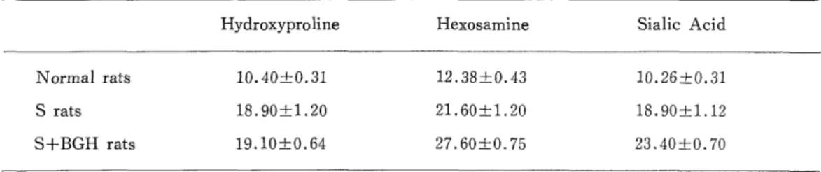
The morphologically characteristic changes were irregular thickening of the base- ment membrane in the streptozotocin induced diabetic group (S rats) and more striking irregular hypertrophy of the same membrane in the group further administered with growth hormone (S+BGH rats). In the outer basement membrane surrounding the pe- ricytes of S rats and S+BGH rats retinal capillary, and deposit of elastine could be sug- gested, which were seen as electron lucid, were proved by tannic acid stainning to be substances with high electron density. Biochemical assay disclosed that the characteristic change was a marked increase in hydroxyproline in the group with diabetes and an in- crease in hexosamine and also in sialic acid in the group further administrated with growth hormone.
In view of the above finding, it was considered that substances of the collagen group are involved in the thickening of the basement membrane in diabetes, and that the loading with growth hormone is influential on glycoproteins deposition.
INTRODUCTION
It was described in one of our previous papers") that diabetic retinopathy, one of the serious complications in diabetes mellitus, was improved by pituitary ablation. How- ever, there remain not a few aspects yet to be elucidated in the mechanism of the ef- fect of the surgery, and moreover little is known about the effects of growth hormone on the retinal capillaries in diabetes mellitus. For the purpose of elucidating the nature of diabetic retinal angiopathy and to reveal the effects of growth hormone, we have re- cently made morphological and biochemical studies of pathological and biochemical stu- dies of pathological changes in the retinal blood vessels of rats with the streptozotocin- induced diabetes, and obtained some interesting findings.
津 田 尚幸,高 久 功
MATERIALS AND METHODS
Female Wister rats, weighing 180-210 g, were injected intravenously through the caudal vein with 65 mg/kg of streptozotocin (The Upjohn Co., U. S. A.) dissolved in 0.3 ml of a citrate buffer (pH 4.5).
One injection was sufficient to induce diabetes. The animals were weighed, and their blood sugar levels were measured every 3 weeks after the injection and their urine sugar levels every 10 days. A simplified blood sugar measuring equipment "The Ames Reflectance Meter" (Ames Co., U. S. A.) (glucose-oxidase method) was used for the measurement of blood sugar levels, and the Tes-Tape (Eli Lilly and Co., U. S. A .) for the test of urine sugar. The rats with blood sugar levels of not less than 150 mg/
dl and with persistent positive urine sugar 10 days after the injection were classified as diabetic (S) , and these rats were breeded for the following 6 months. The group treated with growth hormone (S+BGH) consisted of the S rats that had been reared for six months, then injected intraperitoneally with 3 mg of BGH (Miles Labs. , U. S. A.) (activity: 0.8-1.0 U. S. P. units/mg) daily for 10 days, and observed for one month prior to their use for experimental purposes.
1. For the preparation of electron-microscopic speciments, the eyeballs were enucleated from the animals under ether anesthesia while 3 % glutaraldehyde-phosphate buffer (pH 7.3) solution was being applied dropwise ; each eyeball was immediately fixed in the same 3% glutaraldehyde solution for 3 hours ; its retinal tissue was cut into sec- tions, fixed in 1% osmium tetraoxide for 2 hours, dehydrated with ethanol, and ern- bedded with the Epon 815 resin. Part of the embedded material was stained with 0.1
toluidine blue, and, after the presence of small blood vessels in a histochemical
specimen was confirmed, ultrathin sections were prepared with the Porter-Blum model MT1 ultramicrotome, double stained with uranium acetate and lead citrate, and ex- amined under the JEM model 7 electron microscope. Tannic acid staining') was per-
formed to located that part of the basement membrane which was less electron dense.
2. For the biochemical study, several units of 30 eyeballs each of the rats breeded by the aforementioned method were assigned to the respective experimental groups. After fixation in 10% formaldehyde solution, the retina was separated and immersed in 3%
trypsin solution (by Kuwabara-Cogan's method")) for 8 hours to obtain the retinal blood vessels. The retinal blood vessels thus obtained were homogenized in a Teflon homogenizer, washed and dehydrated with anhydrous acetone, and dried over P205 in
a vacuum desiccator, and dry weight was measured. Part of the sample thus prepared was assayed for hydroxyproline, hexosamine and sialic acid.
1) For the assay of hydroxyproline, part of the dried sample was sealed in a test
tube together with 1 ml of 6 N hydrochloric acid, and allowed to be hydrolyzed 100°C
for 16 hours, after the hydrochloric acid was removed, the hydrolysate was made up to
a given volume with 2 ml of distilled water, and assayed by Neuman-Logan's method
15)
2) For the assay of hexosamine, part of the dried sample was added with 1 ml of 6 N hydrochloric acid. The mixture was sealed in a test tube and hydrolyzed at 100
°C for 6 hours . After removal of the hydrochloric acid, the hydrolysate was made up to a given volume with 2 ml of distilled water, and assayed for hexosamine by Rondle- Morgan's method17>.
3) For the assay of sialic acid, part of the dried sample was digested with pa- pain at 65° for 1 hour. The digest was added to 1 ml of 0.1 N sulfuric acid, and hy- drolyzed at 80°C for 1 hour. The hydrolysate was centrifuged and the supernatant was assayed for slialic acid by the thiobarbituric acid method18).
RESULTS
1 Biological behavior of rats
1) Body weights and blood sugar levels of BGH-treated rats
While normal rats weighed 192±16 g (mean±S. D.) and had the blood sugar levels of 89±9 mg/dl, the animals one month after daily treatment with 3 mg of BGH for 10 days weighed 230±11 g and had the blood sugar levels of 114±13 mg/dl.
Thus, the latter presented increased body weights and increased blood sugar levels, though the urine sugar remained negative. These findings proved BGH to be diabe-
togenic.
2) Body weights and blood sugar levels of S-rats.
The S-rats that had been reared for 6 months prior to BGH administration weighed 207±31.5 g and had the blood sugar levels of 531±39.6 mg/dl.
3) Body weights and blood sugar levels of S+BGH rats.
The above-mentioned S rats were administered daily with 3 mg of BGH for 10 days ; and their body weights and blood sugar levels were measured, 15 days and
30 days after the end of the administrations. The animals weighed 227±17 g, and
had the blood sugar levels of 452±26 mg/dl at termination of the administration, 246
±17 g and 527±46 mg/dl 15 days after the administrations, and 259±24 g and 32 mg/
dl 30 days after the administrations.
The maximum blood pressure measured with the automatic blood pressure re- corder model USM-105 (Ueda Electronic Works) at the end of each experiment was
102±14 mmHg remaining within normal range.
2. Electron-microscopical findings (Figs. 1-5)
Electron-microscopical observation was made for retinal capillaries of which the ba- sement membrane surrounding the endothelium was in direct contact at least in part
with the surrounding glia tissue. The capillary plexuses of the retina in the posterior
pole were divided into 2 layers ; the plexuses in the nerve fiber layer and the ganglion
cell layer were defined as the superficial layer ones, and those in the inner nuclear
layer to the outer plexiform layer as the deep layer ones.
1) Retinal capillaries of normal rats (Fig. 1)
There was no morphologic difference between the superficial and deep layer capillaries. The retinal capillary lumen consisted of a thin monolaminar endothelium without fenestration. The endothelial cells were tightly interconnected. Their nuclei were shaped irregularly and their cytoplasm contained a few mitochondria and poly-
somes and a relatively large number of pinocytotic vesicles in contact with the surface of the internal cavity. The outer basement membrane contacting the pericytes was 500-2500 A thick, and the inner basement membrane contacting the endothelium 500-
2000 A thick. It consisted of amorphous substances with mostly consistent electron density (Fig. 1).
2) Retinal capillaries of S rats (Fig. 2)
Localized irregular thickening of the basement membrane was remarkable so as to measure 700-7500 A in both the inner and outer basement membranes. Electron- lucid, almost homogeneous substances were present in the thickened basement mem- brane. The pericytes showed a decrease of cytoplasmic organelles associated with a marked decrease in electron density.
Changes in the endothelial cells were far less than those in the basement mem- brane and pericytes, but pinocytotic vesicles were increased.
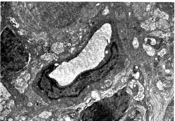
3) Retinal capillaries of S+BGH rats (Fig. 3-5)
The basement membrane of deep capillaries was irregularly and prominently thickened in some localities, being even as thick as 8000 A at some sites.
Fig. 1 Cross section of retinal capillary of normal rat (x 6800)
Fig. 2 Cross section of retinal capillary of S rat, shows an irregular thickening of the basement lamina (x 5800)
Fig. 3 Cross section of retinal capillary of S+BGH rat deposit of low electron density
substance in outer basement lamia (x 10000)
Fig. 4 High magnification of Fig. 3 (x 47000)
Fig. 5 Cross section of retinal capillary of S+BGH rat the electron lucid meterials was
showed as high electron density substances by tannic-acid staining (x 12000)
In the outer basement membrane surrounding the pericytes of the deep capil- laries, there were some parts which were as electron-lucid as those found in the reti- nal capillaries of S rats, and which were proved by tannic acid stainning to be sub- stances with high electron density (Fig. 3, 4 and 5, 6).
3. Biochemical analysis of retinal blood vessels
For the biochemical study, 30 eyeballs each from normal rats, S rats and S+BGH rats collected as a unit were preserved in 10% formaldehyde solution ; after washing the eyeballs, the retinas were separated and the retinal blood vessels were isolated by
Kuwabara-Cogan's trypsin digesting method (8 hours) for use as materials. In order to know the degrees of damages to the blood vessels, preliminary experiments were made by electron-microscopic observation of the retinal blood vessels after the diges- tion. Further several units of 30 normal retinas were assayed at different times for hydroxyproline, hexosamine, and sialic acid to confirm their contents to be mostly con- stant.
The results of assays of 30 retinas each from the three groups after 8-hour digestion were as shown in Table 1.
Table 1. Hydroxyproline, Hexosamine and Sialic acid content of normal, S and S+BGH rats retinal vascular system