Acta Med. Nagasaki 30:195-203
A Retrospective Study in the Diagnosis of 301 Jaundiced Cases
Takatoshi NODA, Yasuharu OHNO, Tohru OSHIBUCHI, Shungo NAKAGO, Shigetoshi MORITA, Akimasa MIZUTANI, Tsutomu TOMIOKA, Toshimitsu MIYAMOTO,
Takashi YAMAGUCHI, Kensuke YAMAMOTO,
Kunihide IZAWA, Tsukasa TSUNODA,
Ryozo YOSHINO, Noboru HARADA, and Ryoichi TSUCHIYA The Second Department of Surgery, Nagasaki University School of Medicine,
Nagasaki, Japan
Received for publication, June 11, 1985
In the past 12 years, 301 patients with a total serum bilirubin over 2 mg/dl (re- ference interval 0.2-0.8 mg/dl) were admitted to the Second Department of Surgery, Nagasaki University School of Medicine, Japan.
The purpose of this paper is to analyse the jaundiced cases and to evaluate the di- agnostic accuracy of the following imaging techniques: Computed Tomography (CT), Ul- trasonography (US), Drip Infusion Cholangiography (DIC), Endoscopic Retrograde Cho- langio-pancreatography (ERCP), Percutaneous transhepatic Cholangiography (PTC) and Selective Celiac Angiography (SCAG). Of the 301 patients, 63 had carcinoma of the bile duct, 48 carcinoma of the pancreas, 26 carcinoma of the gallbladder, 16 hepatoma, 8 carcinoma of the ampulla of Vater, 83 cholelithiasis, 27 parenchymal liver disease, 9 con- genital bile duct disease, 5 chronic pancreatitis, 14 other diseases, and 2 had no final diagnosis. CT was attempted in 33 of 170 patients with malignancy, and 22 of 129 pa- tients with benignancy. A correct finding was obtained in 23 (69.7%) of the 33 patients and 18 (81.8%) of the 22 patients, respectively. US gave a correct finding in 28 (59.6
%) of 47 patients with malignancy, and 36 (69.2%) of 52 patients with benignancy. DIC
gave a correct finding in 1 (5.6%) of 18 patients with malignancy, and 16 (42.1%) of 38
patients with benignancy. ERCP gave a correct finding in 33 (76.7%) of 43 patients with
malignancy, and 38 (74.5%) of 51 patients with benignancy. PTC gave a correct finding in
野 田 剛 稔,大 野 康 治,押 渕 徹,中 郷 俊 五,森 田 茂利,水 谷 明正,富 岡 勉, 宮 本 峻 光,山 口 孝,山 本 賢 輔,井 沢 邦英,角 田 司,吉 野 奈三,原 田 昇, 土 屋 涼 一,114 (91.2%) of 125 patients with malignancy, and 33 (86.8%) of 38 patients with benig- nancy. SCAG gave a correct finding in 72 (66.7%) of 108 patients with malignancy, and
14 (50%) of 28 patients with benignancy.
From these results, we conclude that in spite of developments of non-invasive pro- cedures, it is still necessary to use invasive diagnostic techniques (PTC & ERCP) in the assessment of jaundiced patients.
Key words: Jaundiced cases, Imaging techniques, Noninvasive procedures, Invasive pro- cedures
INTRODUCTION
Many cases of jaundice are seen in hospitals to which patients with gastrointestinal disease are admitted. Generally speaking, . the vast majority of these patients require early treatment. It is important, therefore, to ascertain as early as possible whether the jaundice is obstructive or nonobstructive. If obstruction is present, the site and cause of the obstruction must be identified immediately.
In recent years, new invasive and noninvasive techniques for differentiating ob- structive and nonobstructive jaundice have been developed.
In our department, drip infusion cholangiography (DIP), gray scale ultrasonography (US) and computed tomography (CT) have been used as noninvasive procedures, and endoscopic retrograde cholangiopancreatography (ERCP), percutaneous transhepatic chol- angiography (PTC) and angiography (Angio) as invasive ones.
The aim of this paper is to analyse 301 patients with jaundice and to evaluate the diagnostic accuracy of imaging techniques in those cases.
MATERIAL AND METHODS
Three hundred and one jaundiced patients with total serum bilirubin over 2 mg/dl (reference interval ; 0.2-0.8 mg/dl) were studied retrospectively between October 1969 and December 1981.
One hundred and seventy patients were male and 131 female, mean ages being 59.0 and 58.1 years, respectively.
A final diagnosis was made in all on the basis of histopathological finding but two patients had no final diagnosis.
Abbreviations of techniques used in this study are shown in Table 1.
Table 1.
Patient Details (Oct. 1969-Dec. 1981) Number of Patients : 301
(Male 170; meanage 59.0 years) (Female 131; mean age 58.1 years)
Malignant disease : 170 cases (56.5%)
Benign disease: 129 cases (42.9%)
Unknown 2 cases ( 0..6%)
Total serum bilirubin (reference
interval : 0.2-0.8mg/dl) : Mean 14, Range 2-44 Abbrevations used in this study
DIC = Drip Infusion Cholangioraphy
US = Ultrasonography
CT = Computed Tomography ERCP = Endoscopic Retrograde
Cholangiopancreatography
PTC = Percutaneous Transhepatic Cholangiography
Angio = Angiography
RESULTS
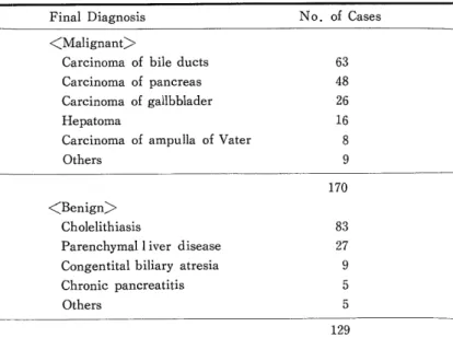
Table 2. shows the causes of jaundice in 299 cases.
One hundred and seventyy cases (57%) had malignant disease, and 129 (43%) benign disease.
Of the 170 cases, 63 were carcinoma of the bile ducts, 48 carcinoma of the pan-
Table 2. Cause of Jaundice in 299 Patients
Final Diagnosis No. of Cases
<Malignant>
Carcinoma of bile ducts 63
Carcinoma of pancreas 48
Carcinoma of gallbblader 26
Hepatoma 16
Carcinoma of ampulla of Vater 8
Others 9
170
<Benign>
Cholelithiasis 83
Parenchymal 1 iver disease 27
Congentital biliary atresia 9
Chronic pancreatitis 5
Others 5
129
creas, 26 carcinoma of the gallbladder, 16 hepatoma, 8 carcinoma of the ampulla of Vater and 9 other malignant lesions.
Of the 129 benign cases, 83 had cholelithiasis, 27 parenchymal liver disease, 9 congenital billiary atresia, 5 chronic pancreatitis and 5 other benign diseases.
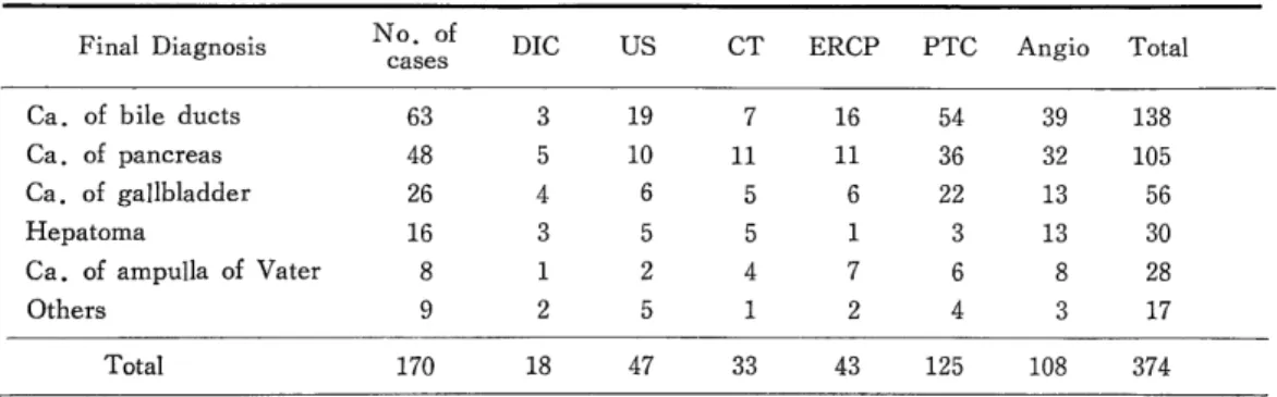
Table 3. summarizes the frequency of each technique attempted in the 170 jaundiced cases with malignancy.
Table 3. Frequency of each technique in patients with jaundice
Final Diagnosis No. of cases DIC US CT ERCP PTC Angio Total
Ca. of bile ducts 63 3 19 7 16 54 39 138
Ca. of pancreas 48 5 10 11 11 36 32 105
Ca. of gallbladder 26 4 6 5 6 22 13 56
Hepatoma 16 3 5 5 1 3 13 30
Ca. of ampulla of Vater 8 1 2 4 7 6 8 28
Others 9 2 5 1 2 4 3 17
Total 170 18 47 33 43 125 108 374
It ranged from 18 to 125 times with an average of 2.2 for each case.
Among the various techniques, invasive procedures such as ERCP, PTC and Angio were employed more often than noninvasive procedures in the malignant cases. Among the invasive procedures, PTC was by far the most popular in the cases with malignancy (125/170; 73.5%).
In the benign disease, there was no significant difference between the use of non- invasive and invasive procedures, and an average of 1.9 techniques were performed for each case.
Both US and ERCP were employed more often than the other procedures in the cases with benign disease.
Nine cases with congenital biliary atresia were excluded because of rare cause of jaundice (Table 4).
In evaluating the capability of imaging technique in cases with jaundice, the diag- nostic criteria had to be indicate because they varied according to each examiner.
We deemed the result positive if it revealed a satisfactory finding for the cause of
Table 4. Frequency of each technique in patients with jaundice
Final Diagnosis No, of cases DIC US CT ERCP PTC Angio Total
Cholelithiasis 83 28 36 12 35 32 14 157
Parenchymal liver disease 27 5 13 6 12 2 11 49
Chronic pancreatitis 5 5 1 1 3 2 1 13
Others 5 0 2 3 1 2 2 10
Total 120 38 52 22 51 38 28 229
jaundice.
Table 5. shows the accuracy of noninvasive procedures in jaundiced cases with malignancy.
Table 5. Accuracy of non-invasive procedures in jaundiced patients with malignancy Final Diagnosis No. of cases DIC US CT
Ca. of blie ducts 63 0/ 3( 0) 13/19( 68) 4/ 7( 57)
Ca. of pancreas 48 0/ 5( 0) 4/10( 40) 7/11( 64)
Ca. of gallbladder 26 1/ 4(25) 2/ 6( 33) 4/ 5( 80)
Hepatoma 16 0/ 3( 0) 5/ 5(100) 5/ 5(100)
Ca. of ampulla of Vater 8 0/ 1( 0) 1/ 2( 50) 3/ 4( 75)
Others 9 0/ 2( 0) 3/ 5( 60) 0/ 1( 0)
Total 170 1/ 18(6) 28/47( 60) 23/33( 70)
DIC was attempted during a period when jaundice was not clinically apparent, but the positive rate was very low.
US was carried out in 47 of the 170 cases (27.6%). It gave accurate information in hepatoma (100%), but the degree of accuracy was not high in carcinoma of the gall- bladder (33%) and of the pancreas (40%). The overall positive rate was 60%.
CT determined the cause of obstruction in 70% of the 33 cases, and was effective in cases with hepatoma (100%) and those with carcinoma of the gallbladder (80%).
The accuracy of invasive procedures in malignant cases is shown in Table 6.
Table 6. Accuracy of invasive procedures in jaundiced patients with malignancy
Final Diagnosis No. of cases ERCP PTC Angio Ca. of bile ducts 63 14/16( 88) 48/ 54( 89) 14/ 39(36)
Ca. of pancreas 48 9/11( 82) 34/ 36( 94) 29/ 32(91)
Ca. of gallbladder 26 3/ 6( 50) 21/ 22( 96) 10/ 13(77)
Hepatoma 16 0/ 1( 0) 3/ 3(100) 12/ 13(92)
Ca. of ampulla of Vater 8 7/ 7(100) 6/ 6(100) 5/ 8(63)
Others 9 0/ 2( 0) 2/ 4( 50) 2/ 3(67)
Total 170 33/43( 77) 114/125(91) 72/108(67)
The cause of obstruction was confirmed in 77% of 43 cases by ERCP, and in 91 of 125 cases by PTC.
ERCP was very useful in carcinoma of the bile ducts, the pancreas and the ampulla of Vater, and PTC in all the malignant cases even without the help of other procedures.
With Angio, however, the detection rate was relatively low (67%).
Similarly, each technique was evaluated in the benign disease.
The results of noninvasive procedures are presented in Table 7.
Table 7. Accuracy of non-invasive procedures in jaundiced patients with no malignancy
Final Dagnosis No. of cases DIC US CT
Cholelithiasis 83 15/28(54) 27/36(75) 9/12( 75)
Parenchymal liver disease 27 1/ 5(20) 9/13(69) 5/ 6( 83)
Chronic pancreatitis 5 0/ 5( 0) 0/ 1( 0) 1/ 1(100)
Others 5 0 0/ 2( 0) 3/ 3(100)
Total 120 16/38(42) 36/52(69) 18/22( 82)
DIC was attempted in 31.7% of the 120 cases, but the positive rate was poor (42%) .
US elucidated the cause of obstruction in 27 (75%) of 36 cases with cholelithiasis.
In the remaining 13 cases, gallstones, especially intrahepatic stones, could not be de- tected.
CT accuracy in the benign disease was relatively high (82%o), but the ability to detect intrahepatic stones was sometimes deteriorated.
Table 8. shows the accuracy of invasive procedures in the benign disease.
Table 8. Accuracy of invasive procedures in jaundiced patients with no malignancy
Final Diagnosis No. of cases ERCP PTC Angio
Cholelithiasis 83 28/35(80) 27/32( 84) 3/14( 21)
Parenchymal liver disease 27 8/12(67) 2/ 2(100) 9/11( 82)
Chronic pancreatitis 5 2/ 3(67) 2/ 2(100) 1/ 1(100)
Others 5 0/ 1( 0) 2/ 2(100) 1/ 2( 50)
Total 120 38/51( 75) 33/38( 87) 14/28( 50)
ERCP determined the cause of obstruction in 80% of the 35 cases with cholelithiasis and revealed an overall result of 75%. In three cases with parenchymal liver disease, ERCP findings were misinterpreted as malignancy.
PTC revealed the etiology of jaundice with a high accuracy (87%) in each benign disease.
Angio could not provide a definite diagnosis of jaundice in cases with cholelithia-
sis.
DISCUSSION
Diseases showing the clinical features of obstructive jaundice are malignant in many casesij2). Our cases were no exception, that is, 170 of 272 patients with jaundice (62.5%), excluding those with non-obstructive jaundice and those with unknown diagnosis, proved to have malignant disease. However, when our patients were studied according to each disease, it was a characteristic finding that the number of patients with cholelithiasis was the largest, i. e. , 83 (27.8% of all patients). In addition, 27 of all patients (9%) who were admitted because of surgical indications had jaundice induced by parenchymal liver disease, and this is considered to be one of the problems that should be noted in daily practice.
Concerning the evaluation of various kinds of examination, a first examination such as drip infusion cholangiography (DIC) is believed not permit qualitative diagnosis'') in spite of the severity of jaundice.
Ultrasonography (US) has spread rapidly because of the marked development of its equipment and of its noninvasiveness.
The diagnostic accuracy of US in our patients was 60% for malignant disease and 69% for benign disease, indicating that the results of US were not so favorable. As mentioned in the criteria for evaluation, this is because the result was defined to be negative in cases in which the cause of jaundice was not identified, even if expansion etc.
of the intrahepatic bile ducts was observed.
However, it is a very useful method, as reported by other authors' 2)4), in determining whether jaundice is obstructive or if a tumor is present in the parenchymal organs.
Therefore, it is thought that US will be widely applied in the future and will be used as an adjunct of PTCD, as described later.
Just like US, computed tomography (CT) is a noninvasive method and, in recent years, has been employed in many medical institutions. CT was introduced in our hospital in 1979 and has been in use since then. The diagnostic accuracy of CT was 70% for maignant cases. This low accuracy is because qualitative diagnosis was carried out in only 57% of cases of carcinoma of the bile duct and 64% of carcinoma of the pancreas.
In particular, it was difficult for CT to identify a tumor in the intrapancreatic bile ducts or in the lower bile duct region.
On the other hand, the diagnostic accuracy of CT was favorable, 82%, for benign changes, and it was especially useful in jaundiced patients with parenchymal liver disease as a diagnosis by exclusion. However, CT met with several difficulties in identifying gallstones in patients with cholelithiasis, in particular intrahepatic lithiasis, and this is considered to indicate a limitation of CT efficiency.
Among invasive mehtods, the diagnositc accuracy of endoscopic retrograd echolangio-
pancreatography (ERCP) has remained within the level of 70% both for malignancy and
benign disease. This accuracy of ERCP was greatly affected by the fact that the evaluation
was held back in six patients with malignancy and seven with benign lesions because of
insufficient contrast. In addition, four of 12 patients who had benign changes, but had jaundice due to parenchymal liver disease, were misdiagnosed by ERCP as having malignan- cy, etc. Thus, over-reading in patients without expansion of the bile ducts was suggested.
Percutaneous transhepatic cholangiography(PTC) showed a high degree of diagnostic accuracy in both malignant and benign disease. PTC was the most frequently performed as compared with other examinations. To be specific, PTC was performed in 125 of 170 patients (73.5%) with malignant disease. This is because PTC combines with drainage, which is associated with treatment, and, under recent conditions in which PTC uses US as an adjunct method' M, the number of punctures has decreased. As a result, compli- cation, including hemorrhage and the leakage of bile, have also markedly declined.
Angiography was carried out in 108 of 170 patients (63.5%) with malignant disease, and this was the greatest number next to that of PTC. However, the diagnostic accuracy was 67% for malignancy and 50% for benign changes, both of which were low. It was particularly low, 36% (14 of 39 patients), for carcinoma of the bile ducts. This is considered to be attributable not only to anatomical specificity including the distribution of blood vessels to the bile ducts, but also to poor vascularization in carcinoma of the bile ducts.
The evaluation of various kinds of examination for disease showing jaundice was carried out in this study, and it was found that the diagnostic accuracy varies with each disease even if the same examination is employed. However, as suggested by our cases and other reports4>5>s>, these seems to be no controversy that, when a patient is diagnosed with jaundice, US is first performed to distinguish whether the jaundice is obstructive or not, and then if it is obstructive, PTC and PTCD if necessary, are immediately undertaken, after which the obstructive region is ascertained and biliary drainage carried out. And, as successive methods, it is believed that after causative disease is suggested to some extent, ERCP, CT and angiography should be selected as links in the treatment plans (Fig. 1).
Fig. 1. Diagnostic Flow Chart