Acta Med. Nagasaki 33 :196-202
Adamantinoma of the Tibia.
- Report of a Case with Findings of Ultrastructural and Immunohistochemical Studies ,
Shingo KINOSHITA, Takeshi MATSUO, Isao SHIMOKAWA, Noriyuki KOSE, Takayoshi IKEDA, and Yuji OHSATO*
First Department of Pathology, Nagasaki University School of Medicine , Nagasaki
* Department of Orthopedics . The Mitsugi General Hospital, Hiroshima Received for publication, June 22, 1988
Running title : ADAMANTINOMA OF THE TIBIA
KEY WORDS : Adamantinoma, Tibia , Immunohistochemistry, Electron microscopy ABSTRACT : A case of tibial adamantinoma in a one-year and ten-month old girl is reported. She had gait disturbance and her roentogenogram showed a well circumscribed radiolucent area in the tibia. Light microscopic examination showed the epithelial component of nests and pseudoglandular arrays in the loose fibrous connective tissue. We finally diagnsed the tumor as adamantinoma of the tibia based on roentogenographical and histological findings. Moreover, the nature of the neo- plastic cells was studied by the immunohistochemical and electron microscopic methods. The adamantinoma is regarded as a unique neoplasm capable of differen - tiating into epithelial elements as well as mesenchymal ones . Detection of epithelial component is important to differentiate from similar disorders. The fibrous dys- plasia-like lesion in stroma is interpreted as a part of the spectrum of mesenchymal differentiation.
INTRODUCTION
Adamantinoma of the long bones is an un- common neoplasm. The histogenesis of the tumor has been debated since Maier first de- scribed it in 1900. The two major proposals, endothelial or epithelial origin have been advo- cated. More recently, however, ultrastructural and immunohistochemical studies have demon- strated convincingly the epithelial nature of nests and islands seen in the tumor. On the other hand, attention has been drawn to the peculiar features of stroma in the tumor such as a fibrous dysplasia-like lesion. We report a case of tibial adamantinoma in a one-year and ten-month-old girl, and review its histogenesis.
CASE REPORT
A one-year and ten-month-old female infant was admitted to The Mitsugi Gereral Hospital on May 27, 1986, because of gait disturbance.
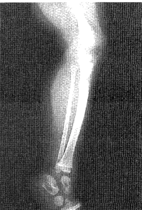
About twenty days prior to her admission, she had fallen down stairs. Roentogenogram show- ed a large radiolucent lesion in the diaphysis of the tibia (Fig. 1). The lesion was well cir- cumscribed, osteolytic or cystic, and located predominantly in the cortex (Fig. 2 ). There were no particular findings on other laborato- ry examinations. A local excision was perform- ed. The excised tumor was elastic hard , measur- ing 3.0 X 1.5 X 1.5cm. The cut surface was yel- lowish white and solid (Fig. 3 ) . An auto-
Mailing address : Shingo Kinoshita, (木 下 真 吾), First Department of Pathology, Nagasaki Uni- versity School of Medicine, 12-4 Sakamoto-cho, Nagasaki 852 , JAPAN.
genous bone graft was bridged in the osseous defect. The postoperative course was unevent- ful and the patient remained well after 10 mon- ths
Fig. 1. This roentogenogram shows a well cir- cumscribed radiolucent area in the prox-
imal portion of the left tibia.
Fig. 2. This is a lateral view of the same lesion.
Periosteal reaction is not evident.
Fig. 3. Cut surface of this tumor is firm or rub- bery in consistency, and pale gray to yel-
lowish-white in colour.
MATERIALS AND METHODS
Light microscopic study
The specimen from the lesion was fixed in a solution of 10% formalin and embedded in par- affin without prior decalcification according to standard procedures. Sections were stained with hematoxylin-eosin, PAS, Hale colloidal Iron, Masson-trichrome, and silver stains.
Immunohistochemistry
Details of the antibodies and sera employed were shown in Table 1. Paraffin sections were stained by the peroxidase-anti peroxidase (PA P) method with some modifications. Briefly, sections dewaxed were treated with 0.3% solu- tion of hydrogen peroxide in absolute methyl alcohol at room temparature to block endoge- nous peroxidase activity. After exposure to normal swine or rabbit serum to reduce non- specific staining, the sections were incubated with the first antibodies overnight at 4 °C.
Then the sections were washed three times with 0.01 M PBS for 5 minutes and incubated with second antibodies for three hours at room temperature. They were washed again and incu- bated with peroxidase-anti peroxidase complex for one hour at room temperature. Then the sections were washed and treated for five mi- nutes with 0.05% 3-3' diaminobenzidine and 0.005% H202 in tris-saline buffer solution at pH 7.4.
Negative controls included replacement of the primary antibodies with PBS, normal rab- bit and normal mouse serum.
Electronmicroscopic study
Formalin-fixed blocks were dissected into one millimeter cubes, fixed again in 2.5% phos- phate buffered glutaraldehyde,then washed with phosphate buffer for 12 hours, postfixed in 1.0% osmium tetroxide for one hour, dehy- drated in a graded ethanol series, and embed- ded in Epon 812. Ultrathin sections were stain- ed with uranyl acetate and lead citrate, and examined with a JEOL 100-B electron micro- scope.
Table 1. List of Antibodies and Sera used in This Study and Their Optimal Dilution.
1) Primary antibodies
$ Rabbit antiserum to human factor VIII related antigen, 1:100, DAKO.
Rabbit antiserum to human keratin,
KIT, Kyowa.
# Mouse antiserum to human viment- in, 1:100, DAKO.
2) Secondary antibodies
# Swine anti-rabbit, 1:50, DAKO, : for the PAP methods.
# Rabbit anti-mouse, 1: 5 0, DAKO, : for the PAP methods.
3) PAI
# PAP, rabbit, 1:100, DAKO.
# PAP, mouse, 1:100, DAKO.
4) Others
$ Normal swine serum, 1:20*, DAKO.
# Normal rabbit serum, 1:20*, 1:100", DAKO
# Normal mouse serum, 1:100**, CL.
* : for reduction of non-specific background staining.
* * : for negative control staining.
DAKO : DAKOPATTS, Copenhagen,
Denmark
Kyowa : Kyowa Medix Inc. Tokyo CL : Cedarlane Laboratories, Ltd.,
Ontario, Canada.
RESULT Light microscopic features
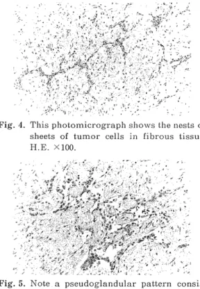
The tumor was composed of looose fibrous connective tissues with nests or sheets of epi- theloid cells (Fig. 4). And a part of the epi- thelial component represented pseudoglandular arrays. A few large nests had irregular cleft- like lumina containing papillae of epithelial
cells (Fig. 5 ). Also vessels were scattered in the fibrous matrix.
Fig. 4. This photomicrograph shows the nests or
sheets of tumor cells in fibrous tissue.
H.E. X100.
Fig. 5. Note a pseudoglandular pattern consis- ted of epithelial cells with an intervening
fibrous stroma. II.E. X100.
Cytologically the cells were oval or round in shape and were not markedly pleomorphic. The nuclei had a uniform vesicular chromatin pat- tern with single nucleolus. The cytoplasm was narrow and faintly eosinophilic. Hale colloidal iron and PAS stains were negative for intracy- toplasm.
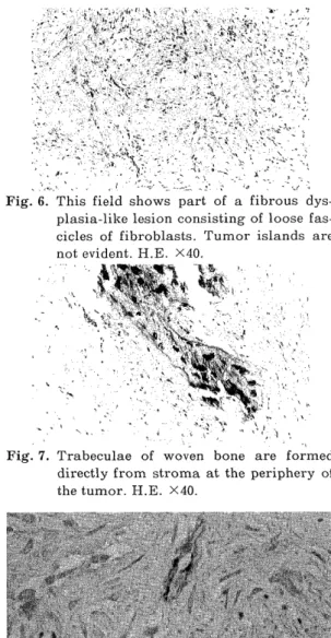
Some lymphocytes aggregation around peri- vascular lesion scattered in the abundant stroma. A fibrous dysplasia-like lesion was also present in the stroma, which was consist- ed of loose fascicles elongating in a vague striform pattern around blood vessels (Fig. 6).
Trabeculae of woven bone were formed at the periphery of the tumor (Fig. 7). However. the lesion differed from the typical fibrous dys- plasia in the presence of the epithelial islands.
These epithelial islands gradually blended with fibrous background. In a part of the tumor, the stroma was abundant of collagen showing a sclerotic appearance.
Fig. 6. This field shows part of a fibrous dys- plasia-like lesion consisting of loose fas-
cicles of fibroblasts. Tumor islands are not evident. H.E. X40.
Fig. 9. Positive staining for keratin in the cyto- plasm of the epithelial cells. PAP stain.
X 400.
Fig. 7. Trabeculae of woven bone are formed directly from stroma at the periphery of
the tumor. H.E. X40.
Fig. 8. Positive staining for factor VIII-related
antigen in the endothelial cells of stromal vessels. The adjacent epithelial cells are
negative. PAP stain. X400.
Immunohistochemical features.
Stain for factor VIII-related antigen showed strong positivity in most of the endothelial cells lining the capillaries and venules in the stroma (Fig. 8 ), while all of the epithelial cells forming nests and channels were uniform- ly negative for this marker:-
Fig. 10. Weakly positive staining for vimentin in the epithelial and stromal cells. PAP
stain. X200.
Keratin was stained intensely in the cyto- plasm of all the cells presented in the nests and channels (Fig. 9), while the endothelial cells of blood vessels and other stromal ele- ments remained negative. Furthermore, keratin was stained in the perinuclear narrow cyto- plasmic rim. On the other hand, the stain for vimentin was weakly positive in the epithelial and stromal elements (Fig. 10).
Electronmicroscopic features
Some details were obtained by electron- microscopic observation. The tumor cells had irregularly folded nuclear membranes with une- venly dispersed chromatin. The cytoplasm of these cells had numerous cytoplasmic projec- tions and tightly interdigitated microvilli, and small intercellular lumina were formed by these cells (Fig.11). Desmosome-like structures were also seen (Fig. 12). The striking feature
Fig. 11 Microvillous projections are seen in the intercellular lumen(L) bounded by cells.
Intercellular junction apparatuses are
seen(arrow). Uranyl acetate and lead
citrate. X23,000.
Fig. 12. Desmosome-like junction connecting two tumor cells is seen (arrow ). Uranyl ace-
tate and lead citrate. X 11, 5 0 0. Higher
magnification. X62,000.
Fig. 13. Incomplete-formed basal lamina (short arrows ), tonofilaments (arrowheads )
and cell processes (long arrows) were
indicated. Uranyl acetate and lead
citrate. X 18,400.
was the presence of tonofilaments and tono- fibrils, which were composed of microfilament bundles with frayed ends (Fig. 13). Cytoplasm contained a few of disturbed mitochondrias, rough endoplasmic reticulums and small vesi- cles. Basal lamina was found out. In the stroma, fibroblast-like cells which contained numerous filamentous materials were obserbed.
Capillaries were seen as well there.
DISCUSSION
Adamantinoma of long bone is a rare distinc- tive tumor, which occurs principally in the tibia. Since this neoplasm was first described by Maier in 1900 1 2 ), approximately over 160 cases have been reported in the English and Japanese literatures. The detailed statistics and clinical events has been gradually eluci dated`) 1'1) 15)22) Nevertheless, the histogenesis of this neoplasm is still controversial. Vascu- lar'1)6)s)11), synovial 10 )16) and epithelial struc- tures 2)9)17)1 s)19)20)24 ) have been proposed as cel- lular origin.
Histologically, small epithelioid islands are found in fibrous stroma. Relative amount of two components vary considerably. The epithe- lioid islands vary in structure and shape case by case. Four different epithelioid patterns are present in adamantinoma')2') : spindled, basal-
oid, squamoid, and tubular. These various histologic features have led to the continuing controversy over its histogenesis. Currently, it is believed that this tumor is an epithelial ori- gin by ultrastructual findings or immunohisto-
chemistry. Our immunohistochemical and ul- trastructural studies suggested the different- iation into the epithelial element. Our findings
confirmed those of RosAi et al. 18) or KNn1'1' et al.9), that is to say, keratin was stained strong-
ly in the almost all of the epitheloid cells. Our ultrastructual features demonstrated cyto- plasmic processes and projections of micro- villous membrane. In addition, basal lamina, desmosome-like structures and tonofilaments with frayed ends could be seen. We are unable to support a vascular origin for adamant- inoma, because we could not find out Weibel- Palade bodies and proliferation of tumoral vessels.
It is also well known that keratin proteins are more wildly distributed in biologic system than formerly thought21), and it does not nec- essarily express squamous character only. Cer- tainly the presence of highly differentiated keratinizing squamous epithelium as previously reported should not be used as irrefutable evi-
dence of epithelial origin, since synovial sar- coma, a tumor generally accepted as mesenchy- mal, may also have areas of keratinizing squa- mous epithelium and nesothelium may undergo squamous metaplasia in respose to various
stimuli23)
The epithelial component is a clue to diagno- sis of adamantinoma, but investigation of stromal component composed of spindle cells is also important in view of histogenesis of the tumor. The fibrous areas either may repre- sent a part of the tumor or may be a reaction.
Areas that suggest transition between the fi- brous and epithelial areas are indicative of the former possibility. The stroma in our case was consisted of fibroblast-like cells and it resem-
bled fibrous dysplasia. Therefore, the tumor appears to differentiate along both mesenchy- mal and epithelial lines. Our results were consistent with the features of synovium or mesothelium7). We believe that origin of ada- mantinoma would be the cell having the biphasic potentiality. Nevertheless, it is inter- esting that, recently, BA:MBTRIIA et al.') and MiLi,s et al. 1 3) described some tumors morpholo- gically identical to tibial adamantinoma lo- cated not in the bone but in the skin and soft tissue over-lying the anterior surface of the tibia. Moreover, EINSTEIN et al.5) suggested the idea that tibial adamantinoma was a form of eccrine carcinoma by using enzyme histochemis- try. Their explanation was different from ours, because they doubted whether the stromal component would be neoplastic.
In any event, although several thories on histogenesis have been proposed, it would be premature to limit the origin to peculiar tis- sues such as epidermis or skin appendages. To determine the correct origin and the exact me- chanism of growth on tibial adamantinoma, additional cases of this entity should be studi- ed in detail.
REFERENCES
1. BAMIIIRRA, E.A., NOGI:IrlA, M. M. F., and MIRANDA, D., ; Adamantinoma of the Soft
Tissue of the Leg. Arch Pathol Lab Med 107:
500-501, 1983.
2. BRAIDwooD, A. S. and McrouGAt_r., A., : Ada- rrantinorm of the tibia. Report of Two Cases.
JBone joint Surg 56-B : 735-738, 1974.
3. CA\4PANACCr, M., LADS, M., and GruN-•rr, A., : Adamantinoma of the long bones. The expe-
rience at the Istituto Ortopedico Rizzoli. Am
J Surg Patho 15 : 533-542, 1981.
4. CILANGUS, G. W., SPEED, J. S., and STHWART, F.
W., : Malignant angioblastoma of Bone-A
Reappraisal of adamantinoma of Long Bone.
Cancer 10 : 540-559, 1957.
5. Ersr:csTi:IN, W., and PITcocI., J. A., : Adaman- tinoma of the tibia. An eccrine Carcinoma.
Arch Pathol Lab Med 108: 246-250, 1984.
6. ELLIOT, G. B., : Malignant angioblastoma of long bone. So-called tibial adamantinoma.
JBone Joint Surg 44-B•: 25-33, 1962.
7 . GI-IADIALLY, F. N., : Diagnostic Electoron Mi- croscopy of Tumors. Second Edition, Butter-
worths : 96-105, 467-480, 1985.
8. Hums, H. G., and MAracovE, R. C.,: Adaman- tinonaa of Long Bone. A Clinico-pathological
Study of Fourteen Cases with Vascular Ori- gin Suggested. J Bone Joint Surg 57-A :148-
154,1975.
9. KNAPP, R. H., WICK, M. R., and ScIIEITIIAUGrl,
B. W., : Adamantinoma of Bone. An Elec- tron Microscopic and Immunohistochemical
Study. Virchows Arch (Pathol Anat) 398: 75- 86, 1982.
10. LI:D1,RER, H., and SINCLAIR, A. J., : Malignant
synovioma simulating Adamantinoma of the tibia. J Path Bact LX V I I : 163-167, 1954.
11. LLoMo3Auv-Boscr-I, A., and OIZTUNO-PACHE:CO, G., : Ultrastructural findings supporting the
angioblastic nature of the so-called adaman-
tinorm of the tibia. Histopathology 2 :189-200,
1978.
12. MAIER, C., : Ein primares myelogenous Plat- tenepithelcarcinom der Ulna. Bruns Beitr
Klin Chir 26: 553, 1900.
13. MILLS, S. E., and Rosnr, J., : Adamantinoma
of the Pretibial Soft Tissue. Clinicopatho-
logic Features, Differential Diagnosis, and Possible Relationship to Intraosseous Dis-
ease. Am J Clin Pathol 83: 108-114, 1985.
14. MOON, N. F., : Adamantinoma of the Appen-
dicular Skeleton. A Statistical Review of Re- ported Cases and Inclusion of 10 New Cases.
Clin Orthop 43: 189-213, 1965.
15. More, H., SIGMA, R., and NAIci\SI 'vlA, H., Adamantinoma of the tibia. A Case Report
and a Statistical Review of Reported Cases.
Acta Pathol Jpn 31 :701-709, 1981.
16. NAJt, A. F., MURRY, J, A., and STASNEY, R. J., : So-called adamantinoma of long bone. J
Bone Joint Surg 46-A : 151-158, 1954.
RlosAI, J., : Adamantinoma of the tibia. -Elec- tron Microscopic Evidence of Its Epithelial
Origin. Am J Clin Patho151 : 786-792, 1969.
18. RosAI, J., and PINKUS, G. S., : Immunohisto-
chemical demonstration of epithelial differen- tiation in adamantinoma of the tibia. Am J
Surg Pathol 6 : 427-434, 1982.
19. RYRIE, B. J., : Adamantinoma of the tibia.
-aetiology and pathogenesis . Br Med J 2 :
1000-1003, 1932.
20. SAACEBRA, J. A. GuTn RRI:'/. D. D. and DIMAS, M. D., : Adamantinoma de la tibia. Observa-
tiones ultrastructurales. Revista medica del
Hospital general, Mexico 31 :241-252, 1968.
21. Scut, GR.1., R., BANKS-SCIH.EGEL, S., and PIxKuS, G. S.,: Imm>lnohistochemical Localization
of Keratin in Normal Human Tissue. Lab Invest 42: 91-96, 1980.
22. UNNI, K. K., DAI-ILI\, D. C., and Br•.AE3ouT, J.
W., : Adamantinoma of Long Bone. Cancer
34 : 1796-1805, 1974.
23. WI,Iss, S. W., and DRFNIA:, H. D., : AD-
AMANTINOMA OF LONG BONE. An Anal-
ysis of Nine New Cases with Emphasis on
Metastasizing Lesions and Fibrous Dysplasia
-like Change . Hum Pathol 8:141-153, 1977.
24. YONIEYAMA, T., WEN•neR, W. G., and Mn.sow, L., : Tibial Adamantinoma-Its Histogenesis
From Ultrastructual Studies. Cancer 40:1138- 1142,1977.