Title
圧迫による脳障害に関する基礎的研究( 第2報:脳循環
と微小血管構築の変化 )
Author(s)
佐久田, 治
Citation
: 1-37
Issue Date
1991-3
URL
http://hdl.handle.net/20.500.12000/25395
Rights
1
.はじめに庄迫に
よる脳障害に関する基礎
的研究
一 脳 循 環と微小血管構築の変化 ー 硫球大学医学部脳神経外科佐 久 出 治 ・ 六 川
二
郎
頭叢内で、mass
l
e
s
i
o
n
により頭蓋内圧が上昇する場合、脳血流は局所的にもまた全体的 にも減少すること、またこの減少がいわゆるa
u
t
o
r
e
g
u
l
a
t
i
o
n
の下限より高し、脳濯流j王で 起こることが報告されている25刷。 この現象に頭蓋内圧上昇とともに圧迫に伴う微小血管 構築の障害が関与することが推測されている叩。しかし、圧迫により微小血管構築がし、か に変化するかについては未だ明らかではない。 私どもは、硬膜外金属球挿入によるネコ脳圧迫モデノレを作製し、庄迫による脳組織の経 時的変化について既に報告したがへ今回は同モデルにおける脳循環動態と微小血管構築 の変化について検討したので報告する。1
実験方法1
圧迫脳の作製 体重2
.
7
-
3
.
4
k
g
(平均3
.
1kg
)の成熟ネコ2
3
匹を用いた。pe
n
t
o
b
a
r
b
i
t
a
l
sodium
20m
g
/
k
g筋注後経口挿管し
、東大脳fr
}f式頭部固定装置に固定し室内空気で調節呼!汲を行いな がら以下の操作を行った。 心屯図監視ドで大腿動)11(¥および静脈にカテーテノレを挿入した。動脈側からは圧力トラン スデューサ ‘(P23
l
D
.
Gould)
を接続し全身動脈圧、平均動脈圧(MABP)
を測定した。またガス分析
(Model1
7
0
,CORNING
)を適時行い、生理的条件下(PaC02
32-37mmH
g)
で実験を維持した。静脈問JIからはpan
c
uroniumbromide 0
.
1
mg/ml を含む生理食~1ül7}c を4ml
/h
l
で輸液し、また必要に応じてメイロンの注入を行った。実験中はヒートバネ ル(TK
司4
0
,朝日電子工業)を用いて直腸温を一定範囲内(3
7
-
3
8
0C
)に保った。右側頭骨-20-に直径
5mm
の骨窓を開け、硬膜外に圧測定用センサー(TCT
/
b
.
Gae
,t]e
c
、)を挿入し頭蓋内圧
CICP)
を測定した。左右の頭頂骨に直径5mm
の宵-窓を聞け、レ4 ザー血流計(ALF2
1
0
0,
ADV
ANCE
)のプロープをノイランサー(MB
-
P
B
. KANETSU
)で保持し 手術用顕微鏡下で脳表に設置した。これらの骨窓はb
oncwax
とde
n
t
a
l
c
e
ment
を悶いて 注意深く密閉した。脳血流(CBF)
の測定はD
i
r
n
a
g
l
ら1)の方法に準じた。 冠状縫合の正中から左側方15mm
、後方15mm
の側頭骨に直径5mm
の'1
'1"窓を│掛け、そ こから直径5mm
の金属球(DAD
A
8
-
2
9
9
-
5
、以下ボーjレと略す)1
2
個(容制効果:約1.4
m1
)を1
0
分毎にl
個ずつ硬膜外に挿入して脳を圧迫(
F
i
g
.
1
A)
し、そのi
擦のMr
¥B
P
.
l
CP.
CBF
をポリグラフで連続記録した。2
.
脳循環動態の分析 各ボーノレ挿入9
分後のMABP,ICP,
CBF
の測定値を比較した。脳謹流E
E
(
C
J:>f>)、Il出 血管抵抗(
CVR)
を以下の式から算出した。CPP
=MABP -ICP
CPP(%)
=
(CPP
/
CPP(control))
X1
0
0
CBF(%)
三(C
BF/
CBF(control
)
)
X
1
0
0
CVR(%)
=
(CPP(%)
/
CBF(%))
X1
0
0
CPP
c
e
r
e
b
r
a
l
p
e
r
f
u
s
i
o
n
p
r
e
s
s
u
r
e
MABP
mean a
r
t
e
r
i
a
l
blood pressure
I
CP
i
n
t
r
a
c
r
a
n
i
a
l
pressure
CVR
cerebrovasc
u
l
a
r
r
e
s
i
s
t
a
n
c
e
CBF
c
e
r
e
b
r
a
l
blood
f
1
0w
統計学的検定は時系列データーに対してDunn
e
t
t
の多重比較、左右差に対してWilcoXO
Jlr
a
n
l
心sumt
e
s
t
を用い、危険率0
.
0
5
以下を有意とした。3
.
微小血管構築の観察 脳の圧迫に引き続き以下の操作を行った。へノfリン2
,0
0
0
単位を腹腔内に投i
7
・し開胸し た。右心耳を切断した後、左心室を介してカニュ句レを大動脈起始部に挿入し固定したわ また左腕頭動脈より遠位部の大動脈弓を結殺した。カニュ レの挿入と同時に5
%
s
u
crose
を含む生理食塩水1
,0
∞
ml
を120mm
Hg
圧で注入し血液を流出させた。号│き続き5%
s
ucro
s
e
,2
% g
l
u
t
a
r
a
J
d
ehyde
を含むO
.
l
I
v
I
リン酸緩衝液(pH
7
.
4
)
1
,0
0
0
ml
で、瀧流閲定を行った。 ついでカニューレに圧トランスデュ サを接続し120m
ll1HgLE
を越えないように手動でメ す2
1
-チノレメタクリレート樹脂(
Mercox
,大日本インキ )約75mlに硬化剤( M
A
1
.
5
g)
を加え てよく混合した後注入した。樹脂注入1
時間後に関頭して脳を取り出し、大脳脚起始部と その前方5m
m
で‘冠状断を行いブロックを作製した(F
i
g
.
1
B)
。このブロックから圧迫の 中心部、圧迫の辺縁部および反対側の皮質からそれぞれ1
cnf程度の切片を採取した。 次に村上の方法1川こ準じて切片を6
0
0C
,20%NaOH
溶液に2
日間浸演し、染色寵中で3
時間流水洗縦して樹脂以外の脳実質組織を除去した。約2
週間この操作を繰り返し血管鋳 型標本を作製した。構造および形態を保持するために真空凍結乾燥機を用いて蒸留水中で 氷結させ、水の表面張力を消却しながら乾;爆させた21)。その後、鋳型標本をイオンコーター も(IB
-
5
、エイコー・エンジニア リング)で白金イオン蒸着し、走資電子顕微鏡(
8
-
4
5
0
, 目立:以下8EM
とi
絡す〉で三次元的に観察した。 なお正常ネコ5
旺から同様の血管鋳型標本を作製し対照群として8EM
による観察を行っf
こ。 盟 . 結 果1
.頭蓋内圧、脳循環動態の変化ICP
のボーjレ挿入前の値(以下前値と略す)は8
.
8
士
2
.
2mmHg
(mean
士
8E)
であった。I
CP
はボーノレ数8
個以上で急速に上昇し1
2
偲挿入時には3
3
.
3
:
:
t
3.2mmHg
となった。統計 学的には8
個以上での上昇は有意であった(p
く
0
.
0
5
,Fig
.
2
)
。MABP
の前値は1
0
7
.
3
士
4.3mmH
g
で‘あった。MABP
はボー/レ挿入にもかかわらずほと んど変化せず、12
個挿入時でも1
0
4
.
0
土4.5mmHgであった(F
i
g
.
2
)
。CPP
の前値は9
8
.
3
士4
.7rnmHg
であった。CPP
はICP
の上昇に対 応 し て 低 下 し1
2
個 挿 入時には70
.
7
土4.
8
mmHg
となった。8
個以上での低下は有意であった(p
く
0
.
0
5
,Fig
.
3
,u
p
p
e
r
)
。
圧迫慎I
J
CBF
はCPP
がほとんど変化しないボール数5
個の時点から有意に減少した(p
<
0
.
0
5
)
。その後はCPP
の低下に対応して減少し、1
2
個挿入時には前値比5
4
.
3
土4.1%
となっ た。反対側CBF
はボーノレ挿入初期から、Cpp
の低下に対応して減少し1
2
個挿入時には6
8
.
5
土2
.4%となった。7
個以上で、の減少は有意で、あった(p
<
0
.
0
5
,F
i
g
.
3
,l
o
w
e
r
)
。両側CBF
を比較すると、圧迫側CBF
が常に低植を示し、4
個以上で有意の左右差が認められた(
p
<
0
.
0
5
)
。
-
22-圧迫側CVRはボ
ール
挿入に伴い急速に増大し
5個で 1
3
9
士1
2%となった
。
8個以上での
増大は有意であった(
p
<
0
.
0
5
)。それに対して反対側
CVR
は有意の変化を示さなかった
(
F
i
g
.
4)
。
両側CVRを比
較すると
、
庄迫側CVRが常に高値を示し
、
ボ
ー
ル数
3個以上で
有意の左右差が認められた
(p
く
0
.
0
5
)。
2
.
微
小血
管構築
の
変化
a
)対照群
脳表面を走行する中大脳動脈枝は直径約
3
0
母7
0
μ の制Tl動脈を分岐していた
。
事
1
1
1
動脈
の表
面に
は、内皮細胞の核に
相当する
5
x
1
0
μ の紡錘形の陥没
(nuclear
i
n
de
n
L
a
t
i
o
n
21 })グ認められた
(
F
i
g
.5
A)。知
l
動脈
はさら
に網目状に
分岐し
、明状
に走行する直径
5
-
1
0
μ
の
毛細血管に移行していた
(
F
i
g
.5
B)。毛細
血管は
主とし
て脳
表へむけ走行する
i
直径3
0
-
6
0
μの細
静脈に移行して
いた
(Fig5
C)
。
血管鋳
型標本の特に表問において細動
脈や
毛布
f
J1血管の中断が認められたが、これらは血管径の変化を伴わないこ
と
、また断面が
鋭角であ
ることから切片採取時に生じた人工的所見と考えられた
。
b
)圧迫側皮質
圧迫中心部では、細動脈の屈曲や毛細血管の消失が認められた
(
F
i
g
.
6
A)
。
さらに強
拡では毛細血管の表面の不整を伴う
狭
小化、それに続く中断が
認められた(Fig.6B)
。
こ
の部位では、あ
き
らかに細静脈と同定しうる血管は認められなかった
。
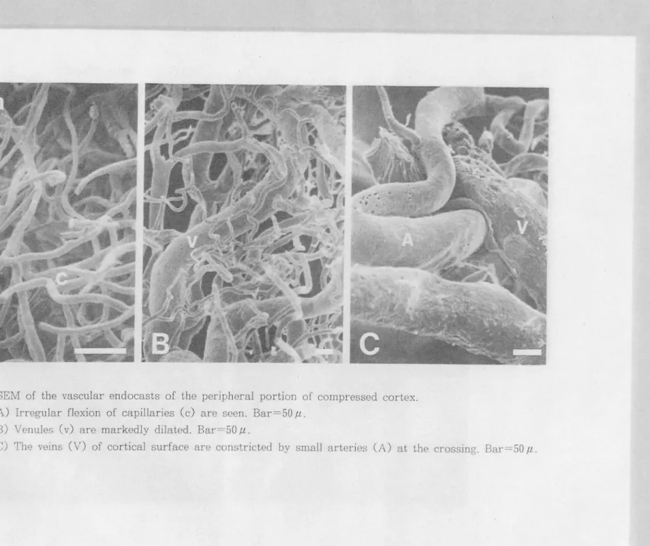
圧迫辺縁部では、細動脈および毛細血管が弧状
の走行を失い不自然に開
i出していた
(F
i
g
.
7
A)
。
細静脈は直径7
0
・1
5
0
μ と著しく拡張し、かっ蛇行していた(-Fi
g
.
7
B)
。
交叉
した細動脈により細静脈が
圧迫されていた(
Fig
.
7
C)o
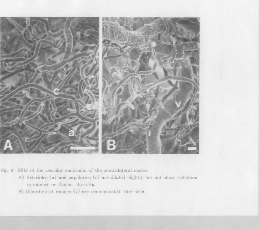
c
)反
対側皮質
中大脳動脈から細動脈までに
変
化
は認められなかった。毛細血管は軽度に拡張していた
が
、
屈曲や狭小
化、また
あきらかな数の減少は認められなか
っ
た (F
i
g
.
8
A)。こ
れらの
血管が吻合する細静脈は拡幅
してい
た
(Fig.8B)。これは圧迫仰j
の圧迫辺縁部に見られ
た細静脈の所見と類似していた
が、交文圧
迫現
象は認められなかった
。
日 . 考 察
脳循環は
、頭
蓋
内圧が冗進
した場合
、
庄の上昇以外にも原因病態に付随した種々の要因
により影響される日間
819L脳がm
ass
l
e
s
i
o
nにより圧迫された場合の脳血流の減
少には、
脳の偏位に
よる微小血管構築
の障害が関与することが推定されている
幻
9.18.1九
Hasuoら
5}-
2
3
-は硬膜下ノ幻レ
ー
ンを用いて脳を圧迫した実験での脳血流の測定から、ま
こ
f
た
Tazawaら
i頭蓋内庄冗進を伴伴
ニ
わない脳出血例での局所脳血流の測定から、脳血流の減少に微小血管構
築の障害が関与すると推定している。
この実験では、圧迫による脳の変形が脳循環や微小血管構築にどのように影響するかに
ついて検討した。
脳血流は圧迫側で瀧流圧がほとんど変化していない時点から減少した
。
また脳血管抵抗
は圧迫側でポ
ー
ル数に対応して増大した
。
これらの結果は、圧迫側において
au
t
o
r
e
g
u
J
a
-i
i
o
nが障害されていること、また圧迫時の脳血流の減少に血管抵抗の増大が関与している
ことを示す
。
圧迫側の微小血管構築でみられた細動脈や毛細血管の屈曲や狭小化に続く中断は
、
対照
群や反対側ではみられないことから、圧迫による脳の変形が原因であると考えられる
。
す
なわち、脳実質の変形に伴う血管系の偏位により屈曲が、また血管系の捻れや閉塞により
血管内腔の血栓化が
生じ狭小化に続く中断が生じたと考えられる。圧迫の
辺縁部と中心部
の微小血管構築を比較すると、前者では血管の屈曲、後者では狭小化に続く中断が主要な
所見であ
った。この部位による所見の差は、圧迫による脳の変形の程度
に応じて微
小
血管
構築が障害されることを
示すものと考えられる。
この細動脈や毛細血管の障害は、浮腫を伴う脳梗塞モデノレの皮質
2')、実験的水頭症ラッ
トの白質と基底核部
14)で観察されているが、これらの報告においても浮腫や脳室の拡大が
あることから、脳の変形がその原因であると考えられる。
細静脈の拡張は、圧迫田
JI周辺部と反対側でみられたことから、頭葦全般にわたる頭葦内
庄冗進により静脈瀧流が!障嘩害
即
迫
の
司
ヰ
中
恥
山
:
コ
1心
,
L
、部で紐細
i
静脈が
1
祥消手向i
失していること、また辺縁部で細静脈が細動脈と交文し圧迫され
ていることから、静脈瀧流の障害はより高度であると考えられる
。
このような圧迫側でみられた紐
i
動脈や毛細血管の屈曲や狭小化に続く中断、また細静脈
の交叉圧迫現象が脳血管抵抗を増大させ、脳血流を減少させたと考えられる
。
これに対して反対側では、微小血管構築は細静脈を除いてほとんど障害されず、血管抵
抗も変化しなかった
。
それにもかかわらず脳血流は瀧流庄の低下に対応しで減少
し
、
a
u
t
o
-r
e
g
u
l
a
t
i
o
nの障害がみられた
。
この障害は圧迫による脳幹の障害
3.1仰のや両側半球間
神経連
絡を介する機序
8,11却によって生じたと推定される
。
以上より圧迫脳では全般的に
au
t
o
r
e
g
u
l
a
t
i.onが障害され脳瀧流庄の低下に対応して脳
-24-血流が減少し、これに加えて圧迫の中心部を最大とする脳の変形により微小血管構築が陣
宝され脳循環を
さらに障害すると考えられる
。
従来
、
頭蓋内massl
e
s
i
o
nを有する患者に対しては、頭蓋内圧や脳瀧流圧を指標として
治療が行ーわれてきた
12I叩
}
。
しかし
mass
l
e
s
i
o
nによる脳の変形が直接脳循環を障害するこ
とを示すこの実験結果からみると、脳の変形を伴う
massl
e
s
i
o
nを有する患者においては
早期に除去手術を行い変形による脳障害を防ぐことが必要であると考えられる
。
V
.
ま
とめ
硬膜外からの圧迫による脳循環動態およひ。微小血管構築の変化について実験的に検討し
た
。
成熟ネコ
2
3匹を用いて頭蓋内圧、平均動脈庄、脳血流を測定しながら、左側頭骨に開
けた骨窓から、 直径 5mm の金属球 12倍!を 10分~ij: に l 個ず、つ挿入し JI出を圧迫した。 引続きメチ
jレメタクリレ
ー
ト樹脂注入による血管鋳型標本を作製し走査電子顕微鏡で観察した
。
1.圧迫脳では全般的(こ
autoreg
u
l
a
t
i
on
が障害された
。
圧迫側では脳血管抵抗の増大お
よび脳瀧流庄の低下に対応して
、
反対側では脳謹流庄の低下に対応して脳血流が減少した
。
2
. 圧迫側の微小血管構築で細動脈や毛細血管の周曲や狭小
化
に続く中断、また細静脈の
交文圧迫現象が認められた
。
これらの微小血管構築の変化が圧迫側の脳血管抵抗を明大さ
せ、脳血流をさらに減少させたと考えられた
。
以上より、圧迫脳における脳循環動態には脳の変形による微小血管構築の障害が大き
く
影響することが証
明
され
た
。
脳の変形を伴う
mass
l
e
s
i
o
nを有する患者にお
いては早期に
除去手術を行うことが必要であると考えられる
。
稿を終えるにあたり、御指導を頂いた
.
琉球大学病理学新政輝男教慢、同麻酔学教授奥出
佳朗教授に深甚なる謝意を現します
。
R M n L文 献
1) Dirnagl U, Kaplan 8, Jacewicz M and PulsinelliW: Continuous measurement of cerebral blood flow by Laser-Doppler FlowmeLry 1n a rat stroke model. J Cereb Blood Flow Metab 9:589-596.1989
2)藤本俊一郎、久山秀幸、西本健、秋岡達郎、長尾省吾、西本詮 脳箆によるI1出圧迫の基礎的研究 とくに
術後に神経税務hE状を起こさないための術中の治様について NeurolMed Chir(Tokyo) 22: 893-900
1982
3) Goodman SJ and Becker DP・ Vascular pathology of the brain stem due to
experimentally increased intracranial pressure. Changes noted in the micro- and macroclrculation.J Neurosurg 39: 601時609,1973
ε1) Grubb JRL, Raichle ME, Phelps ME and Ralcheson RA・Effectsof increased intl'acranial
pressure on cerebralblood volume, bloodflowand oxygen utilization in monkeys. J Neurosurg 43: 385-398, 1975
5)llasuo M, Furuse M, Kuchiwaki H and Kageyama N: Cerebralblood flow associated with intracranial pressur巴 /volume relationship. Acta Neurol Scand 60 (Suppl 72): 378-379,
1979
6) l-IuberP, Meyer JS,トlandaJ and lshikawa S: Electromagnetic flowmeler study of carolid and vertebral blood flow during intracranial hypertension. Acta Neurochir 13: 37-63, 1965 7)Johnston IH and Rowan JO:lntracranial pressuregl'adientsand cerebralblood f1ow. 238・
240 (LangfitlTW, et a1:Cerebral Circulation& Metabolism. Springer-Verlag, BerIin -BeidelbergNew York, 1975)
8)しcnziGL, Franchowiak JRS and Jones T: Cerebraloxygen metabolism and blood flow in hurnan ccrebralischemic infarcLion. J Cereb Blood Flow Metab 2・321-335.1982
9)しewis MP and McLallrin Rし Regionalcerebral blood flow in increased inll'acramal pressure produced by increasedcerebrospinalf1uid volume, intracranialmass and cerebral ederna.160-164 C Brock M and Dietz H:TntracranialPressure. Springer-Verlag, Berl in -Heidelberg-New York, 1972)
10) McNealy DE und Plurn F: Brain stem dysfunction with supratentorial mass lesions. Arch curol(Chicago) 7:26-48, 1962
11) Meyer JS, Shinohara Y, Kanda T, Fukuuchi Y, Ericsson AD and Kok NK: Diaschisis resultingfrom aculeunilateralcerebralinfarction, Arch Neurol 23: 241・247,1970
12)Miller JD and Ledingham TM: Reduction ofincreased intracranial pressure. Comparison bclween hypcrbaric oxygen and hyperventilation. Arch Neurol 24: 210-216, 1971
lS)村上宅郎;微小i血管分布機械のための鋳型走資電子顕微鏡法.細胞 7:11-18, 1975
14 )悶 伸 夫、中間籾一、主主藤俊郎、高久 晃、篠原治道、森沢佐歳.実 験 的 水 頭 症 に お け る 血 管 構 築 .
NeuI'ol Med Chir(Tokyo) 25・701-706,1985
15)PlurnF and Posner JB: Blood and cerebrospinal fluidlactate during hypervemilation Arn J Physiol 212・864‘870,1961
16)Sakuta 0, Mukawa J, Takara E, Nakata M, Kinjo T and Kuda H: HistologicaJchanges ofthebrain by experimentaJ extradllraJC'ompression. 654-656( Hoff JT and BeizAし.
p o n d
InlracranialPressureV1J. 8pringer・Verlag,serlin-Heldelberg, 1989)
17) Shapiro HM, Wyre SR and Loeser J:Barbitllrale-allgmented hypothernlla for reductionof
persisもentintracranialhypertension.J Neurosurg 40: 90-100, 1974
18)鈴木幹男圧迫悩における病理組織学的および酵紫組総化学的検討 Neurol !¥led Chir (Tokyo) 21:
221-232, 1981
19)Tazawa T, Mizukami M, KawaseT and Usami T: The relationshipbelween tnlracranial
pressure and reglonalcerebralblood f10w in hyperlensivelnlracerebral hemorl'hage. J
Cerb Blood Flow Metab 1(8uppl1):8545-S546, 1981
20)筒 井 巧 、 本 間 混、角南典生、門間文行、土本正治、長尾省吾、西本詮:lITI管緊張におよlます脳幹部j凶
血管巡勤中枢の役割ー第l幸f1:特に脳血流蛤およびifJi室内圧の変化について.脳外 12:581-589, 1984
21)堤健二 脳梗t.liに関する実験的研究血管鋳型走資1filll員法を中心に.Neurol MedChil・t1'okyo) 26・
595-600. 1986
22) Wise RJ8, Bernardi 8, Franckowiak R8J, Legg NT and Jones ↑ Serial obsenatlOns on
the pathophysiology ofacuteslroke. The transilionfrom ischemla lo inf且rct1un as
reflected inregionaloxygen extraction. Brain106:197-222, 1983
23)Yada K, Nakagawa Y and Tsuru M: Circulatory disturbance of lhe venOllSsystcm during experimentalintracranialhyperlension. J Neurosurg 39:723-i29, 1973 司 , z 円 L
---
-A
B
Fig-.
1A) Lateral
view of
a
compressed
brain.
8) Corona
l
se
ti
on of
the
compressed bra
in
.
Marked
deformity
ISfound
at
the
compressed side, whereas the
con
tr
a
I
a
t
eml side
r
e
m
ai
1s
almost u
n
chang-
d
excepL
for
midline
shift.
28-120
mm
H
g
f
t
~
~
t
ti
~
~
f
~
~
f
100
._....
MAB
P
D
unnett : N.
S
.
IC
P
----80
m
ean
±
S
E
60
40
20
0
D
unnett (P
<
0. 05
)
pr
e 1
2
3 4
5
6 7
8
9 10 11 12
Number of balls
Fig. 2 Mean
arterial
blood pressur
( MABP) and
intr
acrania
l
pr
ssure
(ICP)
dur
in
g
ball insertion.
29-120
mean± SE 'OJ)100
I E5
80
0.. 0.. 060
Dunnett (P<
0. 05) .._. ..et. CBF40
o--o rt. CBF * P( 0.05 * *P(O.Ol100
Dunnett(P( 0.05) (v.s. rt. CBF)*
80
LL CD60
040
o:f
Dunnett (P<
0. 05) pre1
2
3 4
5
6
7
8
9
1 0 11 12
Number of ballsF
i
g
.
3
Ce
r
e
bral p
e
rfu
s
i
o
n
pr
ess
ur
e
(CPP.
upp
er) and cerebra
l
bl
ood
fl
ow
(CBF,
l
ower)
durin
g
ball
in
ser
ti
o
n.
-180
160
*
140
0:::>
120
0
100
*
*
**
*
*
Dunnett: N.S
.
pre 1 2
3
4 5 6 7 8 9 1 0 11 12
Number of balls
mean
+
SE
--....Qt.CVR
o--o rt
.CVR
*
p
<
0.05
* *P(O.Ol
(v.s.rt.CVR)
F
i
g
.
4 Cerebrovascular r
es
istan
ce
(CVR)
durin
g
ball
insertion.
-Fig
.
5 Scanning electron m1crograms (SEM) of th
e
vascular endocasts in
the
control.
A) Nuclear indentations (n) of endothelial cells are found
in
the arteriole (a). Bar
=
50
f.L.B) Capillary (c) network. Bar
=
50
/.L.c..v c..v
Fig. 6
SEM of the vascular endocasts of the central
portion
of compressed cortex
.
A) Capillary
network and
venous system disappear, and the vascular endocasts
become·
verysparse.
Bar
=
SO
/1.B)
Interruption
of the capillary (c) with
narrowing
and irregular surface 1s
w
..,.
Fig. 7 SEM of t
h
e vascular e
nd
ocasts o
f
t
h
e per
i
phera
l
portion of compressed cortex
.
A) Irregular
flexion
of capillaries (c) are
see
n
.
Bar=50
f.l..B) Venules ( v) are
m
arkedly dilated. Bar
=
50
f.l..c.-:
c.n
I
Fig.
8
SElv!
of the vascular endocasts of the
contralateral
cortex.
A) Arterioles
(a) an
d
cap
illaries
(c) are dila
ed slightly but
not show reduction
in number or flexion. Ba=50
f.l.Experimental
st
udy
on brain
dama
ge
by
extracere
bral
compression
- Alteration of cerebral circulation
and
microvascular architectures
-Osamu Sakuta
,
Jiro Mukawa
Department of Neurosurgery, UniversiLy of the Ryukyus, School of Medicine, Okinawa, Japan