九州大学学術情報リポジトリ
Kyushu University Institutional Repository
腸炎ビブリオ由来RelE/ParEスーパーファミリートキ シンの生化学的解析
张, 晶
https://doi.org/10.15017/1866256
出版情報:Kyushu University, 2017, 博士(システム生命科学), 課程博士 バージョン:
権利関係:
Biochemical characterizations of a RelE/ParE superfamily toxin in Vibrio parahaemolyticus
2017
ZHANG JING
Contents
Abbreviations ... iii
Chapter 1 ... 1
General introduction ... 1
1-1. Toxin-antitoxin (TA) systems ... 1
1-2. Vibrio parahaemolyticus and a viable but non-culturable (VBNC) state ... 5
1-3. TA systems discovered in V. parahaemolyticus ... 9
Chapter 2 ... 13
Biological activity of Vp1843 ... 13
2-1. Introduction ... 13
2-2. Materials and Methods ... 16
2-2-1. Materials ... 16
2-2-2. Plasmids ... 16
2-2-3. Strains ... 18
2-2-4. Protein purification ... 18
2-2-5. E. coli Gyr inhibitory activity ... 19
2-2-6. DNA nicking endonuclease activity ... 19
2-3. Results ... 20
2-3-1. Purification of Vp1843 ... 20
2-3-2. Gyr inhibitory activity ... 21
2-3-3. Vp1843 has a DNA nicking activity ... 23
2-3-4. Characterization of the nicking activity of Vp1843 ... 25
2-3-5. Essential residues in Vp1843 ... 30
2-4. Summary ... 33
Chapter 3 ... 34
Involvement of Vp1842/Vp1843 in the VBNC state . 34
3-1. Introduction ... 343-2. Materials and Methods ... 36
3-2-1. Stains ... 36
3-2-2. Plasmids ... 36
3-2-3. Mediums and reagents ... 36
3-3. Results ... 41
3-3-1. Knock out of vp1842/vp1843 genes from the V. parahaemolyticus genome by conjugation. ... 41
3-3-2. Involvement of vp1842/vp1843 in the VBNC state of V. parahaemolyticus .... 42
3-3-3. Expression level of vp1842/vp1843 in the VBNC state ... 43
3-4. Summary ... 45
Chapter 4 ... 46
Physiological function of Vp1843 ... 46
4-1. Introduction ... 46
4-2. Materials and Methods ... 47
4-2-1. Stains ... 47
4-2-2. Plasmids ... 47
4-2-3. Mediums and reagents ... 47
4-2-4. Flow cytometry ... 47
4-2-5. Fluorescence microscopy ... 48
4-2-6. Pulse-field electrophoresis ... 48
4-3. Results ... 50
4-4. Summary ... 54
Chapter 5 ... 55
General considerations ... 55
References ... 61
Acknowledgements ... 68
Abbreviations
A280 : absorbance at 280 nm Amp : ampicillin
ATP : adenosine triphosphate
bp : base pair
BSA : bovine serum albumin cDNA : complementary DNA CFU : colony forming unit CFX : ciprofloxacin
DAPI : 4’, 6-dianidino-2-phenylindole DNA : deoxyribonucleic adic
DTT : dithiothreitol
EDTA : ethylenediaminetetraacetic acid
Gyr : DNA gyrase
GyrA : A subunit of DNA gyrase GyrB : B subunit of DNA gyrase
IPTG : isopropyl-β-D-thiogalactopyranoside kbp : kilo base pairs
kDa : kilo dalton LB : Luria-Bertani OD : optical density ORF : open reading frame PCR : polymerase chain reaction PDB : protein data bank
PFGE : pulsed-field gel electrophoresis PSK : post-segregation killing
RNA : ribonucleic acid RNase : ribonuclease rpm : rounds per minute
SDS-PAGE : sodium dodecyl sulfate-polyacrylamide gel electrophoresis
SI : superintegron
TA : toxin/antitoxin
TAE : Tris-acetate-EDTA buffer TBE : Tris-borate-EDTA buffer
TE : 10 mM Tris-HCl, pH 8.0, containing 1 mM EDTA Tris-HCl : Tris (hydroxymethyl) aminomethane hydrochloric acid UV : ultra violet
V/cm : voltage/centimeter VBNC : viable but nonculturable β-ME : β-mercaptoethanol
Chapter 1
General introduction
1-1. Toxin-antitoxin (TA) systems
Toxin-antitoxin (TA) systems are composed of a toxin, which causes growth arrest by interfering with a vital cellular process, and a cognate antitoxin, which neutralizes the toxin activity during normal growth conditions (Page et al., 2016). TA systems were initially discovered, more than 30 years ago, as a plasmid-borne factor of post- segregational killing (PSK) (Ogura 1983; Gerdes et al., 1986b). Namely, the products of the toxin genes selectively kill the plasmid-free cells in the population, so as to promote plasmid maintenance in the daughter cells (Fig. 1-1). The selective activation of a toxin in cells that lost the TA-encoding plasmid is based on the instability of an antitoxin (Aakre, 2015). In the well-known TA systems, the antitoxins are more unstable than their toxins, and as a result the antitoxins must be continually transcribed in order for providing enough antitoxins to neutralize the toxins. For example, the first discovered TA system ccdA-ccdB was located in a mini-F plasmid. The antitoxin CcdA is more unstable than its toxin CcdB, and as a result ccdA must be continually transcribed in order to produce enough CcdA to neutralize CcdB. Cells that lose the mini-F plasmid can no longer produce CcdA, and the more stable CcdB is then freed to kill the plasmid-free cells (Van Melderen et al., 1994). This is applicable to another plasmid-born TA system hok-sok, only that the selectivity is based on RNA stability.
Under normal station, the antitoxin sok produce an unstable antisense RNA to inhibit the translation of the toxin gene hok. Loss of the plasmid R1 triggers a rapid degradation of sok RNA, thus frees the production of the Hok toxin, leading to the killing of the plasmid-free cells (Thisted et al., 1992).
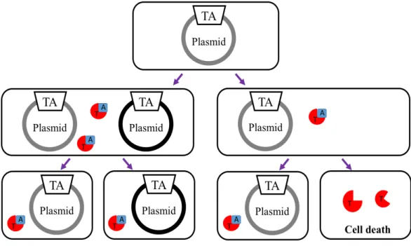
Fig. 1-1. Schematic representation of post-segregational killing (PSK) by plasmid-borne TA systems.
When a plasmid is correctly replicated and divided in the cells, an antitoxin forms a complex with a toxin to block its toxicity. If the plasmid fails to be given to the progeny, the antitoxin is selectively degraded by proteases, and then the activated toxin kills the cell. TA, T and A indicate toxin-antitoxin genes, toxin and antitoxin, respectively.
Shortly after the discovery of TA systems on plasmids, their identification on chromosomes was reported (Gerdes et al., 1986a). Since then, thousands of TA operons have been discovered not only in plasmids, but also on the chromosomes of most free- living bacteria (Aizenman et al.,1996; Masuda et al., 1993). Of all these TA systems, toxins are proteins, while antitoxins are either RNAs or proteins. The TA systems are so far classified into six classes, based on the mechanisms by which the antitoxins block the toxicity of their toxins (Fig.1-2). In the well- known type II TA systems, antitoxin and toxin mRNAs are synthesized from the same promoter and translated into proteins (Fig. 1-3). An antitoxin immediately forms a stable complex with a toxin to block its function. The antitoxin-toxin complex and the antitoxin also bind to the promoter to
Fig. 1-2. Toxin-antitoxin (TA) systems.
Toxins are shown in red and antitoxins in blue. Type I: the sRNA antitoxin base pairs with toxin mRNA to inhibit translation; when activated by stress, the toxins function to destroy the cell membrane and disrupt ATP synthesis. Type II, both antitoxin and toxin are proteins; under growth conditions, the toxin is bound by the antitoxin, which inhibits its toxicity. Both the antitoxin and, in most cases, the TA complex bind the TA promoter to repress transcription. Under stress conditions, cellular proteases such as Lon and ClpXP are activated that preferentially cleave the antitoxins, freeing the toxins to inhibit growth by inhibiting translation or replication. Type III: the antitoxin sRNA is processed by the endoribonuclease (RNase) toxin, resulting in the formation of RNA pseudoknot-toxin complexes, which inhibit toxin activity. Type IV: the protein antitoxin stabilizes bacterial filaments, while the protein toxin disrupted them; in the absence of the antitoxin, this toxin-mediated defilamentation inhibits cell division. Type V: the antitoxin GhoS is an RNase specifically cleave the toxin ghoT mRNA; under conditions of stress, the mRNA of the antitoxin GhoS is degraded.
This figure was cited from the ref. reported by Page et al (Page et al., 2016).
degraded by ATP-dependent proteases such as Lon, ClpXP, or ClpAP, leading to rapid growth arrest and cell death due to the cellular effects of toxins (Christensen et al., 2004;
Fineran et al., 2009). The type II toxins inhibit cell growth by interfering with vital cellular functions, including DNA replication, protein synthesis, cell-wall biosynthesis and ATP synthesis (Short et al., 2013; Masuda et al., 2012).
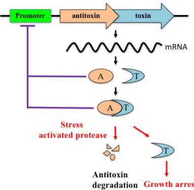
Fig. 1-3. Schematic representation of an operon encoding a type II toxin and antitoxin.
Antitoxin and toxin mRNAs are synthesized from the same promoter and both are translated into proteins. An antitoxin immediately forms a stable complex with a toxin to block its function. Under stress conditions, the antitoxin is degraded so that the toxin is activated, resulting in growth arrest.
Recent findings suggest that chromosomally encoded copies of toxins and antitoxins function as metabolic stress response elements to manage their metabolism to cope with different sources of stress. That is, chromosomally encoded TA systems have been suggested to be a genetic element involved in induction into dormancy, such as a viable
under debate (Wang et al., 2012; Aakre et al., 2013; Christensen et al., 2001; Wang et al., 2013).
1-2. Vibrio parahaemolyticus and a viable but non-culturable (VBNC) state
Vibrio parahaemolyticus is a Gram-negative, halophilic asporogenous rod that is straight or has a single, rigid curve. It was first discovered by Fujino et al. following the large outbreak of food borne disease in Japan, which caused 272 illnesses and 20 deaths in 1950s (Fujino et al., 1953). Since then, it has been recognized as the leading causal agent of human gastroenteritis associated with seafood consumption all over the world (Letchumanan et al., 2014). V. parahaemolyticus grows in the presence of 0.5 ~ 8% NaCl (optimum 3 ~ 6%) at 10 ~ 42°C (optimum is 35 ~ 37°C). It is also tolerant to a pH range of 5.6 ~ 9.6 (optimum is 7.6 ~ 8.0). Notably, the organism has an extremely short generation time (8 ~ 12 min) than Escherichia coli (25 ~ 30 min), under appropriate conditions (3% NaCl, 37°C). V. parahaemolyticus is able to exist in multiple cell types according to a variable circumstance (Fig. 1-4). It can either be a swimmer cell in liquid cultures or a swarmer cell in highly viscous environments (McCarter, 1999).
A B C
Fig. 1-4. Viable identities of Vibrio parahaemolyticus.
(A) Electron micrograph of a swimmer cell (McCarter,1999); (B) Electron micrograph of a swammer cell (McCarter,1999); (C) scanning electron micrographs of a cell entered into the VBNC state (Hino et al., 2014).
Genome sequencing of V. parahaemolyticus RIMD2210633, one of the pandemic strains (Kanagawa phenomenon positive, serotype O3:K6), showed that it contains two circular chromosomes (Chromosome I and II). The larger chromosomeI consists of 3,288,558 bp, encoding for 3,079 proteins and the chromosome II is 1,877,212 bp, producing 1,752 proteins. When compared the V. parahaemolyticus genome sequence with that of V. cholerae, another diarrhoea-causing Vibrio with two circular chromosomes, a super-integron (SI) was identified on the chromosome I in V.
parahaemolyticus (Makino et al., 2003). SI is a large gene-capture and excision system found on chromosome II in V. cholera, and it is characterized by a site-specific integrase gene closely associated with a cognate recombination site attI and a promoter Pc followed by a large array of gene cassettes (Rowe-Magnus et al., 2001). In addition, a large number of the genes located in SI have unknown functions (Mashimo, 2006).
The SI in V. parahaemolyticus (48 kb) contains 78 ORFs, as shown in Fig. 1-6, while that in V. cholerae (126 kb) includes 179 genes, and their gene organization and sequence are perfectly distinct from each other. Nevertheless, it is known that SIs are highly conserved during evolution of each species, and however, the molecular basis of the conservation remains unclear.
Another characteristic feature of V. parahaemolyticus is that it can even enter into the viable but non-culturable (VBNC) state to escape harsh environmental conditions such as temperature and salinities downshift, nutrient starvation. (Bates and Oliver., 2004; Wong and Wang., 2004; Hino et al., 2014). The VBNC state is a unique state distinct from death. Cells in VBNC state are still alive and possess a lot of physiological function as a viable cell. Yet, they have no ability to develop into colonies on suitable media as viable cell. When the cell exists in a VBNC state, the cellular morphology, cell wall, membrane composition, physical and chemical resistance, gene expression, adhesion properties, virulence potential and metabolism are different to those of the viable cells (Li et al., 2014). The resuscitation property of the VBNC state can regain its virulence under suitable conditions (Du et al., 2007). This characteristic of VBNC
Fig. 1-5. The two chromosomes in V. parahaemolyticus.
V. parahaemolyticus contains two circular chromosomes. The larger chromosome I is 3,288,558 bp and the chromosome is 1,877,212 bp. This figure was cited from the ref. reported by Makino et al. (Makino et al., 2003). SI stands for super-integron. The organization of SI is depicted in Fig. 1-6.
. Gene organization in the super-integron (SI) in the V. parahaemolyticus chromosome I. zation of genes in the SI. Color in green, red, gray, blue, light purple, pink, yellow, orange and magenta stand for transposase, Vp intIA, sis protein, toxin-antitoxin gene, threonine efflux protein, a protein involved in cell-cycle regulation, acetyltransferase, PnuC protein, /spermidine acetyltransferase BltD individually. This was created by SnapGene (http://www.snapgene.com).
induction into the VBNC state to effectively prevent bacterial infections and cure infected patients.
Since Colwell and coworkers first reported on the VBNC state (Xu et al., 1982), this phenomenon has now been described for over 50 bacterial species using various criteria for viability and discussed in relation to dormancy and persistency (Oliver et al., 2010).
Although the VBNC state has been believed to be a survival strategy in response to certain harsh environmental stresses, no specific factors have been identified because many environmental conditions induce the VBNC state in different bacterial species.
Recently, there is a growing evidence that activation of TA systems is one of the mechanisms known to trigger such a state with low metabolic activity (Hayes et al., 2009).
1-3. TA systems discovered in V. parahaemolyticus
In a previous study (Hino et al., 2014), we found three gene clusters, vp1821/vp1820, vp1829/vp1830 and vp1842/vp1843, in the V. parahaemolyticus genome database have sequence homology to those encoding the E. coli type II TA proteins, YefM/YoeB, DinJ/YafQ and DinJ/YafQ, respectively. Expression of the putative toxin gene, vp1820 or vp1843, in E. coli strongly inhibited cell growth, while co-production of the putative antitoxin gene, vp1821 or vp1842, neutralized this effect. In contrast, the expression of vp1830 in the presence of 0.2% arabinose had no influence on cell growth. These results suggested that vp1842/vp1843 serve as a TA system in V. parahaemolyticus. It was further found that although Vp1820 has protein synthesis inhibitory activity, Vp1843, unlike the E. coli homologue YafQ, has neither protein synthesis inhibitory activity nor ribonuclease activity. Rather, the expression of vp1843 in E. coli resulted in a morphological change in the cells, while co-expression with vp1842 had no influence on the cell shape.
During the course of the previous study, we found that Vp1843 has sequence homology not only to E. coli YafQ, but also to Caulobacter crescentus ParE (Fig. 1-7) (Dalton and Crosson., 2010). Phylogenetic studies have found that YafQ and ParE belong to the RelE/ParE superfamily (Fig. 1-8), folding into a similar fold, despite the
Fig. 1-7. Alignment of the amino acid sequence of toxic proteins belonging to the RelE/ParE superfamily proteins.
The amino acids were aligned by the computer program Clustal W to maximize the homology for all proteins. Vp1843 (UniProKB Q87NM5) and Vp1830 (UniprotKB Q87NN8) are gene products in V. parahaemolyticus (Hino et al., 2014). RK2ParE, VcParE, EcParE2, EcParE3, CcParE, MtParE1, MtParE2, EcYafQ, EcRelE, EcYoeB, and PhRelE indicate amino acid sequences of RK2 encoded ParE (UniProtKB Q79EC5), V. cholera ParE2 (UniProtKB Q9KMJ0), E. coli ParE2 (UniProtKB Q8X366), E. coli ParE3 (UniProtKB Q8XE95), C. caulobacter ParE (UniProtKB Q9A9T8), M. tuberculosis ParE1 (UniProtKB P9WHG7), M.
tuberculosis ParE2 (UniProtKB P9WHG5), E. coli YafQ (UniProtKB Q47149), E.
coli RelE (UniPtotKB P0C077), E. coli YoeB (UniProtKB P69348), and Pyrococcus horikoshii RelE (UniProtKB O73966), respectively. Amino acid residues are numbered according to the V. parahaemolyticus Vp1843. Amino acids identical to those in Vp1843 are in yellow. * indicate different amino acids between Vp1843 and Vp1830. The essential amino acids Lys37 and Pro45 for Gyr inhibitory activity of Vp1843 is indicated in red.
fact that the toxins belonging to this family share a low similarity in primary structure and display distinct biochemical activities. Namely, the RelE type toxins, such as YafQ and YoeB, stall the ribosome by cleaving mRNA in a translation-dependent fashion (Masuda et al., 2012). Recent structural studies of the ribosome in complex with RelE or YoeB provided insight into the structural basis for ribosome-dependent ribonuclease activity and suggested catalytic residues in individual toxins (Aakre et al., 2013;
Christensen et al., 2001). On the other hand, ParE was originally found on plasmid RK2 with ParD being the antitoxin, and DNA gyrase (Gyr) was identified as its target molecule (Wang et al., 2013). Subsequently, several ParE genes have been found on bacterial chromosomal DNA, and biochemical studies reported that the ParE type toxins strongly poison Gyr. However, the molecular mechanism by which ParE toxins
Fig. 1-8. Evolutionary relationship of RelE/ParE superfamily toxins.
Based on the amino acid sequence comparison shown in Fig.1-7, an evolutionary tree for the RelE/ParE superfamily toxins was constructed. This evolutionary tree shows that the ParE toxins (red), such as Vp1843, are clearly separated from the RelE toxins (blue), such as YafQ and YoeB, with protein synthesis inhibitory activity.
inhibit Gyr has not yet been elucidated (Sterckx et al., 2016; Yuan et al., 2010; Gupta et al., 2016).
In this study, to gain insight into biological properties of Vp1842/Vp1843, inhibitory potency of Vp1843 was first investigated with E. coli Gyr. As described in Chapter 2, I found that it, unlike other ParE toxins, had little inhibitory activity toward Gyr. Rather, Vp1843 exhibited DNA endonuclease activity. Next, I examined whether vp1842/vp1843 is involved in induction into the VBNC state by preparing a mutant strain (∆vp1842/vp1843), in which vp1842/vp1843 in the V. parahaemolyticus genome was knocked out by homologous recombination. As described in Chapter 3, Δvp1842/vp1843 entered into the VBNC state at a comparable rate with that of the wild- type. Furthermore, a preliminary transcriptome analysis using next generation sequence showed that the vp1842/vp1843 transcript in the VBNC state was comparable with that in normal growth conditions. It is thus likely that vp1842/vp1843 is not involved in the induction into the VBNC state. In order to gain insight into physiological functions of vp1842/vp1843, E. coli cells, in which vp1843 was expressed, were characterized in terms of phenotypic properties using flow cytometry and microscopic analysis. I found that expression of vp1843 caused extreme degradation of the chromosomal DNA, as described in Chapter 4. Finally, I discuss a possible physiological role of vp1842/vp1843 in V. parahaemolyticus on the basis of biological information obtained in this study, as described in Chapter 5.
Chapter 2
Biological activity of Vp1843
2-1. Introduction
Phylogenetic studies have found that Vp1843 belongs to the RelE/ParE superfamily.
Toxins belonging to this family fold into a similar fold while they display distinct physiological functions (Fig. 2-1). That is, the RelE toxins, such as RelE and YafQ, inhibit protein synthesis by cleaving mRNA (mRNA inteferase), while the ParE toxins have an inhibitory activity towards DNA gyrase (Gyr). However, their molecular mechanism remains unclear. Namely, the crystal structure of the E. coli RelE in complex with the Thermus thermophilus ribosome revealed that Arg81 and Tyr87 play an essential role in mRNA interferase activity as a general acid and base, respectively (Neubauer et al., 2009). On the other hand, structural and biochemical studies on YafQ found that His50, His63, Asp67 and His87 participate in acid-base catalysis during mRNA hydrolysis and further suggested that Phe91 plays an important role in mRNA positioning (Maehigashi et al., 2015). Collectively, catalytic residues in RelE and YafQ are not conserved, and their catalytic mechanism has not been fully understood.
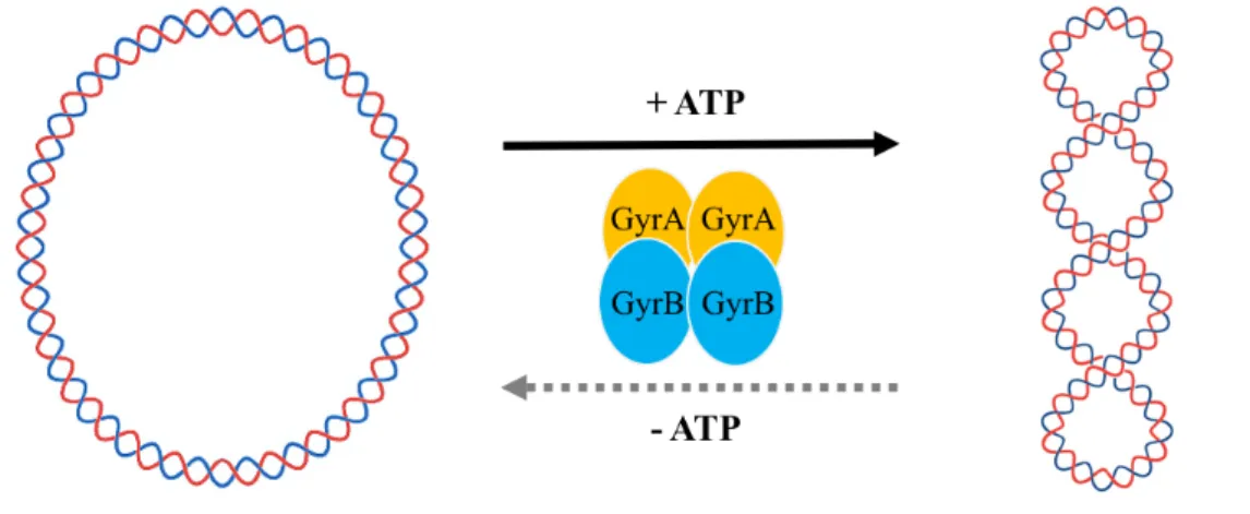
As for ParE toxins, it has been reported that they have an inhibitory activity towards DNA gyrase (Gyr). Gyr is the only type II topoisomerases capable of introducing negative supercoils at the expense of ATP hydrolysis (Gubaev et al., 2016; Gellert et al., 1976). In addition, Gyr is able to relax supercoiled DNA in the absence of ATP (Fig.
2-2) (Gellert et al., 1976). Gyr consists of two subunits, GyrA and GyrB (97 kDa and 90 kDa, respectively, in E. coli) and forms a heterotetramer GyrA2GyrB2 in the active enzyme. The ParE toxins from different bacteria inhibit DNA gyrase in a different mechanism. First, RK2 plasmid ParE and V. cholerae ParE2 stabilize the cleavage complex by interacting with Gyr A subunit (GyrA), despite having distinct binding sites (Jiang et al., 2002; Yuan et al., 2010). Second, Mycobacterium turbaclosis ParE inhibits Gyr by interacting with Gyr B subunit (GyrB), the C-terminal residues being suggested
to be involved in the inhibitory activity (Gupta et al., 2016). Third, E. coli ParE2 interacts with neither GyrA nor GyrB, although its Gyr inhibitory activity has not been experimentally verified (Sterckx et al., 2016). As described above, the molecular mechanism by which ParE toxins inhibit Gyr has not been fully elucidated.
Fig. 2-1. Functions of toxins belonging to the RelE/ParE superfamily.
Toxins which belong to the RelE/ParE superfamily fold into a similar fold, while they display two distinct biochemical activities. The RelE family toxins, such as YafQ and YoeB, stall the ribosome by cleaving mRNA (mRNA interferase), while the ParE family toxins have been reported to interfere with DNA replication by inhibiting DNA gyrase.
Fig. 2-2. Catalytic activities of DNA gyrase.
In the presence of ATP, Gyr (GyrA2GyrB2) is able to convert relaxed DNA into supercoiled DNA. However, in the absence of ATP, Gyr (GyrA2GyrB2) relaxes the supercoiled DNA in a relatively low activity.
Our previous study showed that Vp1843 has neither mRNA cleavage activity nor protein synthesis inhibition activity (Hino et al., 2014). To gain more insight into the biological properties of Vp1842/Vp1843, I investigated the inhibitory potency of Vp1843 with E. coli Gyr.
2-2. Materials and Methods
2-2-1. Materials
Restriction endonucleases and DNA modifying enzymes were purchased from MBI Fermentas. The E. coli Gyr was supplied by New England Biolabs. Oligonucleotides used in this study were purchased from Operon. Ciprofloxacin (CFX) was obtained as hydrochlorides from Enzo Life Sciences. All other chemicals were of analytical grade for biochemical use.
2-2-2. Plasmids
The relaxed plasmids, pBR322 and pUC19, were supplied by Inspiralis and New England Biolabs. The plasmid vectors pET-15b, pET22b, and pBAD/Myc-HisA used for expression of vp1842/Vp1843 in E. coli cells were from Novagen and Invitrogen, respectively.
Plasmids for expression and purification of Vp1843-His and Vp1843 mutants (K37N, P45L, K37N/P45L) were constructed as follows. First, the vp1842/vp1843 fragment was amplified using primer 22b-1843-NdeI-F and XhoI-1843-R and ligated into pET22b after treatment by Nde I and Xho I. Second, the resulting plasmid pET22b- vp1842/vp1843 was used as a template for mutagenesis PCR to obtain pET22b- vp1842/vp1843-K37N (Primers: pET22b-4243-K37N-F and pET22b-4243-K37N-R), pET22b-vp1842/vp1843-P45L (Primers: pET22b-4243-K37N-F and pET22b-4243- K37N-R). pET22b-vp1842/vp1843-K37N/P45L was constructed with a template of pET22b-vp1842/vp1843-P45L and primers of pET22b-vp1842/vp1843-K37N-F and pET22b-vp1842/vp1843-K37N-R. Third, a vp1843-K37N/P45L fragment was amplified using pET22b-vp1842/vp1843-K37N/P45L as a template with primers 22b- 1843-NdeI-F and XhoI-1843-R, and ligated into pET22b with Nde I and Xho I digestion.
The resulting plasmid pET22b-vp1843-K37N/P45L was used for expression and purification of K37N/P45L. Primers were listed in Table 2-1.
Plasmids for examination of toxicity of Vp1843 and its mutants (K37N, P45L, K37N/P45L) were constructed as follows. Gene fragments of vp1843, vp1843-K37N, vp1843-P45L and vp1843-K37N/P45L were amplified using the primer pair of pBAD- 1843-NcoI-F and pBAD-1843-HindIII-R with their corresponding templates pET22b- vp1842/vp1843, pET22b-vp1842/vp1843-K37N, pET22b-vp1842/vp1843-P45L and pET22b-vp1843-K37N/P45L. These fragments were ligated into pBAD-Myc-HisA in the aid of Nco I and Hind III digestion, resulting in three plasmids pBAD-vp1843, pBAD-vp1843-K37N, pBAD-vp1843-P45L and pBAD-vp1843-K37N/P45L.
Sequences of the primers were listed in Table 2-1.
Table 2-1. Oligonucleotide used in this study
Oligonucleotide Sequence (5’-3’)
22b-1843-NdeI-F 5’-TCGAGGTAACATATGATTTTATGGGAAGA
A-3’
XhoI-1843-R 5’-AAGAAGCTCGAGATCCGTAGGGAATTT-3’
pET22b-4243-K37N-F 5’ATTGAAGCAAACGTAGAAAACTTGCTTAA ACAACC-3’
pET22b-4243-K37N-R 5’-GTTTTCTACGTTTGCTTCAATGAGGTTGTC AGTTT-3’
pET22b-4243-P45L-F 5’-AAACAACTTTTAATGGGTGTGCAACGAGA TGGCAT-3’
pET22b-4243-P45L-R 5’-ACACCCATTAAAAGTTGTTTAAGCAAGTT TTCTAC-3’
pBAD-1843-NcoI-F 5’-GTAATCGACCATGGATATGATTTTATGGG AAG-3’
pBAD-1843-HindIII-R 5’-AGAGGAAGCTTTCAGTGGTGGTGGTGGT G-3’
2-2-3. Strains
The E. coli strain JM109 was used as a host cell for cloning, and E. coli strains BL21(DE3) Codon Plus RIL (Stratagene) and LMG194 (Invitrogen) were used as host cells for the expression of recombinant proteins.
2-2-4. Protein purification
For purification of Vp1843 or Vp1843-His, the His-Vp1842/Vp1843 or Vp1842/Vp1843-His complex was purified by affinity column chromatography on a COSOMOGEL His-Accept column. Then, they were treated by 6 M guanidine hydrochloride and loaded onto a COSOMOGEL His-Accept column to separate His- Vp1842 from His-Vp1842/Vp1843 or Vp1843-His from Vp1842/Vp1843-His. The Vp1843 protein solution was dialyzed against 50 mM Tris-HCl, pH 8.0, containing 500 mM NaCl and 400 mM arginine hydrochloride at 4°C overnight and finally against 50 mM Tris-HCl, pH 7.5, containing 100 mM NaCl. Then, the protein solution was concentrated using Amicon® Ultra Centrifugal Filters Ulteracel-3K (Merck Millipore Ltd). Vp1843-His was refolded in a HisTrap column (GE healthcare) by 20 column volumes buffer containing 50 mM Tris-HCl, pH 8.0, 500 mM NaCl, 10% glycerol and 0.02% tween20 in a liner gradient from 0% to 100%. After eluting the refolded Vp1843- His by 50 mM Tris-HCl, pH 8.0, 500 mM NaCl, 10% glycerol, 0.02% tween20, and 400 mM Imidazole, it was further diluted and applied to a HitrapQ column (GE healthcare). The HitrapQ column was washed by 50 mM Tris-HCl, pH 8.0, 50 mM NaCl, 10% glycerol and 0.5mM DTT and then protein was eluted by increasing NaCl concentration from 50 mM to 1.0 M in the same buffer. After collecting the fractions containing Vp1843-His and checking by 15% SDS-PAGE, the protein was dialysis against 50 mM Tris-HCl, pH 7.5, containing 100 mM NaCl. Finally, the protein solution was concentrated using Amicon® Ultra Centrifugal Filters Ulteracel-3K (Merck Millipore Ltd). Mutant proteins were purified in the same manner, as described above.
2-2-5. E. coli Gyr inhibitory activity
Gyr inhibitory assays were carried out as described previously (Sterckx et al., 2016;
Yuan et al., 2010). Inhibitory activity toward Gyr supercoiling was carried out as follows. Relaxed pBR322 (400 ng) or pUC19 (400 ng) was mixed with 2 units of Gyr in the presence or absence of increasing amounts of Vp1843 (1 ~ 5 µM) or CFX (1 ~ 5 µM) in a reaction buffer containing 35 mM Tris-HCl, 24 mM KCl, 4 mM MgCl2, 2 mM DTT, 5 mM spermidine, 1.75 mM ATP, 6.5% glycerol, and 0.1 mg/mL BSA. As for inhibitory activity toward Gyr relaxation, the supercoiled DNA was incubated in the same manner as those described above, except that the reaction buffer without ATP was used. After incubating the reactions at 25°C for 4 h, the mixtures were treated by a stop buffer containing SDS (0.2%) and proteinase K (1 mg/mL) at 37°C for 30 min. The DNA products were analyzed by electrophoresis in a 1% agarose gel in TAE buffer and visualized by ethidium bromide.
2-2-6. DNA nicking endonuclease activity
Relaxed or supercoiled pUC19 (100 ng) or pBR322 (100 ng) was incubated with Vp1843-His (0 ~ 5 µM), its mutants (0 ~ 5 µM), or Vp1842/Vp1843-His (0 ~ 5µM), in a reaction buffer containing 25 mM Tris-HCl, 20 mM NaCl, 4 mM MgCl2, 2 mM β- ME, and 6.5% glycerol. The total reaction volume was adjusted to 10 µl. After incubating the reactions at 25°C for 4 h, the DNA products were released by 0.2% SDS and 1 mg/mL Protease K treatment at 37°C for 30 min and then analyzed by electrophoresis in a 1% agarose gel in TAE and visualized by ethidium bromide. For separation of relaxed form DNA from open-circular DNA with nicks, the reaction products were analyzed by electrophoresis in a 1% agarose gel in TBE, as described previously (Lee et al., 2015). To examine divalent metal ion preference, the nicking activity was measured in the same manner as those described above, except that MgCl2
was replaced by MnCl2 or CaCl2 in the reaction buffer.
2-3. Results
2-3-1. Purification of Vp1843
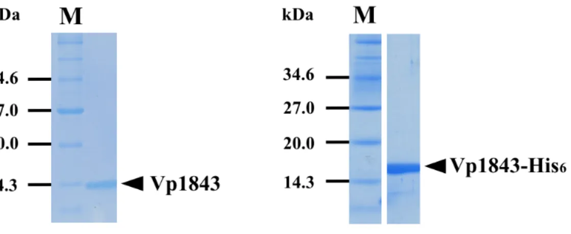
E. coli strain BL21(DE3) Codon Plus RIL harboring pET15b-vp1842/vp1843 or pET22b-vp1842/vp1843 was cultured to log-phase in LB medium (1L). Expression of vp1842/vp1843 was induced by 1 M IPTG at 25°C for 20 h. Cells (5 ~ 6 g) were collected and re-suspended in 100 ml sonication buffer containing 50 mM Tris-HCl, pH 8.0, 500 mM NaCl. After sonication at a mode of (pulse-on for 10 seconds following by pulse-off for 40 seconds with an amplitude of 50) in a progress of 6 min, the supernatant containing total proteins was obtained by centrifuging at 4°C in a speed of 35,000 g for 30 min. Vp1842/Vp1843-His, Vp1843 and Vp1843-His were purified as described in Materials and Methods. The purified samples were applied to 15% SDS- PAGE and electrophoresis at 30 mA for 1 h (Fig. 2-3). The yields of Vp1843 and Vp1843-His were 0.5 mg/L and 4 mg/L, respectively.
Fig. 2-3. Purification of Vp1843 without His6 (left) or with His6 (right).
The toxin Vp1843 was purified as described in Materials and Methods and analyzed by 15% SDS-PAGE. Arrows indicate the location of Vp1843 without His6 (left) or with His6 (right). M indicates the protein markers, bands of 14.3 kDa, 20.0 kDa, 27.0 kDa and 34.6 kDa stand for lysozyme (chiken egg white), trypsin inhibitor (Soybean), triosephosphate isomerase (E. coli) and thioredoxin reductase (E. coli), respectively.
2-3-2. Gyr inhibitory activity
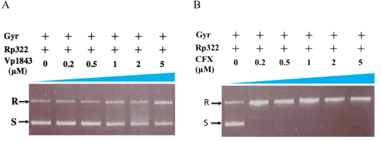
Vp1843 has sequence homology not only to E. coli YafQ, but also to C. crescentus ParE, as shown in Fig. 1-7. Both YafQ and ParE toxins belong to the RelE/ParE superfamily, despite distinct biological activities; YafQ is a protein synthesis inhibitor by cleaving mRNA (Griffin et al., 2013), while ParE has inhibitory activity toward Gyr (Jiang et al., 2012). Since Vp1843 exhibited neither protein synthesis inhibitory activity nor ribonuclease activity (Hino et al., 2014), I tested whether Vp1843 could have any inhibitory activity toward E. coli Gyr using ciprofloxacin (CFX) as a control. In this assay, mixtures containing relaxed pBR322 and Gyr were incubated with an increasing amount of Vp1843 (0 ~ 5 µM) or CFX (0 ~ 5 µM) at 25°C for 4 h, and then, the reaction mixtures were treated with SDS and Proteinase K to detect the Gyr-DNA cleavage
A B
Fig. 2-4. Influence of Vp1843 (A) or CFX (B) on the supercoiling activity of Gyr.
Relaxed pBR322 (200 ng) was mixed with 2 units of Gyr in the presence of Vp1843 (0 ~ 5 µM) (A) or CFX (0 ~ 5 µM) (B) in a reaction buffer containing 35 mM Tris- HCl, 24 mM KCl, 4 mM MgCl2, 2 mM DTT, 5 mM spermidine, 1.75 mM ATP, 6.5%
glycerol, and 0.1 mg/mL BSA. After incubating the reactions at 25°C for 4 h, the mixtures were treated with stop buffer containing SDS (0.2%) and Proteinase K (1 mg/mL) at 37°C for 30 min. The DNA products were analyzed by electrophoresis on a 1% agarose gel in TAE buffer and visualized by ethidium bromide. Rp322, relaxed pBR322; R, relaxed pBR322; S, supercoiled pBR322.
complex on agarose gels, as described previously (Sterckx et al., 2016; Yuan et al.,
Gyr slightly at 25°C for 4 h, accumulating relaxed form DNA in a dose-dependent manner (Fig. 2-4A). However, its inhibitory activity was considerably weaker than CFX (Fig. 2-4B).
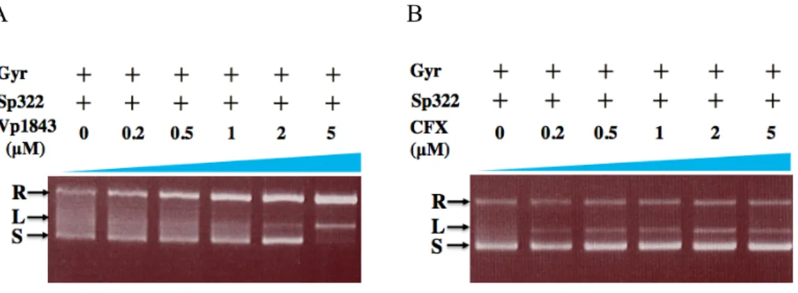
Since it is known that Gyr catalyzes not only DNA supercoiling in the presence of ATP, but also DNA relaxation of supercoiled DNA in the absence of ATP (Collin et al., 2011), I tested whether Vp1843 could inhibit the DNA relaxation activity of Gyr using supercoiled pBR322 as a substrate. In this analysis, the supercoiled pBR322 treated with Gyr was incubated with an increasing amount of Vp1843 (0 ~ 5 µM) or CFX (0 ~ 5 µM) at 25°C for 4 h and then, the reaction mixtures were analyzed as described above. Interestingly, Vp1843 appeared to enhance the Gyr relaxation activity, showing that the dose-dependent decrease of the supercoiled DNA was concomitant
A B
Fig. 2-5. Influence of Vp1843 (A) or CFX (B) on the relaxation activity of Gyr.
Supercoiled pBR322 (200 ng) was mixed with 2 units of Gyr in the presence of Vp1843 (0 ~ 5 µM) (A) or CFX (0 ~ 5 µM) (B) in a reaction buffer containing 35 mM Tris-HCl, 24 mM KCl, 4 mM MgCl2, 2 mM DTT, 5 mM spermidine, 1.75 mM ATP, 6.5% glycerol, and 0.1 mg/mL BSA. After incubating the reactions at 25°C for 4 h, the mixtures were treated with stop buffer containing SDS (0.2%) and Proteinase K (1 mg/mL) at 37°C for 30 min. The DNA products were analyzed by electrophoresis on a 1% agarose gel in TAE buffer and visualized by ethidium bromide.
with the increase of the relaxed form DNA (Fig. 2-5A). In contrast, CFX inhibited the
supercoiled DNA to the relaxed DNA (or open-circular DNA) with nicks by a DNA nicking endonuclease activity.
2-3-3. Vp1843 has a DNA nicking activity
Next, I tested if Vp1843 could convert supercoiled DNA to relaxed DNA (or open- circular DNA) in the absence of Gyr. As expected, Vp1843 alone completely converted supercoiled DNA to relaxed DNA (or open-circular DNA), giving rise linear DNA in a dose-dependent manner (Fig. 2-6).
Fig. 2-6. DNA nicking endonuclease activity of Vp1843.
Supercoiled pBR322 (100 ng) was incubated with Vp1843 (0 ~ 5 µM) in a nicking reaction buffer containing 25 mM Tris-HCl, pH 7.5, 20 mM NaCl, 4 mM MnCl2, 2 mM β-ME, and 6.5% glycerol in 10 µl reaction system. After incubating the reactions at 25°C for 4 h, the DNA products were released by 0.2% SDS and 1 mg/mL Protease K treatment at 37°C for 30 min and then analyzed by electrophoresis on a 1% agarose gel in TAE buffer at 100 V for 50 min and visualized by ethidium bromide. R, L, S stand for relaxed, linear and supercoiled DNA, respectively.
To further corroborate the nicking endonuclease activity of Vp1843, the reaction products were analyzed on 1% agarose gel in TBE buffer instead of TAE buffer, because this gel electrophoresis in TBE buffer is known to separate open-circular DNA
with nicks from relaxed form DNA without nicks, as described previously (Lee et al., 2015). In this electrophoresis, nicked open-circular DNA with one strand cut, relaxed circular DNA without nicks, and linear DNA with free ends are the slowest to the fastest in order of electrophoretic mobility (Lee et al., 2015). As shown in Fig. 2-7, Vp1843 could convert both relaxed form DNA (lane 3) and supercoiled DNA (lane 4) to open- circular DNA, accumulating linearized DNA. I further found that prolong incubation of supercoiled DNA with Vp1843 did not increase the linear products (data not shown), suggesting that the dose-dependent appearance of liner DNA is caused by single- stranded cuts at close sites on opposite strands.
Fig. 2-7. Agarose gel electroporesis of open-circular DNA and relaxed DNA.
Supercoiled pUC19 (100 ng) and relaxed pUC19 (100 ng) was incubated with 5 µM Vp1843 at 25°C for 4 h in the same buffer as in Fig. 2-5, and analyzed by electrophoresis on a 1% agarose gel in TBE buffer at 100 V for 90 min. Lane1, linearized pUC19 by EcoRI; lane 2, relaxed pUC19; lane 3, relaxed pUC19 incubated with Vp1843; lane 4, supercoiled pUC19 incubated with Vp1843; lane 5, supercoiled pUC19. Sp322 indicates supercoiled pBR322, and S, L, R, OC stand for supercoiled, linear, relaxed and open circular DNA, respectively.
2-3-4. Characterization of the nicking activity of Vp1843
As described previously, the E. coli cell growth inhibition by Vp1843 was neutralized by expression of the antitoxin gene vp1842 (Hino et al., 2014). Therefore, I tested whether the antitoxin Vp1842 could influence the Vp1843 nicking activity. The result showed that Vp1842 could neutralize the nicking activity of Vp1843, although linearized DNA slightly accumulated in a dose dependent manner (Fig. 2-8).
Fig. 2-8. Vp1842 neutralize the DNA nicking endonuclease activity of Vp1843.
Supercoiled pBR322 (100 ng) was incubated with Vp1843 or Vp1842/Vp1843 (0 ~ 5 µM) in a nicking reaction buffer containing 25 mM Tris-HCl, pH 7.5, 20 mM NaCl, 4 mM MnCl2, 2 mM β-ME, and 6.5% glycerol in 10 µl reaction system. After incubating the reactions at 25°C for 4 h, the DNA products were released by 0.2%
SDS and 1 mg/mL Protease K treatment at 37°C for 30 min and then analyzed by electrophoresis on a 1% agarose gel in TAE buffer at 100 V for 50 min and visualized by ethidium bromide. O, L, S stand for open circular, linear and supercoiled DNA, respectively.
This finding is consistent with the fact that the co-expression of vp1842 and vp1843 slightly influenced the E. coli cell growth as compared with that of E. coli cells ontaining pBAD/Myc-HisA (Hino et al., 2014). Nevertheless, the result indicates that the strong toxicity of Vp1843 in E. coli is attributable to DNA damage by its nicking
It is known that almost all DNA nicking endonucleases exhibit catalytic activity in the presence of a divalent metal ion (Pingoud et al., 2014; Xu., 2015). The reaction buffer used for the nicking activity for Vp1843 contained 4 mM Mg2+. I first examined whether Vp1843 requires Mg2+ ion for its enzymatic activity. As shown in Fig. 2-9, although Vp1843 converts supercoiled pBR322 to relaxed DNA in the presence of
Fig. 2-9. Vp1843’s nick activity needs divalent Mg2+.
Vp1843 (5 µM) was incubated with 100 ng supercoiled pBR322 in different buffers, containing Na+ or K+ or Mg2+. Reaction conditions are the same as in materials and methods. Buffer A: 25 mM Tris-HCl, pH 7.5 and 20 mM NaCl; B1: 25 mM Tris-HCl, pH 7.5, 20 mM NaCl and 20 mM MgCl2; B2: 25 mM Tris-HCl, pH 7.5 and 20 mM KCl; B3: 25 mM Tris-HCl, pH 7.5, 20 mM KCl and 20 mM MgCl2; C: 25 mM Tris- HCl, pH 7.5, 20 mM NaCl and 20 mM MgCl2, 2 mM β-ME; D: 25 mM Tris-HCl, pH 7.5, 20 mM NaCl and 20 mM MgCl2, 2 mM β-ME, 6.5% glycerol; E: 25 mM Tris- HCl, pH 7.5, 20 mM KCl and 20 mM MgCl2, 2 mM β-ME, 6.5% glycerol. Sp322, supercoiled pBR322; OC, open circular DNA; L, linearized DNA; S, supercoiled DNA.
Mg2+, no conversion from supercoiled DNA to relaxed DNA was detected in the absence of Mg2+, demonstrating requirement of Mg2+ for the enzymatic activity of
Next, we tested preference of divalent metal ions for the Vp1843 activity using MnCl2 and CaCl2 in place of MgCl2. Vp1843 completely converted supercoiled pBR322 to open-circular DNA generating a small amount of linear DNA in the presence of Mn2+, while supercoiled DNA was partially converted to open-circle DNA in the presence of Ca2+, indicating that Vp1843 efficiently nicks DNA in the presence of a divalent ion in the order of Mn2+ > Mg2+ > Ca2+ (Fig. 2-10).
A
B
Fig. 2-10. Ion preference of Vp1843.
(A) Supercoiled pBR322 (100 ng) was incubated with Vp1843 (5 µM) or without Vp1843 (as a control) in a nicking reaction buffer containing either MgCl2, MnCl2
or CaCl2, and the DNA products were analyzed in the same manner as described in Materials and Methods. Sp322 indicates supercoiled pBR322, and S, L, OC stand for supercoiled, linear, and open circular DNA, respectively. (B) The relative conversion from supercoiled DNA to open-circle DNA and linear DNA were used to evaluate the activity of Vp1843. Image J software was utilized to digitalize the results of electrophoresis.
I further examined the nicking activity of Vp1843 at different pH from 5.7 to pH 8.5.
The appearance of bands standing for open-circle DNA (OC) indicated that Vp1843 was active in these conditions. This result showed that the optimum pH value for Vp1843 is pH 7.0 to pH 8.5. (Fig. 2-11).
A
B
Fig. 2-11. pH dependency of the nicking activity of Vp1843.
(A) Supercoiled pBR322 (100 ng) was incubated with Vp1843 (0 µM, 25 µM) in the nicking reaction buffer with Mn2+ at different pH as indicated above. To exclude the possible effect of pH buffer, three kinds of pH buffer (Bis-Tris buffer, Tris buffer, Bicine buffer) were selected and examine the nicking activity of Vp1843. After incubating the reactions for 1 h, the DNA products were analyzed in the same manner as described in Materials and Methods. (B) The conversion rate of supercoiled DNA into open-circle DNA and linear DNA were used to evaluate the activity of Vp1843.
The Image J software was utilized to digitalize the results of electrophoresis.
To find the optimum temperature for Vp1843, I also examined the nicking activity of Vp1843 at a temperature range from 10°C to 50°C. The result indicated that Vp1843 was active in these conditions. DNA nicking activity of Vp1843 was about 70% at 10°C, while 90% from 25°C to 50°C (Fig. 2-12), indicating the optimum temperature of Vp1843 is 25 ~ 50°C.
A
B
Fig. 2-12. Vp1843 is active at a variety of temperature.
(A) Supercoiled pBR322 (100 ng) was incubated with Vp1843 (0 µM, 5 µM, 25 µM) in the nicking reaction buffer with Mn2+ at different temperatures as indicated above.
After incubating the reactions for 1 h, the DNA products were analyzed in the same manner as described in Materials and Methods. (B) The conversion rate of supercoiled DNA into open-circle DNA and linear DNA were used to evaluate the activity of Vp1843. The Image J software was utilized to digitalize the results of electrophoresis.
2-3-5. Essential residues in Vp1843
Previously, we found that the gene cluster vp1829/vp1830, a paralog of vp1842/vp1843, has significant homology to that encoding DinJ/YafQ of E. coli (Hino et al., 2014). However, the expressions of vp1830 in the presence of 0.2% arabinose had no influence on cell growth, indicating that the protein Vp1830 has no toxicity, even though there are only 9 amino acid replacements between Vp1830 and Vp1843 (Fig. 1-7). The subsequent mutational analysis showed that replacing Asn37 or Leu45 in Vp1830 with the corresponding residue Lys37 or Pro45 in Vp1843 significantly
Fig. 2-13. Inhibition of E. coli cell growth by expressing vp1843 or its mutant genes.
E. coli cells harboring the pBAD/Myc-HisA plasmid containing the vp1843 gene were cultured at 37°C in the presence of 0.2% glucose (upper panel) or 0.2%
arabinose (lower panel), and the cell growth was monitored at absorbance of 590 nm.
inhibited the E. coli growth, suggesting that Lys37 and Pro45 in Vp1843 play an essential role in the toxicity toward E. coli cells (Hino et al., 2014). Therefore, mutant genes encoding K37N, P45L and K37N/P45L, in which Lys37 and Pro45 in Vp1843 were individually and simultaneously replaced by the corresponding residues Asn and Leu in Vp1830 respectively, were prepared and their influence on the E. coli growth in the presence of 0.2% arabinose was tested. As shown in Fig. 2-13, P45L or K37N/P45L expression had little influence on the cell growth, although the K37N expression slightly reduced the cell growth as compared with that of E. coli cells with the plasmid vector. This result demonstrated the crucial role of Lys37 and Pro45 in Vp1843 toxicity and suggested that the latter is more essential for the toxicity than the former.
Consequently, K37N, P45L, and K37N/P45L were purified as described in materials
Figure. 2-14. Purification of Vp1843 mutants.
Toxins were purified as described in Materials and Methods and were analyzed by 15% SDS-PAGE. Lane 1, Vp1843-K37N/P45L-His6; lane 2, Vp1843-P45L-His6; lane 3, Vp1843-K37N-His6; lane 4, Vp1843-WT-His6. M stands for protein molecular weight marker: bovine serum albumin (66.4 KDa), Glutamic dehydrogenase bovine liver (55.6 KDa), maltose-binding protein from E. coli (42.7 KDa), thioredoxin reductase from E. coli (34.6 KDa), triosephosphate isomerase from E. coli (27.0 KDa), trypsin inhibitor from soybean (20.0 KDa), lysozyme from chiken egg white (14.3 KDa).
and methods (Fig. 2-14). The enzymatic potency of K37N, P45L, and K37N/P45L was examined using supercoiled pBR322 as a substrate. The two mutants exhibited little DNA nicking activity toward the supercoiled DNA, as shown in Fig. 2-15, demonstrating that Lys37 and Pro45 in Vp1843 play a crucial role in DNA nicking activity. This result further supported that the DNA nicking activity of Vp1843 is responsible for its high toxicity in E. coli cells.
Fig. 2-15. Nicking endonuclease activity of Vp1843 mutants K37N, P45L and K37N/P45L.
Supercoiled pBR322 (100 ng) was incubated with either WT, K37N, P45L, or K37N/P45L of Vp1843 in the nicking reaction buffer. After incubating the reactions at 37°C for 1 h, the DNA products were analyzed in the same manner as described in Materials and Methods. Sp, supercoiled pBR322; OC, L and S indicate open-circular, linear and supercoiled DNA, respectively.
2-4. Summary
The TA system vp1842/vp1843 found in V. parahaemolyticus, has sequence homology to that encoding the E. coli TA protein, DinJ/YafQ, However, the toxin Vp1843, unlike its E. coli homologue YafQ, has neither protein synthesis inhibitory activity nor ribonuclease activity. Rather, the expression of vp1843 in E. coli resulted in a morphological change in the cells, which could not be seen in the cell expression of YafQ, indicating that Vp1843 has a distinctive activity with that of YafQ.
Meanwhile, we found that Vp1843 has sequence homology not only to E. coli YafQ, but also to C. crescentus ParE (Fig. 1-7) (Dalton et al., 2010). Phylogenetic studies showed that YafQ and ParE belong to the RelE/ParE superfamily (Fig. 1-8). Toxins belonging to this family share a low similarity in their primary structures, but fold into a similar fold, despite the fact that they display distinct biochemical activities. YafQ is a representative of RelE toxins, performing as a mRNA interferase, while ParE from Caulobacter crescentus stands for the ParE toxins, poisoning towards Gyr.
I investigated the inhibition activity of Vp1843 towards Gyr and I found that Vp1843, unlike ParE toxins, exhibits a nicking endonuclease activity. Further characterization on the nicking endonuclease activity of Vp1843 showed that a divalent metal ion is essential for the nicking activity of Vp1843, and the ion preference for Vp1843 is in the order of Mn2+ > Mg2+ > Ca2+. It was also proved that the antitoxin Vp1842 is able to block the nicking activity of Vp1843. I found further that Vp1843 exhibits its nicking activity with an optimum temperature at 25 ~ 50°C and an optimum pH at 7.0 ~ 8.5.
Mutations of Lys37 and Pro45 in Vp1843 abolished its nicking activity, suggesting that they play a crucial role in the nicking endonuclease activity.
Chapter 3
Involvement of Vp1842/Vp1843 in the VBNC state
3-1. Introduction
V. parahaemolyticus, a seafood enteropathogen in coastal countries, causes acute gastroenteritis in humans (Nair et al., 2007). A characteristic feature of V.
parahaemolyticus is that it can become nonculturable at a low temperature in a minimum medium while maintaining at least some metabolic activity, but can be recovered by a temperature up-shift treatment (Kaneko and Colwell., 1975). This phenomenon is termed ‘viable but not culturable’ (VBNC). Since Colwell and coworkers first reported on the VBNC state (Xu et al., 1982), this phenomenon has now been described for over 50 bacterial species using various criteria for viability and discussed in relation to dormancy and persistency (Oliver et al., 2010).Although the VBNC state has been believed to be a survival strategy in response to certain harsh environmental stresses, no specific factors have been identified because many environmental conditions induce the VBNC state in different bacterial species.
Recently, there is a growing evidence that activation of TA systems is one of the mechanisms known to trigger such a state with low metabolic activity (Hayes et al., 2009; Ayrapetyan et al., 2015).
In the previous study (Hino et al., 2014), we found that the vp1843 expression exhibited severe toxic to E. coli cells. In order to examine if the TA system vp1842/vp1843 is involved in the induction of VBNC state in V. parahaemolyticus, I constructed a vp1842/vp1843 deficient strain (Δvp1842/vp1843) by homologous recombination using a suicide vector pYAK1 and investigated if it could still enter into the VBNC state. A gene map of the suicide vector pYAK1 is shown in Fig. 3-1.
Fig.3-1. A gene map of pYAK1.
The suicide vector pYAK-1 contains a counter-selection marker sacB gene, a selection marker cat gene, a multiple cloning site, a mob region of the RP4 plasmid and an R6K ori region from plasmid R6K. The sacB gene encodes levane saccharase that converts sucrose to levans, which is harmful to bacteria (Reyrat et al., 1998). The cat gene encodes chloramphenicol acetyltransferase and confers chloramphenicol resistance. The R6K ori initiates replication of the plasmid in pir strains, while not in V. parahaemolyticus. The mob region mediates transformation of this plasmid to host cells.
3-2. Materials and Methods
3-2-1. Stains
E. coli strain BW19851(F-, RP4-2(Km:Tn7,Tc::Mu-1), ΔuidA3::pir+, recA1, endA1, thiE1, hsdR17, creC510) was used to amplify the suicide vector pYAK1. pYAK1 is a R6K origin plasmid, which cannot self-replicate in the absence of , a functional protein encoded by the pir gene (Kolter et al., 1978). Here, E. coli stain BW19851 provides protein for the replication of pYAK1. V. parahaemolyticus O3:K6 (TDH+
RIMD2210633) wild-type was used to construct a vp1842/vp1843 deficient strain.
3-2-2. Plasmids
The suicide vector pYAK1 was kindly provided by Prof. M. Ito and Associate Prof.
N. Okino (Kyushu University).
3-2-3. Mediums and reagents
E. coli BW19851 was cultured in LB medium (1% peptone, 0.5% Yeast Extract, 1%
NaCl, with or without 20 mg/ml chloramphenicol). V. parahaemolyticus was cultured in NB medium (1% peptone, 0.5% Yeast Extract, 3% NaCl). V. parahaemolyticus cells completed homologous recombination were screened by TCBS medium (EIKEN CHEMICAL, Japan).
3-2-4. Construction of the suicide vector pYAK-Δvp1842/vp1843-flanking
This was done by two-steps of PCR. First, the DNA fragment of vp1842/vp1843 with 856 bp upstream and 635 bp downstream flanking sequences was amplified from the genome DNA of wild type V. parahaemolyticus with Phusion Hot Start Flex DNA Polymerase (New England Biolabs) using primers upstream-1842-BamHI-F and 1843- downstream-SphI-R (Table 3-1). After treating by BamHI and SphI, the resultant was
ligated into the suicide vector pYAK1 (Fig. 3-2), and pYAK-vp1842/vp1843-flanking was used for transformation of E. coli BW19851 strain.
Table 3-1. Oligonucleotide used in this study.
Oligonucleotide Sequence (5’-3’)
upstream-1842-BamHI-F 5’-TGCGGATCCGTCTTAAAGCGTTTGTTAGG-3’
1843-downstream-SphI-R 5’-GCAGCATGCTTCAATTGAAGTTCTCAGAAA
CAAGTC -3’
Δ184243-Primer-F 5’-CTCAAAGCΔCGTGTACAACACCAGAAACAA AAATTCCC-3’
Δ184243-primer-R 5’-GTACACGGCTTTGAGATGGGCAACTAACAA ACAATTTAAGAG-3’
Fig. 3-2. Construction of the suicide vector pYAK-Δvp1842/vp1843-flanking.
The DNA fragment of vp1842/vp1843 with 856 bp upstream and 635 bp downstream flanking sequences was amplified and ligated into the suicide vector pYAK1 as described in Materials and Methods. The confirmed plasmid pYAK1- vp1842/vp1843-flanking was used as a template for the second PCR to delete vp1842/vp1843, resulting in a plasmid of pYAK1-Δvp1842/vp1843-flanking. Up and Down indicate upstream and downstream flanking sequences, respectively.
Colonies grown on LB plate with chloramphenicol (20 µg/ml) were further selected by colony PCR with GoTaq Green Master Mix (Promega). The confirmed plasmid
pYAK1-vp1842/vp1843-flanking was amplified, extracted, and used as a template for the second PCR. Primers Δ184243-Primer-F and Δ184243-primer-R were used to delete the vp1842/vp1843 genes from this plasmid by PrimerSTAR Mutagenesis Basal Kit (TaKaRa), resulting in the plasmid of pYAK1-Δvp1842/vp1843-flanking (Fig. 3- 2). The construction was confirmed by colony PCR and DNA sequencing and the succeeded suicide vector with only the flanking sequences of vp1842/vp1843 was used in the following conjugation experiment.
3-2-5. Knockout of the vp1842/vp1843 genes using homologous recombination
Knockout of the vp1842/vp1843 gene cluster was carried out by the allelic exchange procedure, as shown in Fig. 3-3. The experiment was carried out as reported previously (Whitaker, et al., 2014). A single colony of E. coli stain BW19851 harboring the suicide vector pYAK-Δvp1842/vp1843-flanking and a single colony of wild type V.
parahaemolyticus (RIMD 2210633) were individually grown in a suitable medium (LB with or without chloramphenicol) at 37°C for 14-16 h. Then, the cells centrifuged at 3,500 g for 5 min were re-suspended in 100 µl LB and mixed well. The mixtures were transferred to a 0.2 µm sterile nitrocellulose membrane filter (Toyo Roshi Kaisha, Japan) located on the surface of an LB plate without antibiotics and incubated at 25°C overnight. The cells were transferred from the membrane to 5 ml LB medium containing 20 µg/ml chloramphenicol and keep at 25°C for 2 h. Then, the cells were spread onto a TCBS (EIKEN CHEMICAL, Japan) plate with 20 µg/mL chloramphenicol and incubated at 25°C overnight. Several colonies were selected, streaked on a TCBS plate containing 10% sucrose and incubated at 37°C for 5 h. For each ancestor cell, 3 colonies of subsequent generations were selected and streaked onto a novel TCBS plate containing 10% sucrose. The knockout of the vp1842/vp1843 gene cluster in the anticipated cells, Δvp1842/vp1843, was confirmed by colony PCR as well as by DNA sequencing.
Fig. 3-3. Allelic exchange procedure and Vp-Δvp1842/vp1843 construction.
Step 1: The plasmid pYAK1-Δvp1842/vp1843 integrated into the genome of V.
parahaemolyticus, and the intermediate strain was selected by TCBS medium with 20 µg/ml chloramphenicol. Step 2: Plasmid excised with gene cluster vp1842/vp1843 and the deficient strain were selected by TCBS with 10% sucrose. Step 3: Plasmid removal happened due to the inability of replication in V. parahaemolyticus. Colony PCR was utilized to select the deficient strain.
3-2-6. Induction into the VBNC state
Induction of V. parahaemolyticus wild-type or its mutant Δvp1842/vp1843 into the VBNC state was carried out as described previously (Hino et al., 2014) with some modifications. The cells were cultured individually in NB medium (EIKEN CHEMICAL, Japan) containing 3% NaCl at 37°C until 0.3 of OD660. The cells were gently harvested with 5,000 rpm for 15 min and washed 3 times with 1.85% NaCl. The cells were re-suspended in 1.85% NaCl, and the starting number of cells was adjusted to 1 x 10⁶ CFU/ml. The cells were then incubated at 10°C without shaking for 15 days.
Numbers of total cells, viable cells, and culturable cells were counted every day with a Live/Dead BacLight Bacterial Viability Kit L-7007 (Molecular Probes, Waltham, MA) and viewed by fluorescence microscopy. Viable cells with intact membranes were stained green by the fluorescent SYTO 9, while dead cells with compromised membranes were stained red by propidium iodide.
3-2-7. Transcriptome analysis
Total RNA was extracted from V. parahaemolyticus in both the normal state and the VBNC state using RNeasy Mini Kit (QIAGEN) and precipitated by 100% ethanol.
Then, the RNAs (20µg/100µl) were subjected to next generation sequencing (Macrogen). Functional annotation analysis of the results was performed using DAVID (http://david.abcc.ncifcrf.gov/) based on Gene Ontology (http://geneontology.org/), KEGG (http://www.genome.jp/kegg/ ) and other functional annotation databases.
3-3. Results
3-3-1. Knock out of vp1842/vp1843 genes from the V. parahaemolyticus genome by conjugation.
Previous studies proved that V. parahaemolyticus cells cultured in 1.85% NaCl at 4°C could enter into the VBNC state within two weeks (Hino et al., 2014). In order to investigate whether the TA system vp1842/vp1843 is involved in the induction into the
A
B
Fig. 3-3. Preparation of the V. parahaemolyticus strain ∆vp1842/vp1843.
A; Gene organization neighboring the vp1842/vp1843 gene in V. parahaemolyticus is schematically presented. The genes encoding Vp1842 and Vp1843 are shown in dark grey and flanking genes are in white boxes. F and R primer sets indicated by black arrows were designed in the 5’ and 3’ flanking regions of vp1843 and vp1842 genes, respectively. B; PCR analysis of the gene encoding Vp1843 and Vp1842 confirming deletion of the gene segments in five ∆vp1842/vp1843 strains (lanes 1- 5). DNA size markers were run in lane M.
VBNC state, I knocked out vp1842/vp1843 from the V. parahaemolyticus genome using homologous recombination as described in Materials and Methods. As a result, five transformants termed ∆vp1842/vp1843 strains were obtained, and they were evaluated by PCR using primer sets designed to amplify the target gene segment with the 5’ and 3’ flanking regions, as shown in Fig. 3-3. The PCR amplification of each disruptive clone gave DNA products with predicted sizes around 1,500 bp, corresponding to the lengths of flanking genes (Fig. 3-3). In contrast, the genomic DNA from the parent strain indicated amplification of the expected length of the PCR product, corresponding to the sizes of vp1842/vp1843 and additional flanking regions, as shown in Fig. 3-3. However, when five clones were further cultivated, the clone 5 only gave a single band corresponding to 1,500 bp, while other four clones provided several DNA bands by colony PCR (Data not shown). This finding demonstrated that homologous recombination occurred at the target vp1842/vp1843 gene in the V. parahaemolyticus strain 5 (∆vp1842/vp1843).
3-3-2. Involvement of vp1842/vp1843 in the VBNC state of V. parahaemolyticus
After I obtained the colony (strain5: ∆vp1842/vp1843) with vp1842/vp1843 deficient, I tested to see if it could still enter the VBNC state. The cells were cultured individually in NB medium (EIKEN CHEMICAL, Japan) containing 3% NaCl at 37°C until 0.3 of OD660. The cells were gently harvested after 5,000 rpm for 15 min and washed 3 times with 1.85% NaCl. The cells were re-suspended in 1.85% NaCl, and the starting number of cells was adjusted to 1 x 10⁶ CFU/ml. The cells were incubated at 10°C without shaking for 15 days. Numbers of total cells, viable cells, and culturable cells were counted every day with a Live/Dead BacLight Bacterial Viability Kit L-7007 (Molecular Probes, Waltham, MA) and viewed by fluorescence microscopy. Viable cells with intact membranes were stained green by the fluorescent SYTO 9, while dead cells with compromised membranes were stained red by propidium iodide. As shown
into the VBNC state under stress conditions at a comparable rate with that of the wild- type. Although it is premature on the basis of only a single deletion mutant to draw firm conclusions, this result suggested that the TA system vp1842/vp1843 is not involved in the VBNC state in V. parahaemolyticus. Alternatively, vp1842/vp1843 may be synergistically involved in the VBNC state with other TA systems in V.
parahaemolyticus.
Fig. 3-4. Involvement of the TA system vp1842/vp1843 in the VBNC state.
Induction of V. parahaemolyticus wild-type or its mutant Δvp1842/vp1843 into the VBNC state was carried out as described previously (Hino et al., 2014) with some modifications.
3-3-3. Expression level of vp1842/vp1843 in the VBNC state
The knock-out experiment suggested that the TA system vp1842/vp1843 is not involved in induction into the VBNC state. In order to confirm this assumption, I compared an expression level of vp1842/vp1843 in the VBNC state with that in normal growth condition by transcriptome analysis using next-generation sequencer.
The result showed that the amounts of transcripts for vp1842/vp1843 and
vp1829/vp1830 in the VBNC state were comparable to those in normal growth conditions (Table 3-2). This result further supported that vp1842/vp1843 is not responsible for the VBNC state. Interesting, the deep sequencing analysis showed that the transcript of another TA system vp1821/vp1820 which is a homolog of E.
coli TA system yefM/yoeB located within SI in the V. parahaemolyticus chromosome I increased by 4-folds as compared with that in the normal growth conditions, suggesting its involvement in the VBNC state. Further studies will be required for definition of its implication in the VBNC state.
Table 3-2. Expression level of possible TA systems in the VBNC state and normal state
Gene ID b/a.fc N_a N_b Homologues
in E. coli
Identity of Proteins
vp1842 -1.235 6.776 6.471 dinJ 22%
vp1843 1.295 6.536 6.910 yafQ 17%
vp1829 -1.436 5.207 4.685 dinJ 23%
vp1830 1.034 4.462 4.511 yafQ 18%
vp1821 4.052 7.634 9.652 yefM 45%
vp1820 4.229 7.383 9.463 yoeB 63%
• a and b stand for the normal state and the VBNC state, respectively.
• b/a.fc means fold change between two samples using normalized value. A positive value means that the gene in sample b is up-regulated and a negative value means that it is down-regulated. fc=2^(N_b/N_a).
• N stands for normalized value of gene expression by Quantile Normalization.
3-4. Summary
I knocked out the TA system vp1842/vp1843 from the V. parahaemolyticus genome and examined if the mutant (Δvp1842/vp1843) could enter into the VBNC state. The result showed that vp1842/vp1843 deficient strain-∆vp1842/vp1843, like the wild type V. parahaemolyticus, still be able to enter into the VBNC state. This result suggested that vp1842/vp1843 is not involved in the induction of the VBNC state. To confirm this assumption and also to find genes responsible for the VBNC state, a trancriptome analysis using next-generation sequencer was performed. The result showed that the vp1842/vp1843 transcript was not up-regulated in the VBNC state. But I found that another TA system vp1821/vp1820, which encodes homologues of YefM and YoeB in E. coli, showed 4-fold increase in the VBNC state. The involvement of Vp1820 in V.
parahaemolyticus will be further examined in the future. Further studies will be required to define molecules involved in the VBNC state in V. parahaemolytics.