folA, a New Member of the TyrR Regulon in Escherichia coli K‑12
著者 YANG Ji, OGAWA Yoshito, CAMAKARIS Helen, SHIMADA Tomohiro, ISHIHAMA Akira, PITTARD A.
J.
出版者 American Society for Microbiology journal or
publication title
Journal of bacteriology
number 16
page range 6080‑6084
year 2007‑08
URL http://hdl.handle.net/10114/749
0021-9193/07/$08.00⫹0 doi:10.1128/JB.00482-07
Copyright © 2007, American Society for Microbiology. All Rights Reserved.
folA, a New Member of the TyrR Regulon in Escherichia coli K-12
䌤Ji Yang,
1Yoshito Ogawa,
1,2Helen Camakaris,
1Tomohiro Shimada,
2Akira Ishihama,
2and A. J. Pittard
1*
Department of Microbiology and Immunology, The University of Melbourne, Victoria 3010, Australia,1and Department of Frontier Bioscience and Micro-Nano Technology Research Center, Hosei University, Koganei, Tokyo 184-8584, Japan2
Received 29 March 2007/Accepted 29 May 2007
ThefolAgene was identified as a new member of the TyrR regulon by genomic SELEX. Binding of TyrR to two sites infolA activated its transcription. Mutations in the N-terminal or central domain of TyrR, the␣ subunit of RNA polymerase, or integration host factor all abolished activation of thefolApromoter.
The TyrR regulon ofEscherichia coliK-12 comprises at least eight separate transcription units (6). These various transcrip- tion units have been identified over time by observing fluctu- ations in the levels of particular proteins caused by the pres- ence or absence of the amino acid phenylalanine or tyrosine in the medium or as a result of the introduction of various mu- tations into the regulator genetyrR. In addition to the tyrR gene itself, all of the known genes of the regulon encode proteins which have a role in either the biosynthesis or trans- port of the aromatic amino acids. Flanking or overlapping the promoters of each of these transcription units are 18-bp se- quences to which the TyrR protein selectively binds and which are referred to as TyrR boxes (6). These sequences are related to the palindrome TGTAAAN6TTTACA and vary from one another in both their location and their agreement with the ideal consensus. The only absolutely invariant feature of these boxes is the G, the C, and the spacing of 14 bp between them.
In solution, TyrR protein is a dimer, but in the presence of tyrosine and ATP, it self-associates to form a hexamer (11).
Although phenylalanine is reported not to facilitate hexamer- ization at physiological concentrations, it does facilitate the binding of TyrR dimers to multiple binding sites (12).
In the case of activation, TyrR protein is bound to a TyrR box upstream of the promoter. The addition of one of the aromatic amino acids induces a change in the protein, facili- tating an interaction between the N-terminal domain of TyrR and the␣subunit of RNA polymerase, resulting in enhanced transcription.
Using the method of genomic SELEX (9) with purified TyrR protein to identify TyrR-specific DNA binding sites, we iden- tified seven of the eight known members of the TyrR regulon and five other possible new members.lacZtranscriptional fu- sions were constructed to test whether the transcription of any of these five genes exhibited TyrR-mediated responses in vivo.
Expression of only one of these, namely,folA, encoding the enzyme dihydrofolate reductase, showed clear and unambigu- ous TyrR-mediated effects. This paper reports a genetic anal-
ysis of the interactions between TyrR protein and this new member of the TyrR regulon.
Selection and identification of TyrR-bound fragments by SELEX.A plasmid library carrying randomE. coliK-12 frag- ments of 100 to 300 bp was constructed as described previously (9). Using this library as templates, linear genomic DNA frag- ments were amplified by PCR, and the resulting PCR frag- ments were used in SELEX. The binding assay to detect fragments with TyrR binding sites used a purified TyrR protein carrying six histidine residues at its amino terminal end (H6- TyrR). The selection was carried out under three different conditions: first, with H6-TyrR alone; second, with H6-TyrR and ATP (ATP increases the affinity between TyrR and TyrR boxes [6]); and third, with H6-TyrR, ATP, and tyrosine (ATP and tyrosine facilitate TyrR hexamer formation [11]). Follow- ing incubation at 37°C for 30 min, the H6-TyrR–DNA com- plexes were purified by chromatography on Ni-nitrilotriacetic acid columns. The H6-TyrR-bound DNA fragments were then cloned into vector pT7Blue (Novagen). A total of 250 clones from the three experiments were sequenced. Following an analysis of the sequences, those fragments which covered the promoter region of a coding sequence and contained at least one recognizable TyrR box were identified.
The results in Table 1 are the accumulated results from the three experiments. Except foraroG, which possesses a single TyrR box, all of the known transcription units of the TyrR regulon were identified. The least effective screen was H6- TyrR alone, which yielded only fragments witharoFandtyrP promoters. In the presence of ATP (100M), a much wider range of fragments were selected, in both the absence and presence of tyrosine (100M). Two members of the regulon, tyrB and aroP, were obtained only with the screen including ATP and tyrosine. The most frequent isolates overall (aroF andaroL) contain multiple boxes with strong resemblances to the ideal consensus. In addition to the known members of the TyrR regulon, we identified five new targets,cusC,cyaA,proP, folA, and holE, whose expression could theoretically be af- fected by TyrR. These genes encode a number of unrelated functions. The genecusCencodes a component of a copper transport system,cyaAencodes adenyl cyclase,proPencodes a proline specific permease, folAencodes dihydrofolate reduc- tase, andholEencodes a component of DNA polymerase III.
Furthermore,cyaA,folA, andholEhave also been identified by algorithm as possible members of the TyrR regulon (4, 8).
* Corresponding author. Mailing address: Department of Microbi- ology and Immunology, The University of Melbourne, Victoria 3010, Australia. Phone: 03-8344-5696. Fax: 03-9347-1540. E-mail: aj.pittard
@microbiology.unimelb.edu.au.
䌤Published ahead of print on 8 June 2007.
6080
by Akira Ishihama on August 29, 2007 jb.asm.orgDownloaded from
In order to determine the consequences of TyrR binding for gene expression, we constructed promoter/operator-lacZ fu- sions for each of the genes under consideration using the single-copy vector pMU2385 (7). Each of the promoter/oper- ator-lacZfusions was introduced into strains that were tyrR (tyrR366), haploidtyrR⫹, or multicopy tyrR⫹ with the tyrR⫹ gene present on plasmid pACYC177. Cultures were grown in minimal medium and in minimal medium supplemented with phenylalanine or with tyrosine. The only gene to show signif- icant modulation of expression in a haploidtyrR⫹strain and enhanced activation in a multicopytyrR⫹background wasfolA.
The data for the other four genes whose expression was not regulated by TyrR are not shown. The data forfolAare shown in Table 2, along with results obtained using varioustyrRmu- tants and strains with mutations affecting integration host fac- tor (IHF) production and the␣subunit of RNA polymerase.
The mutant tyrR alleles, each of which has been previously described (6), were cloned on plasmid pSU39, a pACYC177 derivative, and introduced into strain JP8042, which has atyrR null phenotype (tyrR366) and also carries the plasmid with the folA-lacZfusion (single copy).
ThetyrRRQ10allele specifies a protein with an arginine-to- glutamine substitution at position 10 of the TyrR N-terminal domain which inactivates a putative aromatic amino acid bind- ing site involved in activation. The aspartate-to-alanine change at position 103 (tyrRDA103) affects a putative interaction site between the N-terminal domain and the␣ subunit of RNA polymerase. Both mutants have been shown to have a greatly reduced ability to activate expression oftyrP,aroP, andmtr(6).
The host strain with a mutantrpoAallele,rpoADN250,has been shown also to be unable to activate expression oftyrPormtrin tyrR⫹strains (6).
The deletion of residues 226 to 410 in the central domain of TyrR (TyrRD226-410) destroys the ability of TyrR protein to hexamerize and to repress a number of genes specifically re- pressed by tyrosine (6). The more specific change in the central domain,tyrREQ274, has a similar effect on tyrosine-mediated regulation (3).
Because of certain similarities between the regulation of folA and the TyrR-mediated regulation of the gene tpl in Citrobacter freundii (1), we also used a himA himD double
mutant to investigate a possible role for IHF in TyrR-mediated regulation offolA.
Overall, the results in Table 2 clearly show that tyrosine- mediated activation of folA requires first a fully functional TyrR N-terminal domain to carry out aromatic amino acid binding and interaction with the␣subunit of RNA polymer- ase, as is the case fortyrP,aroP, andmtr. Unlike the situation with these genes, activation offolAalso requires a fully func- tional TyrR central domain, which is necessary for the binding of a second molecule of tyrosine and the subsequent self- association to form a hexamer. The failure to observe signifi- cant activation in the IHF-negative strain indicates that in this case, IHF presumably is required to facilitate bending of the DNA to bring the TyrR protein close enough to the promoter to allow interactions with the␣subunit of RNA polymerase.
The weaker phenylalanine-mediated activation shows the same absolute requirement for a fully functional N-terminal domain, but the requirement for a fully functional central domain is less marked.
Specific interaction of the TyrR protein with thefolAregu- latory region.Inspection of thefolAregulatory region revealed the presence of three putative TyrR boxes which are centered at positions⫺106.5 (TyrR box 1),⫺145.5 (TyrR box 2), and
⫺186.5 (box 3) (Fig. 1). DNase I footprinting was carried out to characterize the interaction of the TyrR protein with the folAregulatory region. The32P-labeledfolAfragment spanning the region between positions⫺254 and⫹1 relative to the start site of transcription was generated by PCR. The32P-labeled folAfragment was incubated with various amounts of native TyrR protein (11) in the presence of ATP (100 M) and phenylalanine (100M) or tyrosine (100M). Binding reac- tions were carried out for 15 min at 37°C in a total volume of 25l of transcription buffer (50 mM Tris-HCl [pH 7.8], 50 mM NaCl, 3 mM magnesium acetate, 0.1 mM EDTA, 0.1 mM dithiothreitol, and 25g/ml bovine serum albumin). The sam- ples were then treated with 0.02 unit of DNase I (Boehringer Mannheim) to allow partial digestion of the DNA. After incu-
TABLE 2. TyrR-mediated activation offolA-lacZfusion
Phenotype of host strain
TyrR phenotype of plasmida
-Galactosidase sp act (fold activation) expressed from
folA-lacZfusion in the indicated mediumb
MM Tyr Phe
TyrR⫺ NAc 108 110 (1) 102 (1)
TyrR⫹ NA 103 271 (2.6) 127 (1.2)
TyrR⫺ TyrR⫹ 101 458 (4.5) 267 (2.7)
TyrR⫺ TyrRRQ10 105 155 (1.5) 96 (1)
TyrR⫺ TyrRDA103 100 190 (1.9) 150 (1.5)
TyrR⫺ TyrR⌬226–419 110 188 (1.7) 231 (2.1)
TyrR⫺ TyrREQ274 95 139 (1.5) 201 (2.1)
HimA⫺HimD⫺ TyrR⫹ 36 54 (1.5) 46 (1.3)
RpoADN250 TyrR⫹ 40 45 (1.1) 60 (1.5)
atyrR⫹was carried on plasmid pACYC177, and mutanttyrRalleles were carried on pACYC177 derivative pSU39.
bUnits are those defined by Miller (5). The values of-galactosidase activities are averages from three independent assays, with the standard deviation below 15%. MM, minimal medium; Tyr, MM containing 1 mM tyrosine; Phe, MM containing 1 mM phenylalanine. Fold activation is the specific activity of-ga- lactosidase obtained in the presence of tyrosine or phenylalanine divided by that obtained in MM.
cNA, notyrRgene is present on the plasmid.
TABLE 1. TyrR-bound DNA fragments isolated by SELEX Promoter region
covered by fragment
No. of clones isolated by
SELEX
Identification by algorithm
Regulation experimentally
confirmeda
aroF 62 ⫹ ⫹
aroL 48 ⫹ ⫹
tyrP 25 ⫹ ⫹
mtr 8 ⫹ ⫹
tyrB 6 ⫺ ⫹
tyrR 6 ⫺ ⫹
aroP 1 ⫹ ⫹
aroG 0 ⫹ ⫹
cusC 19 ⫺ ⫺
cyaA 13 ⫹ ⫺
proP 3 ⫺ ⫺
folA 3 ⫹ ⫹
holE 1 ⫹ ⫺
aConfirmed by in vivo studies with promoter-lacZtranscriptional fusions.
VOL. 189, 2007 NOTES 6081
by Akira Ishihama on August 29, 2007 jb.asm.orgDownloaded from
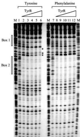
bation for 45 s at room temperature, the reaction was termi- nated by phenol extraction. The resulting DNA fragments were analyzed on a 6% sequencing gel (Fig. 2). In the presence of either tyrosine or phenylalanine, full protection correspond- ing to TyrR box 2 (the strong box) was seen at all TyrR concentrations used (from 12.5 to 250 nM). However, protec- tion of box 1 (the weak box) occurred in the presence of tyrosine only at concentrations of TyrR protein of 50 nM or above, and in the presence of phenylalanine, box 1 was only weakly protected at concentrations of 100 nM or above. In the presence of tyrosine and high concentrations of TyrR, there was also evidence of some protection of the region between boxes 1 and 2. The third putative box centered at⫺186.5 failed to show any protection, and subsequent mutation of this box failed to affect expression (data not shown). Consequently, this third box has been excluded from the discussion.
To investigate the role of each of the two TyrR boxes in regulation, mutations affecting one of the invariant GC pairs were introduced separately into each one (Fig. 1). As can be seen in Table 3, inactivation of either box destroys TyrR- mediated activation offolA.
Discussion.The gene folAencodes the enzyme dihydrofo- late reductase, which carries out the reversible reaction dihy- drofolate plus NADPH⫽tetrahydrofolate plus NADP⫹. Tet- rahydrofolate plays a central role in one-carbon metabolism in the synthesis of glycine, methionine, and purines. The sub- strate of this reaction, dihydrofolate, is a product of a pathway leading from chorismic acid viapara-aminobenzoic acid. Little is known about the regulation of this pathway or the way in which the various pathways to aromatic vitamins compete with the pathways for phenylalanine, tyrosine, and tryptophan bio- synthesis for the important initial substrate chorismic acid. It can be argued, however, that of the three aromatic amino acids, tyrosine has the most significant effect on chorismic acid biosynthesis. The TyrR-mediated repression ofaroF(a gene which encodes a major isoenzyme for the first reaction of the
pathway) andaroL(a gene encoding an enzyme for the fifth reaction of the pathway), coupled with the feedback inhibition by tyrosine of thearoF-encoded DAHP synthase, significantly affects the level of synthesis of chorismic acid. Although ty- rosine also reduces the conversion of chorismic acid to tyrosine by causing TyrR to repress bothtyrAandtyrBand by inhibiting prephenate dehydrogenase, it is possible that the net effect of these interactions is a reduced availability of chorismate for folic acid biosynthesis. If that is so, it may be that TyrR- tyrosine activation offolAexpression in some way balances this effect on chorismate supply. Alternately, it is possible that under conditions in which the cell normally encounters high levels of tyrosine, there is an increased demand for tetrahy- drofolate and one-carbon transfers. Further work will be re- quired to explain the physiological significance of this complex system of TyrR-tyrosine mediated activation of folAexpres- sion. It is worth noting that no other gene of this chorismate- to-folate pathway has been selected by TyrR protein in genomic SELEX or has been revealed in bioinformatic searches (4, 8).
Although there are unresolved questions relating to the physiology, the genetics of the system is fairly clear. TyrR- mediated activation does not occur if thetyrRgene has muta- tions affecting the N-terminal domain’s interaction with the aromatic amino acids or with RNA polymerase. Similarly, a mutation inrpoAwhich affects activation oftyrP,aroP, andmtr also affects folA activation. Unlike the situation with these three genes but similar to what is probably the case for thetpl gene ofCitrobacter freundii(1), activation offolAalso requires a fully functional central domain. Activation offolAalso re- quires IHF, which is not required for the simpler activation of genes such asmtr (data not shown). A putative IHF binding site was found between the TyrR boxes and thefolApromoter (Fig. 1). Furthermore, mutational studies have established that both of the identified TyrR boxes play an important role in activation. As in the case oftpl, the box with the least agree- FIG. 1. Nucleotide sequence of thefolAregulatory region. The numbering of the sequence is relative to the start site of transcription. The⫺35 and⫺10 regions of thefolApromoter (10), the two TyrR boxes, and the putative IHF binding site are indicated. In the TyrR boxes 1 and 2, the positions where mutations were introduced are marked with asterisks above the bases. To inactivate box 1, the three nucleotides CCA were changed to GAT, and to inactivate box 2, the three nucleotides ACA were also changed to GAT.
by Akira Ishihama on August 29, 2007 jb.asm.orgDownloaded from
ment to the consensus is the one closest to the promoter. This box, box 1, has only 8 of the 12 bases in the arms of the palindrome which agree with the consensus and has a central region comprised exclusively of GC pairs. It has all the char- acteristics of a low-affinity binding site, which would require an adjacent stronger binding site for effective binding to occur.
Box 2, with 11 of its 12 bases in the palindromic arms agreeing
with the consensus and with three AT pairs in the central region, appears to play this role. We would hypothesize that the TyrR hexamer binds across both boxes and, with the assis- tance of IHF, produces a complex in which the N-terminal domain of one or more of the TyrR molecules is able to interact with the␣subunit of RNA polymerase and activate transcription (Fig. 3). The DNase I protection experiment showed that, in addition to the protection of boxes 1 and 2, the intervening region between the two boxes was also partially protected by TyrR-tyrosine (Fig. 2). This may reflect the for- mation of a complex structure in this region. In the presence of phenylalanine, the weaker box 1 is protected only at higher TyrR concentrations (100 nM or above), which would explain why phenylalanine-mediated activation as shown in Table 2 is observed only in the presence of enhanced TyrR protein levels (i.e., with multicopytyrR⫹).
This complex system is designed to respond primarily to increased levels of tyrosine by activating expression offolA, in contrast to the well-studied situation intyrP, where tyrosine- induced hexamerization causes repression. It is a good exam- ple of the selective application of the same mechanism to produce opposite outcomes.
The usefulness of the genomic SELEX approach is sup- ported by the identification of seven of the eight known mem- bers of the TyrR regulon. The one which was not detected, aroG, is the only member of the regulon with a single TyrR box, and in this case it is possible that the GC-rich central region of this box generally associated with those called weak FIG. 2. DNase I footprinting of the folA promoter region. The
DNase I protection assay was carried out in the presence of either tyrosine (lanes 1 to 6) or phenylalanine (lanes 7 to 12). The protected regions corresponding to TyrR boxes 1 and 2 are indicated with open bars. The base positions in the intervening region between the two TyrR boxes, which were protected by TyrR only in the presence of tyrosine, are marked with asterisks. The final concentrations of TyrR protein used in each reaction are as follows: lanes 1 and 7, no TyrR;
lanes 2 and 8, 12.5 nM; lanes 3 and 9, 25 nM; lanes 4 and 10, 50 nM;
lanes 5 and 11, 100 nM, and lanes 6 and 12, 250 nM. Lanes M, GA DNA sequence ladders. The protein used was native TyrR protein purified as described by Wilson et al. (11). The method for DNase I footprinting has been described previously (12).
TABLE 3. Effect of TyrR box mutations onfolAactivation
folA-lacZfusion
-Galactosidase sp act in the indicated mediuma
MM Tyr Phe
Wild type 101 458 267
TyrR box 1 mutation 102 80 83
TyrR box 2 mutation 80 107 119
aSee Table 2, footnoteb.
FIG. 3. A hypothetical model for tyrosine-dependent activation of the folApromoter by the TyrR protein. In vivo, in the absence of tyrosine, TyrR exists as a dimer, which binds strongly to TyrR box 2 and weakly to TyrR box 1 (the TyrR dimer is shown by dotted lines).
The presence of tyrosine induces TyrR to form a hexamer, which strengthens the binding of TyrR to box 1. The TyrR molecule bound to box 1 interacts directly with the␣subunit of RNA polymerase, leading to activation of transcriptional initiation of thefolA promoter. The binding of IHF to thefolApromoter is presumably required for cre- ating an ideal DNA conformation, which facilitates TyrR-RNA poly- merase interaction.
VOL. 189, 2007 NOTES 6083
by Akira Ishihama on August 29, 2007 jb.asm.orgDownloaded from
TyrR boxes (6) may cause it to be a less effective binding site.
Baseggio et al. (2) have shown that the affinity of TyrR protein for this single aroG box is about 1/10 of its affinity in the presence of tyrosine for the double box found in genes such as tyrP. The expression ofaroGdoes not appear to be affected by the presence or absence of the aromatic amino acids, and its place in the TyrR regulon is dependent on observations of enhanced expression intyrRmutants and decreased expression in strains with an elevated level of TyrR protein. The single TyrR box ofaroGoverlaps the⫺35 region of the promoter, and regulation appears to involve competition between RNA polymerase and TyrR protein in binding to the same region of DNA. Under these circumstances, it is not surprising to find that a binding site with a relatively low affinity for TyrR has been selected foraroGduring evolution.
The finding that four of the five new genes with TyrR bind- ing sites adjacent to their promoters showed no signs of TyrR- mediated regulation in vivo cautions against a too-ready ac- ceptance of the conclusion that the existence of binding sites for a regulator protein near a promoter automatically implies effective regulation of that promoter by that protein. On the other hand, if such binding occurs in vivo, it will influence the overall concentration of nonbound TyrR protein available to interact with other binding sites associated with known mem- bers of the TyrR regulon. The finding of only one new member of the TyrR regulon, as a result of this latest search, may indicate that the TyrR system of regulation is fairly pathway specific and is unlikely to be involved in more global interac- tions within the cell.
This work was supported by a grant from the Australian Research Council.
We thank J. Praszkier for helpful advice.
REFERENCES
1.Bai, Q., and R. L. Somerville.1998. Integration host factor and cyclic AMP receptor protein are required for TyrR-mediated activation of tpl in Citrobacter freundii. J. Bacteriol.180:6173–6186.
2.Baseggio, N., W. D. Davies, and B. E. Davidson.1990. Identification of the promoter, operator, and 5⬘and 3⬘ends of the mRNA of theEscherichia coli K-12 genearoG. J. Bacteriol.172:2547–2557.
3.Kwok, T., J. Yang, A. J. Pittard, T. J. Wilson, and B. E. Davidson.1995.
Analysis of anEscherichia colimutant TyrR protein with impaired capacity for tyrosine-mediated repression, but still able to activate at sigma 70 pro- moters. Mol. Microbiol.17:471–481.
4.Li, H., V. Rhodius, C. Gross, and E. D. Siggia.2002. Identification of the binding sites of regulatory proteins in bacterial genomes. Proc. Natl. Acad.
Sci. USA99:11772–11777.
5.Miller, J. H.1974. Experiments in molecular genetics. Cold Spring Harbor Laboratory, Cold Spring Harbor, NY.
6.Pittard, J., H. Camakaris, and J. Yang.2005. The TyrR regulon. Mol.
Microbiol.55:16–26.
7.Praszkier, J., I. Wilson, and A. J. Pittard.1992. Mutations affecting trans- lational coupling between therepgenes of an IncB miniplasmid. J. Bacteriol.
174:2376–2383.
8.Robison, K., A. M. McGuire, and G. M. Church.1998. A comprehensive library of DNA-binding site matrices for 55 proteins applied to the complete Escherichia coliK-12 genome. J. Mol. Biol.284:241–254.
9.Shimada, T., Fujita, N., Maeda, M., and A. Ishihama.2005. Systematic search for the Cra-binding promoters using genomic SELEX system. Genes Cells10:907–918.
10.Smith, D. R., J. I. Rood, P. I. Bird, M. K. Sneddon, J. M. Calvo, and J. F.
Morrison.1982. Amplification and modification of dihydrofolate reductase inEscherichia coli. Nucleotide sequence of folgenes from mutationally altered plasmids. J. Biol. Chem.257:9043–9048.
11.Wilson, T. J., P. Maroudas, G. J. Howlett, and B. E. Davidson.1994. Ligand- induced self-association of theEscherichia coliregulatory protein TyrR. J.
Mol. Biol.238:309–318.
12.Yang, J., J. S. Hwang, H. Camakaris, W. Irawaty, A. Ishihama, and J.
Pittard.2004. Mode of action of the TyrR protein: repression and activation of thetyrPpromoter ofEscherichia coli. Mol. Microbiol.52:243–256.
by Akira Ishihama on August 29, 2007 jb.asm.orgDownloaded from